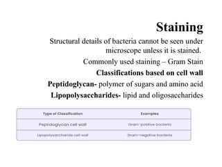
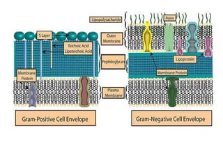
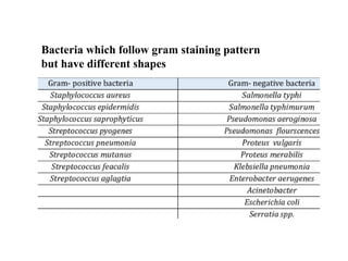
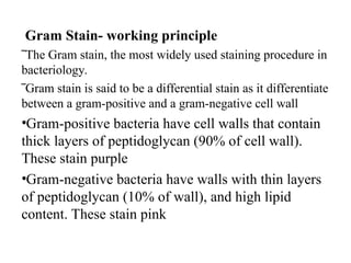
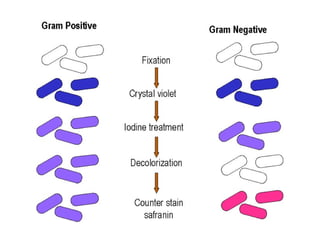
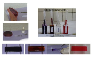
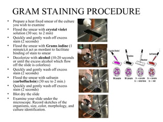
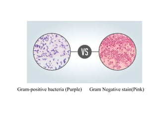
Course Diagnostic Bacteriology
Introduction
Bacteria, the very first organisms to evolve on
earth as a unicellular organism.
They reside in human body as normal flora
Hence it requires special training to diagnose
infectious organism appropriately.
Protocols
• For accurate diagnosis in bacteriology it is
pivotal to collect, transport and diagnose with
protocols
 Collect complete information on request form
- Patient details
- Onset of symptoms / clinical conditions
- Time of collection
- History of medications
- Travel/ contact history
- Sample type
 Collect and Transport sample carefully
- Disinfect the site
- Sterile Container/ swab/ needle
- Add preservative (if required)
- Maintain temperature at 4C(to preserve sample and reduce
multiplication)
- Mention “High Risk” if sample is infectious
- Seal appropriately ( if fluid)
- Tranport ASAP (In case of fastidious microorganism N.
gonorrhoea and H. Influenza die during transit)
Sample Examination (Gross)
 It begins with macroscopic examination
(gross examination) of sample which include
the volume and morphology
- Blood/mucus
- Fluid (clear/ cloudy)
- Pus/ Abcess
- Swab /Aspirate
- urin /stool
Sample Examination
(Microscopic)
• Specimen are received in many forms.
Preparation of direct smears is usually
recommended for fluid/swab/stool
specimen
‾ Urine /mucus/ Pus/ stool samples are analysed for WBCs
and epithelial cells
‾ Nasophyrangeal swabs and cervical swabs are not
examined microscopically because these sites have
bacteria already
Staining
Structural details of bacteria cannot be seen under
microscope unless it is stained.
Commonly used staining – Gram Stain
Classifications based on cell wall
Peptidoglycan- polymer of sugars and amino acid
Lipopolysaccharides- lipid and oligosaccharides
Bacteria which follow gram staining pattern
but have different shapes
• Bacterial Shapes
Bacilli
Cocci
Spiral
Gram Stain- working principle
‾The Gram stain, the most widely used staining procedure in
bacteriology.
‾Gram stain is said to be a differential stain as it differentiate
between a gram-positive and a gram-negative cell wall
•Gram-positive bacteria have cell walls that contain
thick layers of peptidoglycan (90% of cell wall).
These stain purple
•Gram-negative bacteria have walls with thin layers
of peptidoglycan (10% of wall), and high lipid
content. These stain pink
Stain components
GRAM STAINING PROCEDURE
• Prepare a heat fixed smear of the culture
you wish to examine
• Flood the smear with crystal violet
solution (30 sec. to 2 min)
• Quickly and gently wash off excess
stain (2 seconds)
• Flood the smear with Grams iodine (1
minute),it act as mordant to facilitate
binding of stain to smear
• Decolorize with alcohol (10-20 seconds
or until the excess alcohol which flow
off the slide is colorless)
• Quickly and gently wash off excess
stain (2 seconds)
• Flood the smear with safranin
(carbolfuchsin) (30 sec to 2 min.)
• Quickly and gently wash off excess
stain (2 seconds)
• Blot dry the slide
• Examine your slide under the
microscope. Record sketches of the
organisms, size, color, morphology, and
culture identification.
Gram-positive bacteria (Purple) Gram Negative stain(Pink)

Diagnostic bacteriology and classes and bacterial sample collection.ppt

  • 1.
  • 2.
    Introduction Bacteria, the veryfirst organisms to evolve on earth as a unicellular organism. They reside in human body as normal flora Hence it requires special training to diagnose infectious organism appropriately.
  • 3.
    Protocols • For accuratediagnosis in bacteriology it is pivotal to collect, transport and diagnose with protocols  Collect complete information on request form - Patient details - Onset of symptoms / clinical conditions - Time of collection - History of medications - Travel/ contact history - Sample type
  • 4.
     Collect andTransport sample carefully - Disinfect the site - Sterile Container/ swab/ needle - Add preservative (if required) - Maintain temperature at 4C(to preserve sample and reduce multiplication) - Mention “High Risk” if sample is infectious - Seal appropriately ( if fluid) - Tranport ASAP (In case of fastidious microorganism N. gonorrhoea and H. Influenza die during transit)
  • 5.
    Sample Examination (Gross) It begins with macroscopic examination (gross examination) of sample which include the volume and morphology - Blood/mucus - Fluid (clear/ cloudy) - Pus/ Abcess - Swab /Aspirate - urin /stool
  • 6.
    Sample Examination (Microscopic) • Specimenare received in many forms. Preparation of direct smears is usually recommended for fluid/swab/stool specimen ‾ Urine /mucus/ Pus/ stool samples are analysed for WBCs and epithelial cells ‾ Nasophyrangeal swabs and cervical swabs are not examined microscopically because these sites have bacteria already
  • 7.
    Staining Structural details ofbacteria cannot be seen under microscope unless it is stained. Commonly used staining – Gram Stain Classifications based on cell wall Peptidoglycan- polymer of sugars and amino acid Lipopolysaccharides- lipid and oligosaccharides
  • 9.
    Bacteria which followgram staining pattern but have different shapes
  • 10.
  • 11.
    Gram Stain- workingprinciple ‾The Gram stain, the most widely used staining procedure in bacteriology. ‾Gram stain is said to be a differential stain as it differentiate between a gram-positive and a gram-negative cell wall •Gram-positive bacteria have cell walls that contain thick layers of peptidoglycan (90% of cell wall). These stain purple •Gram-negative bacteria have walls with thin layers of peptidoglycan (10% of wall), and high lipid content. These stain pink
  • 13.
  • 14.
    GRAM STAINING PROCEDURE •Prepare a heat fixed smear of the culture you wish to examine • Flood the smear with crystal violet solution (30 sec. to 2 min) • Quickly and gently wash off excess stain (2 seconds) • Flood the smear with Grams iodine (1 minute),it act as mordant to facilitate binding of stain to smear • Decolorize with alcohol (10-20 seconds or until the excess alcohol which flow off the slide is colorless) • Quickly and gently wash off excess stain (2 seconds) • Flood the smear with safranin (carbolfuchsin) (30 sec to 2 min.) • Quickly and gently wash off excess stain (2 seconds) • Blot dry the slide • Examine your slide under the microscope. Record sketches of the organisms, size, color, morphology, and culture identification.
  • 15.
    Gram-positive bacteria (Purple)Gram Negative stain(Pink)