

Recommended
More Related Content
What's hot
What's hot (20)
Viewers also liked
Viewers also liked (20)
Similar to Diabetic retinopathy
Similar to Diabetic retinopathy (20)
Recently uploaded
Recently uploaded (20)
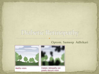
Diabetic retinopathy
- 2. Series of retinal changes that occur in patients with DM Serious sight-threatening complication of DM Most common cause for legal blindness Common in type I DM than type II 11/1/2015DR 2
- 3. Duration of DM: Mostly >10 yrs Poor metabolic control Pregnancy Hypertension Nephropathy Heredity Smoking Obesity 11/1/2015DR 3
- 4. Microangiopathy Microvascular occlusion Retinal ischaemia Arteriovenous shunt formation Neovascularisation Capillary leakage and haemorrhage Microaneurysm Retinal oedema Hard exudates 11/1/2015DR 4
- 5. Early stage: Asymptomatic Advance stage: Seeing spots or floaters in visual field Blurred vision Distorted vision Difficult in seeing well at night 11/1/2015DR 5
- 6. Background Diabetic Retinopathy: Microaneurysms: Tiny, round, red dots located in INL FFA shows tiny hyperfluorescent dots representing non- thrombosed microaneurysms Hard exudates: Waxy-yellowish lesions with distinct margins at OPL FFA shows hyperfluorescence due to blockage of background choroidal fluorescence 11/1/2015DR 6
- 7. Retinal oedema: Initially, located between OPL and INL Best detected by fundus examination with Goldman lens FFA shows diffuse late hyperfluorescence due to retinal capillary leakage Haemorrhage: Intra retinal haemorrhage from venous end of capillaries Dot and blot haemorrhage Retinal nerve fibre layer haemorrhage due to larger pre- capillary arterioles Flame shaped haemorrhage 11/1/2015DR 7
- 8. Management: No treatment required Review annually Associated factors should be controlled 11/1/2015DR 8
- 9. Is BDR that shows the signs of proliferative DR Signs: Cotton wool spots: Local infarcts of RNFL due to blockage of pre-capillary arterioles Small whitish superficial lesions FFA shows hyperfluorescence 11/1/2015DR 9
- 10. Intraretinal microvascular abnormalities(IRMA): Shunts that runs from arterioles to venules by-passing capillary bed FFA shows focal hyperfluorescence at areas of capillary closure Venous changes: Dilatation of veins Venous looping Arterial changes: Narrowing Silver-wiring 11/1/2015DR 10
- 11. Dark blot haemorrhage: Haemorrhagic retinal infarcts located within middle layers of retina Management: Reviewed in regular basis Photocoagulation can be done if follow up is not possible 11/1/2015DR 11
- 12. Involvement of fovea by oedema, hard exudates or ischaemia Classification: Focal exudative Well-circumscribed retinal thickening along with hard exudates FFA shows focal hyperfluorescence due to leakage and macular perfusion Diffuse exudative Diffuse retinal thickening associated with cystoid changes FFA shows widespread spotty hyperfluorescence of microaneurysms and late diffuse hyperfluorescence which shows flower petal pattern , if CMO is present 11/1/2015DR 12
- 13. Ischaemic: Associated with pre-proliferative DR FFA shows capillary non-perfusion at fovea Mixed: Characterized by both ischaemia and exudation 11/1/2015DR 13
- 14. Clinically significant macular oedema(CSME): Diabetic maculopathy can lead to CSME if present with, Retinal oedema within 500µm centre of fovea Hard exudates within 500µm centre of fovea and associated with adjacent retinal thinking Retinal oedema, one disc diameter or larger in size, at least a part of which is within one disc diameter of foveal centre Management: Argon laser photocoagulation Vitrectomy 11/1/2015DR 14
- 15. DR in long run can lead to Proliferative DR Signs: Neovascularisation is hallmark of proliferative DR Neovascularisation may be; NVD- New vessels at disc NVE- New vessels elsewhere FFA detects neovascularisation in early stage and shows late hyperfluorescence due to leakage from new vessels Management: Panretinal photocoagulation 11/1/2015DR 15
- 16. 11/1/2015DR 16
- 17. Serious sight threatening complication of DR Some complications are: Pre-retinal haemorrhage Tractional retinal detachment Rubeosis iridis Management: Pars-plana vitrectomy 11/1/2015DR 17
- 18. What is his major complaints Whether he has blurred vision or not Whether he has distorted vision or not Whether he is seeing floaters or not Whether he can appreciate black spots while seeing Whether he is suffering from DM or not If yes, since when Whether he is suffering from hypertension or any other systemic disease Whether his family members had DR or any similar symptoms His blood sugar report!! 11/1/2015DR 18
- 19. 11/1/2015DR 19