The objectives of this study were to detect and characterize the phytoplasma in tissues of diseased hibiscus plants using Dains’ stain light microscopy and molecular based techniques. Molecular characterization was performed using the DNA sequencing and phylogenetic analysis of the spacer region between 16S and 23S rRNA fragment of the isolated phytoplasma genome. This work concerning phytoplasma associated witches' broom (group 16SrII) diseases of hibiscus plants is achieved for the first time in Egypt.
13 isolation and identification of endophytic fungi from 13 ijtas 93-2018-hu...BIOLOGICAL FORUM
ABSTRACT: The choice of host plant is of critical importance when working with endophytic fungi. The exploration of endophytic fungi is still an emerging field and all plants seem to harbour fungi with some bioactive content and activities. However, there are certain metabolites that are characteristic of certain biotopes. Thus, a rationale for selecting promising plant sources should be established. Of particular interest are the plants that are used as medicinal plants or plants that populate a unique environment. Artemisia is a widely used medicinal plant. In this research work, the endophytic mycota of Artemisia scoparia was studied. In order to isolate endophytic fungi, 155 plant segments from 20 samples of Artemisia scoparia were collected from its natural habitat in Dachigam National. This habitat is a unique environment and a protected area. Six different fungal isolates were obtained from root, leaf and stem plant parts. Among the identified isolates, the most abundant genera were Gliocladium solani followed by Penicillium melinii with a colonization frequency of 62 and 37.5% respectively. The objective of this study was to report new data regarding the endophytic fungi found in medicinal plant Artemisia scoparia. This systematic investigation revealed that traditional medicinal plants are a rich and reliable source of novel endophytic fungi.
Keywords: Endophytic fungi, Kashmir, Medicinal plant
the presentation is about microbial endophytes, discovery of endophytes, their types, isolation methods of different types and identification and the useful impacts of them to the plant ecology.
Exploitation of endophytic fungi for plant disease management
Introduction
Plant- Endophytic fungi interaction
Diversity of endophytic fungi in plants
Colonization
Endophytic fungi : Mechanism
Case studies
Conclusion
Future aspects
Endophytic fungi in disease resistance (Latz et al., 2018)
Antibiotics produced by fungal endophytes
Plant immune defense system
Lytic enzyme secretion
Endophytic fungi in stress tolerance
PREFERENTIAL ASSOCIATION OF ENDOPHYTIC BRADYRHIZOBIA WITH DIFFERENT RICE CULT...Anamika Rana
All of rice oligotrophic endophytic Bradyrhizobia in this study were obtained except SUT-R74.
6 Bradyrhizobial strains were obtained from 98 bacterial strains.
Bradyrhizobium is found only in rice root, with 10% relative abundance of total Alphaproteobacteria.
Endophytic Bradyrhizobia could not be obtained from the monoculture system.
Thai rice cultivars, the Thai Bradyrhizobial strains could promote rice growth better than Japanese strains.
Three rice cultivars (Pathum Thani 1, Kasalath, and Nipponbare), cultivar Pathum Thani 1 responded only to putative Thai rice endophytic Bradyrhizobia.
This phenomenon was not found in Japanese rice cultivars.
Non-PB strains are also capable of forming a natural endophytic association with rice.
Strains SUT-PR9, WD16, RP5, and RP7 displayed non-PB phenotypes but were genotypically close to PB strains.
This presentation is to understand the concepts of endophytes that reside within plants & to explore the applications of endophytes for the management of plant diseases.
Introduction to endophytes and their application to develop commercial productsPrograma TF Innova
Ponencia: Introduction to endophytes and their application to develop commercial products
Autor: Dr. Gary Strobel
Evento TF Innova: Workshop Biotechnology "Isolation and identification of endophytic fungi from vascular plants"
Isolation of endophytes from potato and their antagonist effect against Fusar...Innspub Net
Plant endophytes may be intercellular or intracellular depending upon their location in the plant tissue because they are present inside the cells or in the intracellular space, respectively. Isolation of endophytic bacteria has been reported from both monocot and dicot plants, ranging from woody trees, such as teak and pear, to herbaceous crop plants such as mustard and maize. The aim of this study was the isolation of endophytes from potato and their antagonist effect against Fusarium oxysporum. Endophytic fungi were isolated from leaves, stems and roots of healthy Potato plant derived from Chak No.359/E.B Village, Tehsil Burewala. Isolation of endophytic fungi from plant parts was done according to the method described by Petrini. The media used in the present study was the Potatodextrose agar (PDA) for fungus and nutrient agar medium for maintaining bacterial stains. F.oxysporum was taken from the Plant pathology lab of UAF sub-campus Burewala-Vehari . The results of the experiment clearly revealed that the stems, root and leaf of the potato plants under present investigation had the maximum colonization frequency for fungal endophytes. Fusarium oxysporum showed rapid growth 5-7cm in5 days. Fusarium oxysporum was white and growing rapidly that later produced dark violet pigments in PDA. Erwinia showed light green, circular, shining, slimy, smooth characteristics. The isolate strain of Bacillus showed rodshaped, fuzzy white or slightly yellow circular and irregular characteristics.
Titulo Ponencia: Endophytes Identification: morphological methods
Autor: Dr. Gary Strobel
Evento TF Innova:
Workshop Biotechnology "Isolation and identification of endophytic fungi from vascular plants"
Plant growth-promoting mechanisms of endophytesThe Tiny Domain
The global changes in climate and increasing population have unfortunate effects in food production and will become insufficient to feed the world. The green revolution could alleviate poor crop production by using high yielding varieties and use of chemical fertilizers and agrochemicals. But excessive use of chemical fertilizers and agrochemicals has resulted in the deterioration of soil fertility. Hence, agronomic practices are moving toward sustainable and environment friendly approach.
4068 isolation, identification and characterization of entomopathogenicSheena Prem
Control of white grub using entomopathogenic nematode (Heterorhabdtidae and steinernematidae )and entomopathogenic fungi Isolation of Symbiontic bacteria of antomopathogenic nematode .
In vitro evaluation of Trichoderma viride and Trichoderma harzianum for its e...iosrjce
IOSR Journal of Pharmacy and Biological Sciences(IOSR-JPBS) is a double blind peer reviewed International Journal that provides rapid publication (within a month) of articles in all areas of Pharmacy and Biological Science. The journal welcomes publications of high quality papers on theoretical developments and practical applications in Pharmacy and Biological Science. Original research papers, state-of-the-art reviews, and high quality technical notes are invited for publications.
Identification of Race/strain of Phytopathogenic Fungi through Conventional A...Sarda Konjengbam
Detection and identification of fungi has relied on a combination of microscopy and culture based techniques. Conventional methods often rely on identification of disease symptoms, isolation and culturing of environmental organisms, and laboratory identification by morphology and biochemical tests. These methods, although the cornerstone of fungal diagnostics, can lead to problems in identification, resulting in incorrect interpretation, diagnosis and ultimately treatment. The methods rely on experienced, skilled laboratory staff, the ability of the organism to be cultured, are time consuming, non quantitative, prone to contamination and error and often delay management (Atkins and Clark, 2004). During the last decades, the advent of molecular biology promised to offer radical alternatives in the detection and enumeration of fungal pathogens. Molecular technology increases understanding of the biology and population structures of plant pathogens, provides quick and accurate answers to epidemiological questions about plant diseases, and supports disease management decisions. New, rapid screening methods are being developed and increasingly used in all aspects of fungal diagnostics.
The objectives of this study to investigate the occurrence and etiology of tomato malformation recently observed in Egypt and illustrate the responsibility of associated phytoplasma. Also, the study was extended to characterize associated phytoplasma, based on molecular techniques.
This work aimed to (i) Identify and characterize Onion yellow dwarf virus potyvirus (OYDV) in the onion plants in Egypt. (ii) Clone and sequence the coat protein gene of the Egyptian isolate of OYDV and comparing it with other OYDV isolates reported in the GenBank database. (iii) Study the influence of therapeutic doses of kinetin (6-Furfurylaminopurine) on production of virus-free onion plantlets and improve its regeneration ability through in vitro micropropagation.
The present study has been conducted to perform the following objectives:
1- Study the effect of different temperature degrees and meristem culture technique on elimination of Potato leafroll virus (PLRV) and Potato virus x (PVX), from the most commonly potato cultivars in Egypt (Spunta and Lady Rosette).
2- Production of potato minitubers from direct transplanting of in vitro virus-free plantlets in greenhouse. Also, investigate the effect of different soil mixtures on minitubers production for both cultivars.
Assessment of Endophytic Fungal Flora Responsible for Plant Growth Promotion...Sryahwa Publications
The present paper discusses the highest colonization of fungal endophytes as Alternaria speciesin comparison with Colletotrichumspecies and Fusarium species in all three plants Pongamia pinnata, Securinega leucopyrus and Rhus mysorensis. These endophytic fungi protect these plants from various
environmental factors such as temperature, moisture and other environmental factors.
This study was carried out on the mycoflora associated with seeds of different citrus species. Citrus seed material was collected from districts of Punjab, i.e. Multan, Sargodha and Khanpur. Standard methods were applied for the isolation and identification of fungi. A total of 11 fungi including Aspergillus fumigatus, Aspergillus flavus, Dreschslera tetramera, Alternaria alternata, Curvularia lunata, Macrophomina phaseolina, Aspergillus niger, Fusarium solani, Fusarium moniliforme, Rhizopus and Penicillium spp were isolated from the seeds of citrus. For control of isolated seed-born fungi, 3 recommended fungicides such as Ridomil Gold, Bavistin, Score and two chemical Salicylic acid and Boric acid, were used at 20, 30, 40 mg/10 mL and 5, 6, 7 μL/10 mL, respectively and chemical with 20, 30, 40 mg/10 mL. All these fungicide and chemicals significantly reuced with population of all fungi present in naturally infected seed samples. Ridomil Gold and Salicylic acid were found to be the best for the control of se d-born fungi of citrus seed at 40 mg/10 mL. The isolation and identification of different mycotoxins is essential to study health status of the citrus consumers and to safeguard the standards of WTO.
Detection of fungi associated with water hyacinth Eichhornia crassipes in Ira...Innspub Net
The study was carried out in the laboratories at the Faculty of Agriculture, Baghdad University to isolate and identify the species of fungi that associated with water Hyacinth Eichhornia crassipes. The samples were collected from Tigris river side’s at Al- Kraat area in north of Baghdad, Iraq. The presence ratios of fungi were recorded and their pathogenicity was tested. Results of isolation and identification showed the presence of fourteen fungi associated with water Hyacinth leaves including; Alternaria sp., Aspergillus flavus, Aspergillus niger, Drechslera sp., Chaetomium sp., Cladosporium sp., Fusarium solani, Macrophomina phaseolin, Mucor sp., Mycelia Sterile Fungi, Pythium aphanidermatum, Ulocladium sp., Rhizopus sp. and Trichoderma sp. at different percentages . However, the percentages of presence were deferent and the most frequently were A. alternata and Rhizopus sp. reached 76.33% and 80.40 % respectively. The percentages of the other fungi were ranged between 4.25% -30.60 %. It has been found that nine of these fungi, the more prevalent, showed high capacity of inducing infection on Water hyacinth leaves at percentages ranged between44.4% – 100% compared with zero infection in control. Macrophomina phaseolina, Pythium aphanidermatum and Rhizopus sp. were found to be the more pathogenic with disease severity attained to 100 %. Different symptoms were developed on the leaves inoculated with different fungi as spotting and wilting followed by leaves dryness. This is the first report for fungi associated with water hyacinth leaves at Al-Kraat area in Iraq.
The initial objective of this work was to isolate Tomato spotted wilt virus (TSWV) from asymptomatic infected plants and then identify the virus on the basis of biological properties among the most common or other hosts if possible after inoculation. Phylogenetic analysis was then conducted to gain information about the similar identity of the TSWV-isolate that reported in this study with available TSWV sequences from other parts of the world.
The objective of this study was to examine the antiviral activity of native lactoferrin against Potato virus x, the most important virus that severely affects potato crop and productivity in Egypt, using tissue culture technique and spraying the plants in greenhouse by the aqueous solution of lactoferrin.
Research topic was come from successful inactivation of some plant viruses by gamma irradiation like Citrus tristeza virus, Necrotic ring spot virus and Prune dwarf virus. Gamma irradiation has been also used to sterilize agricultural products in order to increase their conservation time or to reduce pathogen when being traded from a country to another. Gamma radiation is high-energy radiation emitted from certain radioactive isotopes as cobalt 60, these isotopes are potential sources of gamma radiation. Therefore, this research was conducted to find out the inactivation possibility of Hibiscus witches' broom (HibWB)-phytoplasma using gamma irradiation through tissue culture technique with clarify their effect on in vitro growth and survival rate.
PREVALENCE AND CHARACTERIZATION OF VIRULENCE PROPERTIES OF PSEUDOMONAS AERUGI...SUS GROUP OF INSTITUTIONS
Pseudomonas aeruginosa is the epitome of an opportunistic pathogen of humans that cause urinary tract infections, respiratory system infection, particularly in victim of severe burns, cancer and AIDS patient who are immunocompromised. Most Pseudomonas infections are both invasive and toxigenic. The particular bacterial determinants of virulence mediate different stages of infection and are ultimately responsible for the characteristic syndromes that accompany the disease. In the present study P. aeruginosa was found to be more prevalent in burn patients (100%) followed by urinary tract infection samples (71%), sputum samples (66%) and wound samples (59%). 85% isolates recovered from clinical samples were mucoid. A total of 35% isolates were strong siderophore producers, 19% isolates were strong protease producers while 52% were strong phospholipase producers. Isolates from burns, sputum and environment sample were strong rhamnolipid producers. Elevated level of hemolysin production was observed in burn, urine and wound isolates. The prominence of haemagglutination ability in environmental isolates followed by burns isolates provided evidence for its being a nosocomial pathogen. The association between virulence determinants and disease can indicate the precise role played by the determinant in estabilishing the disease. Isolates were maximally sensitive towards lactam antibiotics.
Management of potato virus Y (PVY) in potato by some biocontrol agents under ...Open Access Research Paper
The study was conducted to test the activity of Pseudomonas fluorescens, Rhodotorula sp and fermented neem extract to protect potato plants against potato virusY disease development under field conditions. Infected potato tubers were soaked in P. fluorescens, Rhodotorula sp suspensions and in fermented neem extracts separately and sown in the field in completely randomized block design. The development of virus symptoms and the accumulation of virus in the plant based on Enzyme Linked Immunosorbent Assay (ELISA) were followed. The results obtained showed that the treatment of potato tubers with the three agents have significantly accelerated plant emergence, 5-6 days early than non treated ones, and improved plant growth, the plant dry weights ranged from 120-177 g/plant compared to 42 g/plant in non treated plants. The enhancement of plant growth was found associated with reduction in disease severity based on symptoms development and restriction of virus concentration as proved by ELISA absorbance of 405 nm, 0.14-0.23 compared with 2.50 in non treated plants. The results indicated that the use of bioagent to induce systemic resistance provide an efficient tool, as insecticide alternative to manage potato virus Y in potato. Check out more by following link https://innspub.net/management-of-potato-virus-y-pvy-in-potato-by-some-biocontrol-agents-under-field-conditions/
Similar to Detection and molecular characterization of phytoplasma associated with Hibiscus Witches’-Broom in Egypt (20)
The First Workshop of Plant Pathology Research Institute
The Modern Trends in Controlling Plant Diseases
10-11 February 2019
Under auspices of
Prof. Dr. Eszzaldin Omar Abusteit
Minster of Agricultural and Land Reclamation
Prof. Dr. Mohamed Soliman
Director of Agricultural Research Center
&
President of the workshop
Prof. Dr. Ashraf El Saied Khalil
Director of Plant Pathology Research Institute
In Egypt isolates of Prunus necrotic ringspot virus (PNRSV) were detected in peach and apricot grooves (Abdel-Salam et al. 2008a), Rosa spp. (Abdel-Salam et al., 2008b) as well as on sugarbeet plantations (Abdel-Salam et al., 2006a).
In the present study, incidence of PNRSV is reported on apple (Malus domestica). Severe symptoms mimic infections with PNRSV were recently detected on apple from several orchards in the vicinity of Nubaria city, Beheira governorate. Infected-apple samples were brought to the laboratory for further detection at serological and molecular levels to check the presence of virus. The present study reports the presence of an isolate of PNRSV on apple, viz. PNRSV-Apple.
The present study aims to (I) evaluate the antiviral activity of eugenol oil nanoemulsion (EON) on eliminate Banana bunchy top virus (BBTV) from naturally infected banana plants and produce virus-free banana plants, (II) identify fungal contaminants of in vitro banana cultures and (III) evaluate the potential of EON on the suppression of the identified microbial contaminants and reduce of their occurrence frequency.
The objectives of this study are: (i): To investigate and recognize the internal and abnormalities impacts induced by phytoplasma infection in the tomato host according to recent studies have shown that the association between plants and phytoplasmas can result in anatomical alteration in phloem tissues of infected plants, and great differences between healthy and diseased samples using microscopic examination of longitudinal, cross or ultra-thin sections of leaf blade, leaf petiole and stem. (ii): To determine the efficiency of different techniques toward production of phytoplasma-free tomato plantlets and mitigation of phytoplasma disease.
زراعة الأنسجة النباتية
طرق الإكثار بزراعة الأنسجة النباتية
البيئة الغذائية المستخدمة فى الزراعة
مراحل زراعة الأنسجة النباتية
العوامل المؤثرة فى نجاح زراعة الأنسجة
العقبات التى تنشأ أثناء مراحل زراعة الأنسجة النباتية
مجالات زراعة الأنسجة النباتية
بدأت تقنية زراعة أنسجة النبات بهدف اكثار وإنتاج نباتات خالية من الأمراض النباتية، وتعددت أشكال هذه التقنية مثل زراعة القمم المرستيمية للنبات، وإستخدام العلاج الحرارى أو الكيماوي، وقد واكب هذا ظهور طرق حديثة فعالة تمثل مصادر جديدة للتغايرالوراثى فى برامج التربية وتحسين النبات وتعتبر من وسائل المقاومة الحديثة التى تعرف باسم المقاومة المستحثة وذلك عن طريق تحفيز أو حث جزء من أجزاء النبات لدفعه على مقاومة الأمراض بإستخدام وسائل متنوعه مثل الحث الفيزيائى عن طريق إستخدام الأشعه فوق البنفسيجية أوأشعة جاما.
ومن هنا جاء إستخدام هذه التقنية الجديدة في مجال زراعة الأنسجة النباتية والتي تعتمد على حث البراعم الداخلية على التوالد والتكاثر بدون تكوين الكالس وإنتاج شتلات خالية من الأمراض النباتية مع تجانس النباتات فى النمو والحصول على أعداد كبيرة من النباتات فى أقل وقت، حيث أمكن تطبيقها بنجاح عن طريق إستخدام أشعة جاما ونقلها لمحاصيل مختلفة كالطماطم (Solanum lycopersicon L.) والهيبسكس (Rosa-sinensis L.) للمرة الأولى فى مصر لمقاومة أمراض الفيتوبلازما المختلفة وتقييم جرعات مختلفة من أشعة جاما لتحديد الجرعات المناسبة لكل نبات وكذلك إختيار الأوساط الغذائية المناسبة لزراعة الأجزاء النباتية المشععة وذلك فى معمل زراعة الانسجة بقسم بحوث الفيروس والفيتوبلازما بمعهد بحوث أمراض النباتات – مركز البحوث الزراعية بالجيزة.
أن تطبيق أدوات جديدة تعتمد على تقنية زراعة الأنسجة والتشعيع باستخدام أشعة جاما قد تساعد على إقتراح استراتيجيات فعالة لمقاومة الأمراض النباتية بصفة عامة وأمراض الفيتوبلازما بصفة خاصة وكذلك منع إنتشارها بالإضافة إلى تحقيق التنمية الزراعية ودفع عجلة النمو والتقدم الزراعى عالمياً وزيادة الصادرات عن طريق تحسين إنتاجية المحاصيل المختلفة وزيادة كمية الغذاء التى ينتجها النبات الواحد وتقليل الفقد فى المحصول نتيجة الإصابة بالأمراض وذلك للايفاء بالمتطلبات المتزايدة للبشرية نتيجة الزيادة الهائلة فى أعداد السكان وتقليل معدلات الإستيراد واخيراً تحقيق الاكتفاء الذاتى.
THE IMPORTANCE OF MARTIAN ATMOSPHERE SAMPLE RETURN.Sérgio Sacani
The return of a sample of near-surface atmosphere from Mars would facilitate answers to several first-order science questions surrounding the formation and evolution of the planet. One of the important aspects of terrestrial planet formation in general is the role that primary atmospheres played in influencing the chemistry and structure of the planets and their antecedents. Studies of the martian atmosphere can be used to investigate the role of a primary atmosphere in its history. Atmosphere samples would also inform our understanding of the near-surface chemistry of the planet, and ultimately the prospects for life. High-precision isotopic analyses of constituent gases are needed to address these questions, requiring that the analyses are made on returned samples rather than in situ.
Seminar of U.V. Spectroscopy by SAMIR PANDASAMIR PANDA
Spectroscopy is a branch of science dealing the study of interaction of electromagnetic radiation with matter.
Ultraviolet-visible spectroscopy refers to absorption spectroscopy or reflect spectroscopy in the UV-VIS spectral region.
Ultraviolet-visible spectroscopy is an analytical method that can measure the amount of light received by the analyte.
Richard's aventures in two entangled wonderlandsRichard Gill
Since the loophole-free Bell experiments of 2020 and the Nobel prizes in physics of 2022, critics of Bell's work have retreated to the fortress of super-determinism. Now, super-determinism is a derogatory word - it just means "determinism". Palmer, Hance and Hossenfelder argue that quantum mechanics and determinism are not incompatible, using a sophisticated mathematical construction based on a subtle thinning of allowed states and measurements in quantum mechanics, such that what is left appears to make Bell's argument fail, without altering the empirical predictions of quantum mechanics. I think however that it is a smoke screen, and the slogan "lost in math" comes to my mind. I will discuss some other recent disproofs of Bell's theorem using the language of causality based on causal graphs. Causal thinking is also central to law and justice. I will mention surprising connections to my work on serial killer nurse cases, in particular the Dutch case of Lucia de Berk and the current UK case of Lucy Letby.
A brief information about the SCOP protein database used in bioinformatics.
The Structural Classification of Proteins (SCOP) database is a comprehensive and authoritative resource for the structural and evolutionary relationships of proteins. It provides a detailed and curated classification of protein structures, grouping them into families, superfamilies, and folds based on their structural and sequence similarities.
The increased availability of biomedical data, particularly in the public domain, offers the opportunity to better understand human health and to develop effective therapeutics for a wide range of unmet medical needs. However, data scientists remain stymied by the fact that data remain hard to find and to productively reuse because data and their metadata i) are wholly inaccessible, ii) are in non-standard or incompatible representations, iii) do not conform to community standards, and iv) have unclear or highly restricted terms and conditions that preclude legitimate reuse. These limitations require a rethink on data can be made machine and AI-ready - the key motivation behind the FAIR Guiding Principles. Concurrently, while recent efforts have explored the use of deep learning to fuse disparate data into predictive models for a wide range of biomedical applications, these models often fail even when the correct answer is already known, and fail to explain individual predictions in terms that data scientists can appreciate. These limitations suggest that new methods to produce practical artificial intelligence are still needed.
In this talk, I will discuss our work in (1) building an integrative knowledge infrastructure to prepare FAIR and "AI-ready" data and services along with (2) neurosymbolic AI methods to improve the quality of predictions and to generate plausible explanations. Attention is given to standards, platforms, and methods to wrangle knowledge into simple, but effective semantic and latent representations, and to make these available into standards-compliant and discoverable interfaces that can be used in model building, validation, and explanation. Our work, and those of others in the field, creates a baseline for building trustworthy and easy to deploy AI models in biomedicine.
Bio
Dr. Michel Dumontier is the Distinguished Professor of Data Science at Maastricht University, founder and executive director of the Institute of Data Science, and co-founder of the FAIR (Findable, Accessible, Interoperable and Reusable) data principles. His research explores socio-technological approaches for responsible discovery science, which includes collaborative multi-modal knowledge graphs, privacy-preserving distributed data mining, and AI methods for drug discovery and personalized medicine. His work is supported through the Dutch National Research Agenda, the Netherlands Organisation for Scientific Research, Horizon Europe, the European Open Science Cloud, the US National Institutes of Health, and a Marie-Curie Innovative Training Network. He is the editor-in-chief for the journal Data Science and is internationally recognized for his contributions in bioinformatics, biomedical informatics, and semantic technologies including ontologies and linked data.
(May 29th, 2024) Advancements in Intravital Microscopy- Insights for Preclini...Scintica Instrumentation
Intravital microscopy (IVM) is a powerful tool utilized to study cellular behavior over time and space in vivo. Much of our understanding of cell biology has been accomplished using various in vitro and ex vivo methods; however, these studies do not necessarily reflect the natural dynamics of biological processes. Unlike traditional cell culture or fixed tissue imaging, IVM allows for the ultra-fast high-resolution imaging of cellular processes over time and space and were studied in its natural environment. Real-time visualization of biological processes in the context of an intact organism helps maintain physiological relevance and provide insights into the progression of disease, response to treatments or developmental processes.
In this webinar we give an overview of advanced applications of the IVM system in preclinical research. IVIM technology is a provider of all-in-one intravital microscopy systems and solutions optimized for in vivo imaging of live animal models at sub-micron resolution. The system’s unique features and user-friendly software enables researchers to probe fast dynamic biological processes such as immune cell tracking, cell-cell interaction as well as vascularization and tumor metastasis with exceptional detail. This webinar will also give an overview of IVM being utilized in drug development, offering a view into the intricate interaction between drugs/nanoparticles and tissues in vivo and allows for the evaluation of therapeutic intervention in a variety of tissues and organs. This interdisciplinary collaboration continues to drive the advancements of novel therapeutic strategies.
Earliest Galaxies in the JADES Origins Field: Luminosity Function and Cosmic ...Sérgio Sacani
We characterize the earliest galaxy population in the JADES Origins Field (JOF), the deepest
imaging field observed with JWST. We make use of the ancillary Hubble optical images (5 filters
spanning 0.4−0.9µm) and novel JWST images with 14 filters spanning 0.8−5µm, including 7 mediumband filters, and reaching total exposure times of up to 46 hours per filter. We combine all our data
at > 2.3µm to construct an ultradeep image, reaching as deep as ≈ 31.4 AB mag in the stack and
30.3-31.0 AB mag (5σ, r = 0.1” circular aperture) in individual filters. We measure photometric
redshifts and use robust selection criteria to identify a sample of eight galaxy candidates at redshifts
z = 11.5 − 15. These objects show compact half-light radii of R1/2 ∼ 50 − 200pc, stellar masses of
M⋆ ∼ 107−108M⊙, and star-formation rates of SFR ∼ 0.1−1 M⊙ yr−1
. Our search finds no candidates
at 15 < z < 20, placing upper limits at these redshifts. We develop a forward modeling approach to
infer the properties of the evolving luminosity function without binning in redshift or luminosity that
marginalizes over the photometric redshift uncertainty of our candidate galaxies and incorporates the
impact of non-detections. We find a z = 12 luminosity function in good agreement with prior results,
and that the luminosity function normalization and UV luminosity density decline by a factor of ∼ 2.5
from z = 12 to z = 14. We discuss the possible implications of our results in the context of theoretical
models for evolution of the dark matter halo mass function.
Multi-source connectivity as the driver of solar wind variability in the heli...Sérgio Sacani
The ambient solar wind that flls the heliosphere originates from multiple
sources in the solar corona and is highly structured. It is often described
as high-speed, relatively homogeneous, plasma streams from coronal
holes and slow-speed, highly variable, streams whose source regions are
under debate. A key goal of ESA/NASA’s Solar Orbiter mission is to identify
solar wind sources and understand what drives the complexity seen in the
heliosphere. By combining magnetic feld modelling and spectroscopic
techniques with high-resolution observations and measurements, we show
that the solar wind variability detected in situ by Solar Orbiter in March
2022 is driven by spatio-temporal changes in the magnetic connectivity to
multiple sources in the solar atmosphere. The magnetic feld footpoints
connected to the spacecraft moved from the boundaries of a coronal hole
to one active region (12961) and then across to another region (12957). This
is refected in the in situ measurements, which show the transition from fast
to highly Alfvénic then to slow solar wind that is disrupted by the arrival of
a coronal mass ejection. Our results describe solar wind variability at 0.5 au
but are applicable to near-Earth observatories.
Multi-source connectivity as the driver of solar wind variability in the heli...
Detection and molecular characterization of phytoplasma associated with Hibiscus Witches’-Broom in Egypt
1. Egyptian J. Virol, Vol. 10: 162-171, 2013
Detection and molecular characterization of phytoplasma associated
with Hibiscus Witches’-Broom in Egypt
Mokbel, Samah A.1
,Ahmed K. El Attar1
; Azza G.Farag2
.
1Department of Virus and Phytoplasma Research, Plant Pathology Research Institute, Agricultural Research Center.,
Egypt.
2 Biotechnology Departments, Faculty of Science, Taif University, KSA.
Abstract
Hibiscusrosa-sinensisis a valuable ornamental species widely planted in Egypt. The
Phytoplasma associated witches’-Broom has been detected on symptomatic hibiscus
plants. Samples of hibiscusleaves were collected from El Giza, Alexandria, Qlubia,El-
Fayom, and El-Mansouragovernoratesand analyzed for phytoplasma infection.The
collected hibiscus plants showed characteristic symptoms for Phytoplasma associated
witches’-Broom disease, which is characterized by excessive axillary branching,
abnormally small leaves, and deformed flowers. Dienes’ stain was used for detection of
witches’ broom infection midribs of the symptomatichibiscus plant. The phloem of
infected tissues showed scattered area stained bright blue. Molecular detection utilizing
nested and direct PCR as well as DNA sequencing was used for the diagnosis of the
witches’ Broom infection. Total DNA was isolated from leaf tissues of infected hibiscus
plants. Nestedpolymerase chain reaction(PCR) was performed using the universal -
phytoplasma specific primers; P1/P7, R16F2n/R16R2.Witches’ broom specific primers
SR1/ SR2, at the spacer region (SR), were used for the direct PCR. Amplicons of
expected size characteristic for phytoplasma associated witches’ broom were obtained
from infected hibiscus samples. DNA sequencing and phylogenetic analysis of the 16S
rRNA gene fragment from the hibiscus witches' broom phytoplasma showed 100%
homology with the same region of thehibiscus witches' broom strain thatisolatedin Brazil
(PhytoplasmaBrasiliense). The DNA sequence was submitted for the gene bank as the
first report of the Brasiliensephytoplasma associated witches’ broom affecting hibiscus in
Egypt.
Key words: Hibiscus, Dienes’ stain, 16SRNA, phytoplasma, PCR, Sequencing.
Introduction
Red Hibiscus (rosa-sinensis) is a
widely grown evergreen ornamental
herbs, shrubs and trees of thetropics and
sub-tropics which are grown as landscape
plants, attractive roadside plants, border
plants or as acontainer plants in
greenhouses (Houet al; 2005). It’s a
valuable species widely planted in Egypt.
The Red Hibiscus belongs to
thefamilyMalvaceae and are reported to
possess various medicinal propertieslike,
regulation of menstrual cycle, curing
hypoglycemia, potentiate hair
growth,anti-hypertensive, anti-tumor,
antioxidant and even treatment of
venerealdiseases (Telefoet al.,1998;
Herrera, 2004;Chang et al .,2006and
Temitope, 2010).
The 16SrII group of the Witches’-broom
phytoplasma diseaseis a group of severe
plant disorders caused by obligate, cell
wall-less bacteria, which are responsible
for important yield losses in many crops
worldwide (Lee , et al., 2000 and
Bertaccini, 2007 ) including ornamental
plants and fruit crops. Infected plants
show a wide range of symptoms. The
2. 163 Mokbel et al.
Egyptian J. Virol, Vol. 10:162-171, 2013
phytoplasma associated witches' broom
disease was characterized by leaf
yellowing and malformation, as well as
by short internodes (Hogenhoutet al.,
2008; Arismendiet al., 2010; Bertaccini
and Duduk, 2009).
Phytoplasmas associated with the
witches’ broom, of the 16SrII group, have
been recorded worldwideon different
crops and become a problem in hibiscus
culture as well. It have been found in the
many plants in the Middle East,
Mediterranean region, Australia, Mexico,
Israel, Indonesia, Egypt, and Chile
(Bertaccini and Duduk, 2009; Khan et
al., 2007; and Arismendi et al., 2010).
This disease has become a major problem
in Hibiscus culture in Braziland in
Australia(Helena et al 2001).
Light microscope staining methods have
been described as a quick, simple method
for detecting phytoplasma diseases
(Deeley et al 1979). Those involved often
specialized fluorescent microscopy
(Hibbenet al 1986 and Fránováet al.,
2007).The first approaches were mainly
based on the observation of symptoms
caused by different strains and the
observation of the phytoplasma presence
in sections of phloem tissues when
Dienes’ stained (Musetti, 2013).
However, many issues in phytoplasma
detection can occur making it difficult to
have an accurate phytoplasma diagnosis.
They can include an unbalanced
distribution of the phytoplasma in plant
organs as well as the recovery
phenomenon and a low concentration
(especially in field-collected samples
where the amount of phytoplasma DNA
is less than 1% of the total amount of
plant DNA) (Bertaccini, 2009 andFirrao
et al., 2007).
Although microscopic techniques are still
important for detecting phytoplasmas
(Chapman et al., 2001; Fránováet al.,
2007), PCR based assays using universal
(generic) or broad-spectrum primers
designed based on conserved sequences
of phytoplasma DNA (e.g. 16S rRNA,
16S-23S spacer region, and DNA-
fragments sequences) allow the detection
of a wide array of unknown phytoplasmas
associated with different plant diseases
(Sinclair and Griffiths , 2000). Nested
PCR assays were designed to increase
both sensitivity and specificity of
detection of phytoplasma diseases (Lee et
al., 2000). Universal primers, but specific
for phytoplasmas, based on the regions of
the 16S rRNA gene, intergenic region,
and 23S rRNA gene are used (Smart et
al., 1996;Heinrich et al.,
2001).Furthermore, sequencing of PCR
products helps in the specific
identification and characterization of
phytoplasma groups (Lee at al., 1998;
2000). However, the majorities of
phytoplasma reports in Egypt are based
on electron microscopy of ultrathin
section, light microscopy, graft and
dodder transmission, and DNA
amplification with PCR using universal
primers (Ammaret al., 2005;Om-Hashem,
2007); Only recent work has involved
PCR-using specific primers and DNA
sequencing for a more specific
identification of these prokaryotes.
The objectives of this study were to
detect and characterize the phytoplasma
in tissues of diseased hibiscus plants
using Dains’ stain light microscopy and
molecular based techniques. Molecular
characterizationwas performed using the
DNA sequencing and phylogenetic
analysis of the spacer region between 16S
and 23S rRNA fragment of the isolated
phytoplasma genome. This work
concerning phytoplasma associated
witches' broom (group 16SrII) diseases of
hibiscus plants is achieved for the first
time in Egypt.
3. 164
Detection and molecular characterization of phytoplasma associated with Hibiscus Witches’-
Broom in Egypt
Egyptian J. Virol, Vol. 10:162-171, 2013
Materials and methods:
Plant samples and symptoms of
phytoplasma.
Symptomatic leaves of hibiscusrosa-
sinensiswere collected from five
differentlocations in Egypt; El- Giza,
Alexandria, Qlubia,El-Fayom, and El-
Mansoura. Witches’ broom disease was
observedon hibiscus plants showing the
identical symptoms of phytoplasma
infection.
Light microscopy of Dienes’staind
tissues:
Dienes’ stain was used to detect witches’
broom disease caused by
phytoplasmainhibiscus plants. The used
Dienes’ staincontained 2.5g Methylene
blue, 10g Maltose,1.25g Azure II and
0.25g Sodium carbonate dissolved in
100ml Water.The stain was filtered
through filter paper Whatman No1.Free
hand section, of infected leaves midrib of
Hibiscus rosa-sinensis were prepared.
The prepared sections were transferred
onto 70% ethanol then stained using
Dienes’ stain for 10 min, and washed
with distilled water. After washing, the
section was examined by light
microscope at x 330 times (Hibbenet al
1986 andMusetti 2013).
Nucleic acid extraction and PCR:
The total DNA was extracted from
hibiscus plant tissues using plant DNA
extraction kit (sigma, USA). The DNA
extracted from symptomatic and/or
asymptomatic hibiscus plants was used as
template for PCR. The Universal
phytoplasma-specific primers,P1/P7 and
R16F2n/R16R2 were used to amplify the
16SrRNA and 16S/23, spacer region of
the phytoplasma genome in nested
PCR(Leeet al., 2004, and Bhatet al.,
2006). The primer pair P1/P7 was used -
in the first step PCR- for the
amplification of 1.8 kb product of 16S
rRNA gene. 1 μl DNA extracted from
hibiscus plants was used in 25μl total
PCR mixture contained 25 pmol of each
primer; 200 μM of each dNTP; 1x
polymerase reaction buffer; 2.5 mM
MgCl2; 1.25 U of dream-Taq polymerase
(Fermentas) and sterile water to a final
volume of 25 μl. The DNA amplification
was started with a denaturation step at
94°C for 2 min followed by 35 cycles
consisting of denaturation at 94°C for 30
s, annealing at 55°C for 1 min, and
primer extension at 72°C for 1.5 min. A
final extension step was added for 10 min
72C°.
The primer pair R16F2n, R16R2 used to
amplify a 1.2 kb fragment of 16S rRNA
gene in the second step nested-PCR as
described by (Wang and Hiruki 2001).
One μl of DNA amplified by direct PCR
with primer pair P1/P7 from hibiscus
samples were used at 1:10 dilution as
template for nested-PCR. The nested
PCR was started with a denaturation step
at 94°C for 2 min followed by 35 cycles
consisting of denaturation at 94°C for 30
s, annealing for 2 min at 50°C, and
primer extension at 72°C for 3 min. A
final extension step was added for 10 min
72C°.
Hibiscus witches' broom was detected
through direct PCR using witches' broom-
specific primers SR1: AGGCGGATC
CTTGGGGTTAAGTCGTAA and SR2:
AGGCGAATTCCGTCCTTCATCGGCT
CTT representing the phytoplasma-
specific 16S/23S rRNA (rDNA)
intergenic spacer region (SR) (Smart et
4. 165 Mokbel et al.
Egyptian J. Virol, Vol. 10:162-171, 2013
al., 1996). Amplification was started with
a denaturation step at 94°C for 2 min
followed by 5 cycles at 94°C for 15s,
45°C for 15s, and 72°C for 30s followed
by 25 cycles of 15 s at 94 °C, 15 s at 55
°C and 30 s at 72 °C, with a final
extension of 10 min at 72°C. All PCR
products were stained with gel star
(Lonza, USA) and separated on 1%
agarose gel with 1xTBE buffer then
analyzed using (Gel Doc 2000 Bio.RAD).
The molecular weight of the PCR
products were determined by comparison
with 100 bp and/or 1 kb DNA ladder.
DNA Sequencing:
The 1.2 kb fragment of 16S rRNA gene
in the second step nested-PCR was
purified and directly sequenced using
Automated DNA sequencing. The
forward and the reverse primers
R16F2n/R16R2 were used for DNA
sequencing. The nucleotide sequence was
analyzed using DNAMAN Sequence
Analysis Software (LynnonBioSoft.
Quebec, Canada) and compared with
those of phytoplasma strains available in
GenBank.
Results:
Symptoms:
Symptoms of hibiscus witches' broom
disease caused by phytoplasma were
observed on hibiscusornamental plant
growing in El-, Giza, Alexandria,
Qlubia,El-Fayom, and El-Mansoura
governorates. The infectedplants were
characterized with the following
symptoms, excessive axillary branching,
leaf yellowing, short internodes,
proliferation of shoots, abnormally small
leaves, deformed flowers, and in some
cases, premature flower
dropping.Highlyinfectedplants may die
due to severe infection (Fig.1).
Light microscopy of Dienes’ stained
tissues:
The previously stained hand sections of
hibiscus leaves midrib were examined
using light microscope.Fig.2 shows that,
phloem was colored bright turquoise blue
indicating the presence of phytoplasma in
midrib sections prepared from
infectedhibiscus sample. The phloem of
sections prepared from healthy samples
remained unstained
Nested PCR:
The universal-phytoplasma specific
primer pairs; P1/P7 and R16F2n / R16R2
were used for the nested PCR. The
intensity of the 1.8 kb fragment for the
first round PCR, using P1/P7 primer pair,
was too low to be detected by
electrophoresis analysis. A fragment of
approximately 1.2 kb was amplified by
the second round PCR, using R16F2n /
R16R2 primer pair, from each DNA
sample extracted from infected plants.
Healthy plants used as negative controls
didn’t show any PCR amplified products
(Fig. 3 A). Electrophoresis analysis for
the PCR products showed clear bands at
the expected size, only when the template
DNA was extracted from phytoplasma-
infected hibiscus samples.
Witches’ broom-specific PCR
detection:
Samples showed positive results for the
nested PCR, using the primer pairs
R16F2n/R16R2,were analyzed for
hibiscus witches’ broom. The direct PCR
for detection of the hibiscus witches’
broom was performed using the specific
primer pair SR1/SR2 and yielded a strong
band of approximately 325 bp for DNA
extracted from hibiscus samples showed
witches'-broom symptoms, (Fig. 3 B). No
bands were detected for healthy plant
controls. These findings demonstrated the
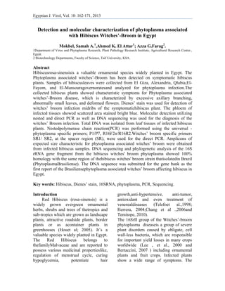
5. 166
Detection and molecular characterization of phytoplasma associated with Hibiscus Witches’-
Broom in Egypt
expected associations of a
phytoplasmawith diseased hibiscus
witches broom exhibiting reduction in
size leaf proliferation of lateral shoots,
and stunting symptoms.
Multiple sequence alignment of
thenucleotide sequence of our PCR
fragment was done with the
corresponding sequences of the
otherphytoplasma strains on GeneBank
(Fig. 4). Nucleotide sequence for our
findings was submitted to the gene bank
and accessioned by GenBank accession
number “KF716175”
Sequence analysis
Nucleotide sequencing of the 1246 kb
purified PCR fragment of 16S rRNA
gene was performed and compared with
sequences of other phytoplasma strains
available in GenBank. The obtained data
were analyzed using DNAMAN software.
Fig. (1):Symptoms of the phytoplasma associatedwitches’ broom on Hibiscus plants. A:
Healthy Hibiscus plant control. B, C and D: infected plants showing leaf yellowing, short
internodes, proliferation of shoots, and premature flower dropping.
Egyptian J. Virol, Vol. 10:162-171, 2013
6. okbel et al.
167
MM
Fig.
tissue
with
Fig.
using
L2 ar
using
differ
(2): Cross s
e stained wi
the healthy p
3: Gel el
gUniversal p
re different s
g witches' bro
rent samples
Egy
section of le
th dark blue
plant control
[A]
lectrophores
phytoplasma-
samples sho
oom-specific
s showed W.
yptian J. Viro
eaf midrib fr
e color after
l; C at magn
is for the
-specific [R1
owed W. B s
c PCR prime
. B symptom
ol, Vol. 10:16
from Hibiscu
r treatment w
nification of
detection
16] PCR pri
symptoms. H
ers. M: 100 b
ms. HPC: He
2-171, 2013
us witches’
with Dienes’
330 X.
[B
of the ph
mers. M: 1 K
HPC: Health
bp-plus, DN
althy Plant C
broom, show
’stain,A and
B]
hytoplasma
Kb DNA La
hy Plant Con
NA Ladder. L
Control [B].
wing phloem
d B compare
in Hibiscu
adder. L1 an
ntrol [A]; an
L1 and L2 ar
m
ed
us
nd
nd
re
7. 8166
Dettection and mmolecular charracterization o
Fig 4
with
tree a
Discu
Phyto
most
the
crops
detec
the
param
preve
phyto
latenc
We
phyto
using
The
succe
prelim
prese
hibisc
Muse
4:Sequence a
the differen
as “Hibiscus_
ussion:
oplasma is c
important p
productivity
s including
ction and ch
diagnosis o
mount imp
ention strateg
oplasmas m
cy period.
detected
oplasma in a
g both classi
main light
essfully for
minary me
ence of phyt
cus tissues
etti, 2013.
Egy
analysis and
nt phytoplasm
_W.B_”.
considered a
plant pathog
y of severa
g hibiscus
haracterizatio
of phytopla
portance fo
gies, particu
may have
the prese
association w
ical and mo
microscop
r the diag
ethod, to
toplasmas in
s which a
Bro
of phytoplasm
yptian J. Viro
the phyloge
ma strains i
as one of th
gens reducin
al economi
s. Sensitiv
on as well a
asmasare o
or effectiv
ularly becaus
avery lon
ence of
with hibiscu
lecular tools
py was use
gnosis, as
assess th
n the infecte
agreed wit
oom in Egypt
ma associatedd with Hibiscuus Witches’-
ol, Vol. 10:16
enetic tree s
n the GeneB
he
ng
ic
ve
as
of
ve
se
ng
a
us
s.
ed
a
he
ed
th
Mole
infec
Phyto
Broo
with
plant
mole
phyto
follow
phyto
ment
sectio
PCR
direc
DNA
neste
phyto
R16F
expec
direc
speci
2-171, 2013
howed rang
Bank. Our i
ecular diag
ction of
oplasma As
m based on
phytoplasm
ts were samp
ecular cha
oplasma.A n
wed for
oplasma in s
tioned in m
on.
amplificati
ct, as well
A extracted f
ed PCR
oplasma- sp
F2/R16R2 g
ctedfragmen
ct PCR usin
ific PCR pri
e of 99-100
solate is ind
gnosis con
hibiscus p
ssociated wi
the symptom
ma diseases
pled for the
aracterization
nested-PCR a
the detecti
suspectedlea
materials a
ions were
as nested P
from Hibiscu
using the
ecific prime
gave amplifi
nt at1.2 kb
ng the witc
imers (SR1/S
%. Similarit
dicated in th
nfirmed th
plants wit
ith Witches
ms associate
s. Suspecte
detection an
n of th
approach wa
ion of th
af samples a
and method
obtained b
PCR for th
us tissue. Th
e universa
ers P1/P7 an
cation of th
b, while th
ches' broom
SR2) showe
ty
he
he
th
’-
ed
ed
nd
he
as
he
as
ds
by
he
he
al
nd
he
he
m-
ed
8. 169 Mokbel et al.
Egyptian J. Virol, Vol. 10:162-171, 2013
a clear band at ~ 325 bpcorresponding to
the 5’-end of the 23S rDNA and the
partial 16S rRNA gene. The first round of
the nested PCR using the P1/P7 primers
didn’t show a band due to the low
phytoplasmatic DNA concentrations as
compared to plant DNA. That agrees with
the fact that, the detection of
phytoplasmatic DNA was achieved only
with a nested-PCR (Hamedet al., 2013).
In the second PCR reaction, conditions
are optimized and the sensitivity of the
technique is increased facilitating
visualization of the ampliconsthat refers
to the use of the PCRproduct of the first
Round PCR as a DNA template for the
second one. DNA amplified from
templateDNA isolated from any of the
healthy, non-symptomatic, plant samples
werefound to be negative for
phytoplasma presence in both direct and
nested PCR.Those PCR results clearly
demonstrated the natural infection of
hibiscus with phytoplasma associated
with Witches’-Broom.
Phylogenetic analysis and homology with
the sequence of the 16S ribosomal RNA
(rRNA) gene, the 16-23S rRNA spacer
region, and the 5`-end of the 23S rRNA
gene identified the phytoplasma as
belonging to the 16Sr II group (97-99%
homology).
Sequence obtained from the1246 bp PCR
product associated with infected Hibiscus
rosa-sinensis was submitted to BLAST
analysis which showed a 100% similarity
with reference strain of hibiscus witches'
broom from Brazil, belonging to 16SrII-
group.
DNA sequencing and phylogenetic
analysis indicated that this phytoplasma
clustered in the 16SrII group. A 1,246 bp
sequence of the 16S rRNA gene from the
hibiscus witches’ broom phytoplasma
showed 100% homology with the 16S
rRNA gene, hibiscus witches' broom in
Brazil (HQ230579-SiWB-B) and peanut
witches'-broom (EU099547-YN02).
Nucleotide sequence determined in this
study was submitted to the gene bank and
accessioned by Gen Bank accession
number “KF716175” as the first report of
phytoplasma infection affecting hibiscus
witches' broom in Egypt.
References:
Ammar, M. I., Amer, M. A. and Rashed,
M. F. (2005).Detection of Phytoplasma
Associated with Yellow Streak
Disease of Date Palms (Phoenix
dactyliferaL.) in Egypt.Egyptian J.
Virol. 2, 74-86
Arismendi, N. S., Andrade, N. S., Riegel,
R. Sch. and Carrillo R. L.(2010).
Presence of a phytoplasma associated
with witches' broom disease in
UgnimolinaeTurcz.
andGaultheriahillyreifolia. Chilean
Journal of Agricultural Research 70
(1):26-33
BertacciniA, and Duduk B. (2009).
Phytoplasma and phytoplasma
diseases: a review of recent research.
PhytopatholMediterr: 48:355-78.
Bertaccini A. (2007). Phytoplasmas:
diversity, taxonomy, and
epidemiology. Front Biosci; 12: 673-
89.
Bhat A.I., Madhubala R., Hareesh P.S. and
Anandaraj M. (2006). Detection and
characterization of the phytoplasma
associated with a phyllody disease of
black pepper (Piper nigrum L.) in
India. ScientiaHortic. 107:200–204.
Chang Y. C., Haung K. X., Haung A. C.,
HO Y. C. and Wang C. J. (2006).
Hibiscus anthocyanins-rich extract
inhibited LDL. Oxidation and
oxLDL-mediated macrophages
apoptosis. Food ChemToxicol,
44:1015–23.
Chapman, G.B., Buerkle, E.J.,
9. 170
Detection and molecular characterization of phytoplasma associated with Hibiscus Witches’-
Broom in Egypt
Egyptian J. Virol, Vol. 10:162-171, 2013
Barrows,E.M., Davis, R.E. and Dally,
E.L. (2001). A light and transmission
electron microscope study of a black
locust tree,
Robiniapseudoacacia(Fabaceae),
affected by witches’ broom, and
classification of the associated
phytoplasma. J. Phytopathol.
149:589-597.
Deeley, J; Stevens, W. A., and Fox,
R.T.V. (1979).Use of dienes, stain to
detect plant diseases induced by
mycoplasma like
organism.Phytopathology. 69: 1169-
1171.
Firrao G, Garcia-Chapa M, Marzachì C.
(2007). Phytoplasmas: genetics,
diagnostics and relationships with the
plant and insect host. Front Biosci;
12:1353-75.
Fránová, J., Petrzik,K. Paprštein,F.,
Kučerová, J., Navrátil, M.andVálová,
P. (2007). Experiences with
phytoplasma detection and
identification by different methods.
Bull. Insectol. 60:247-248.
Hamed, A. H., Ahmed K. El Attar1; and
Om-Hashim M. El-Banna (2013). First
record of a Phytoplasma Associated
with faba bean (Viciafaba L.)
Witches’-Broom in Egypt.
International Journal Of Virology;
online first.
Heinrich, M., Botti, S., Caprara, L.,
Arthrofer,W., Strommer, S. and
Hanzer,V. (2001). Improved detection
method for fruit tree
phytoplasmas.Plant Mol. Biol. Rep.
19:169-179.
Helena G. M., Robert E. D., Ellen L. D.,
Saskia H., Joao P. P. and Paulo S. T.
(2001).
‘CandidatusPhytoplasmabrasiliense’, a
new phytoplasma taxon associated
with hibiscus witches’ broom disease
International Journal of Systematic and
Evolutionary Microbiology 51, 1109–
1118
Herrera A. A., Flores R. S., Chavez-Soto
M. A. and Tortoriello J. (2004).
Effectiveness and tolerability of a
standarized extract from Hibiscus
sabdariffa in patients with mild to
moderate hypertention: a controlled
and randomized clinical trial.
Phytomedicine, 11:375–82.
Hibben C. R., Lewise C. A. and Castello
J. D. (1986).Mycoplasma- like
organisms, cause of Lilac Witches-
Broom. Plant Dis. 70: 312-345.
Hogenhout,S.A.,Oshima,K.,Ammar, E.
D.,Kakizawa,S.,Kingdom,H. N. and
Namba,S.(2008). Phy- toplasmas:
bacteria that manipulate plants and
insects. Mol .Plant Pathol. 9, 403–423.
Hou D. X., Tong X, Terahara N., Lou D.
and Fujii M. (2005). Delphenidin
3-sambubioside, a Hibiscus
anthocyanin, induces apoptosis in
human leukemia cells through reactive
oxygen species-mediated
mitochondrial pathway. Arch
BiochemBiophy, 440: 101–09.
Khan, A.T., Bott, S., Al-Suthi, A.M.,
Gundersen- Rindal D. E. and
Bertaccini, A.F. (2007). Molecular
identification of a new phytoplasma
associated with alfalfa witches’-broom
in Oman. Phytopathology 92: 1038–
1047
Lee I. M., Martini M., Marcone C., Zhu
S. F., (2004). Classification of
phytoplasma strains in the elm
yellows group (16SrV) and
proposition of
10. 171 Mokbel et al.
Egyptian J. Virol, Vol. 10:162-171, 2013
‘CandidatusPhytoplasmaulmi’ for the
Phytoplasma. Int J
SystEvolMicrobiol. Mar;54(Pt
2):337-47.
Lee, I.M., Davis, R.E. and Gundersen-
Rindal ,D.E.(2000). Phytoplasma:
phyto-pathogenic molecules.- Annual
Review of Microbiology, 54: 221-255
Lee, I.-M., Bertaccini, A., Vibio, M. and
Gundersen, D. E. (2000). Detection of
multiple phytoplasmas in perennial
fruit trees with decline symptoms in
Italy. Phytopathology 85:728–735.
Lee, I.-M., Gundersen-Rindal, D.E.,
Davis, R.E. and Bartoszyk, I.M.
(1998). Revised classification scheme
of phytoplasmas based on RFLP
analysis of 16S rRNA and ribosomal
protein gene sequences. Int. J. Syst.
Bacteriol. 48:1153-1169.
Musetti R. (2013).Dienes' staining and
light microscopy for
phytoplasmavisualization.MethodsMol
Biol. 938:109-113
Om-Hashem M. El-banna, M. S. Mikhail
,Azza G. Farag and A. M. S.
Mohammed (2007). Detection of
phytoplasma in tomato and pepper
plants by electron microscopy and
molecular biology based methods
Egyptian J.Virol.4 : 95-113
Sinclair W.A., Griffiths, H.M., and Davis,
R.E. (2000). Ash yellows and Mac
witches’broom: phytoplasmal diseases
of concem in forestry and horticulture.
Plant Dis. 80, 468-475.
Smart, C.D., Schneider, B. Blomquist,
C.L. Guerra, L. J. Harrison, N., and
Ahrens, A. U. (1996). Phytoplasma-
specific PCR primers based on
sequences of the 16S-23S rRNAspacer
region. Appl. Environ. Microbiol.
62:2988-2993.
Telefo P. B. (1998). Effects of an
aqueous extract of Aloe buettneri,
Justiciainsularis, Hibiscus macranthus,
Diclipteraverticillata on some
physiological and biochemical
parameters of reproduction in
immature female rats. J
Ethnopharmacol, 63:193-200.
Temitope J. (2010). Control of
Reproduction in Oreochromisniloticus
(Linnaeus 1758) Using Hibiscus Rosa-
sinensis (Linn.) Leaf Meal as
Reproduction Inhibitor. Journal of
Agricultural Science Vol. 2, No. 4;
Wang K, Hiruki C., (2001). Use of
heteroduplex mobility assay for
identification and differentiation of
phytoplasmas in the aster yellows
group and the clover proliferation
group.Phytopathology. Jun;91(6):546-
52.