
Recommended
More Related Content
Similar to cytokines-131118235323-phpapp01.pdf
Similar to cytokines-131118235323-phpapp01.pdf (20)
More from OsmanHassan35
More from OsmanHassan35 (20)
Recently uploaded
Recently uploaded (20)
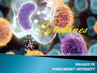
cytokines-131118235323-phpapp01.pdf
- 2. The immune system recognizes the presence of pathogens by several proteins that bind to molecules secreted by the pathogen or carried on their surface. The cells responsible for these immune responses include: a) B-Cells b) T-Cells c) Macrophages d) Neutrophils e) Basophils f) Eosinophils g) Endothelial cells h) Mast cells These cells have distinct roles in the immune system and communicate with other immune cells by cytokines, which control proliferation, differentiation and function of cells of the immune system.
- 4. The development of an effective immune response involves lymphoid cells, inflammatory cells, and hematopoietic cells. The complex interactions among these cells are mediated by a group of proteins collectively designated cytokines to denote their role in cell-to-cell communication. Cytokines / immunocytokines (Greek , cyto =‘cell’ & kinos =‘movement’) are low molecular weight regulatory proteins or glycoproteins secreted by white blood cells and various other cells in the body in response to a number of stimuli.
- 5. Cytokines bind to specific receptors on the membrane of target cells, triggering signal-transduction pathways that ultimately alter gene expression in the target cells. The cytokines and their fully assembled receptors exhibit very high affinity for each other and deliver intracellular signals. The cytokines and their receptors exhibit very high affinity for each other, with dissociation constants ranging from 10 –10 to 10 –12 M. Because their affinities are so high, cytokines can mediate biological effects at picomolar concentrations.
- 6. A particular cytokine may bind to receptors on the membrane of the same cell that secreted it, exerting autocrine action. It may bind to receptors on a target cell in close proximity to the producer cell, exerting paracrine action. In a few cases, it may bind to target cells in distant parts of the body, exerting endocrine action
- 7. Cytokines exhibit the attributes of: Pleiotropy, Redundancy, Synergy, Antagonism, cascade induction, which permit them to regulate cellular activity in a coordinated, interactive way.
- 8. •Two or more cytokines that mediate similar functions are said to be redundant. •Cytokine synergism occurs when the combined effect of two cytokines on cellular activity. • antagonism that is, the effects of one cytokine inhibit or offset the effects of another cytokine.
- 9. Interleukins - that act as mediators between leukocytes. The vast majority of these are produced by T-helper cells. Lymphokines - produced by lymphocytes. Monokines - produced exclusively by monocytes. Interferons - involved in antiviral responses. Colony Stimulating Factors - support the growth of cells blood cell . Chemokines - mediate chemoattraction (chemotaxis) between cells.
- 10. Cytokines have been classified on the basis of their biological responses into pro or anti-inflammatory cytokines, depending on their effects on immunocytes . Major cytokines include: Lymphokines Interleukins (IL) Monokines Interferons (IFN) colony stimulating factors (CSF) Tumor Necrosis Factors-Alpha and Beta (TNF)
- 11. # Type-1 & Type-2 Type-1 cytokines are cytokines produced by Th1 T-helper cells. Include IL-2 (IL2), IFN-gamma (IFN-G), IL-12 (IL12) & TNF- beta (TNF-b). Type-2 cytokines are those produced by Th2 T-helper cells. Include IL-4 (IL4), IL-5 (IL5), IL-6 (IL6), IL-10(IL10), and IL-13 (IL13). # Mediators of natural immunity: TNF-α, IL-1, IL-10, IL-12, type I interferons (IFN-α and IFN-β), IFN-γ, and chemokines. # Mediators of adaptive immunity: IL-2, IL-4, IL-5, TGF-β, IL-10 and IFN-γ.
- 12. # Classified into family groups according to the types of secondary and tertiary structure. IL-6 (IL6), IL-11 (IL11), CNTF (C-NTF), LIF, OSM (Oncostatin- M), EPO (Erythropoietin), G-CSF (GCSF), GH (Growth Hormone), PRL (Prolactin), IL-10 (IL10), IFN-alpha (IFN-A), IFN-beta (IFN-B) form long chain 4 helix bundles. IL-2 (IL2), IL-4 (IL4), IL-7 (IL7), IL-9 (IL9), IL-13 (IL13), IL-3 (IL3), IL-5 (IL5), GM-CSF (GMCSF), M-CSF (MCSF), SCF, IFN- gamma (IFNG) form short chain 4 helix bundles. Beta foil structures are formed by IL1-alpha (IL1A), IL1-beta (IL1B), aFGF (FGF-acidic), bFGF (FGF-basic), INT-2 (INT2), KGF (FGF7). EGF, TGF-alpha (TGF-A), Betacellulin (BTC), SCDGF, Amphiregulin, HB-EGF, form EGF-like antiparallel beta-sheets.
- 13. Lymphokines are a subset of cytokines that are produced by a type of immune cell known as a lymphocyte. They are protein mediators typically produced by T cells to direct the immune system response by signalling between its cells. Lymphokines have many roles, including the attraction of other immune cells, including macrophages and other lymphocytes, to an infected site and their subsequent activation to prepare them to mount an immune response. Circulating lymphocytes can detect a very small concentration of lymphokine and then move up the concentration gradient towards where the immune response is required. Lymphokines aid B cells to produce antibodies.
- 14. Lymphokines include: Colony-stimulating factors (csfs), including GM-CSF. Interferons (ifns) – IFNγ. Interleukins IL-1 to IL-8, IL-10, IL-13. Macrophage inflammatory protein-1 beta (mip-1β). Neuroleukin (lymphokine product of lectin-stimulated T cells). Osteoclast-activating factor. Platelet-derived growth factor (PDGF). Transforming growth factor beta (tgfβ). Tumour necrosis factor-alpha (cachectin) (TNFα). Tumour necrosis factor-beta (tnfβ, lymphotoxin α, LT).
- 15. Actions of lymphokines include: Activates B cells, inhibits macrophage function : IL-10. Activation of neutrophils, eosinophils, and monocyte /macrophages :GM-CSF . Bone resorption : osteoclast activating factor Bone marrow – growth and differentiation of immune cells :IL-3 B cell growth and differentiation :IL-4. B cell differentiation, activates some microphages (pmn) :IL-5 Co-stimulator of T cells, induces growth in B cells :IL-6 Inflammation, fever, catabolism and cachexia, activation of some microphages :TNF Hematopoiesis stimulators :IL-3, IL-7, GM-CSF Macrophage-activating activity (MAF) :INF-γ Stimulates proliferation of activated T and B cells : IL-2 Inhibits T cell growth, activates macrophages :TGFβ
- 16. A monokine is a type of cytokine produced primarily by monocytes and macrophages. Examples include interleukin 1 and tumor necrosis factor- alpha. Other monokines include alpha and beta interferon, and colony stimulating factors.
- 17. a variety of naturally occuring polypeptides that are the family of cytokines which affect functions of specific cell types and are found in small quantities. They are secreted regulatory proteins produced by lymphocytes, monocytes and various other cell types and are released by cells in response to antigenic and non- antigenic stimuli. Consist of IL1 to IL37. IL-1 activates Antigen Presenting Cells and CD4+ lymphocytes; affects the differentiation of the B-Cells and T-Cells and other immunocompetent cells and takes part in the regulation of production of other cytokines and GM-CSF (Granulocyte- Macrophage Colony-Stimulating Factor). IL-2 stimulates the proliferation and activation of B-Cells and T-Cells. IL-4 plays a role in the differentiation of TH2, in allergic responses, and in the switching of antibody types.
- 18. IL-3 is a potent activator of hemopoietic cells. It stimulates NK- Cells and acts as a synergist with IL-4 during the induction of CD4+ lymphocyte activation process. IL-5 stimulates the production and maturation of eosinophils during inflammation. IL-7 is known as the growth factor of the immature B-Cells and T-Cells. It induces apoptosis of tumor cells and causes differentiation of cells from a subgroup of acute myeloblastic leukemia. IL-8 acts as a chemotactic factor that attracts neutrophils, basophils and T-Cells to sites of inflammation. IL-9 stimulates the excretion of IL-2, IL-4, IL-6, IL-11, and takes part in a stimulation of cytotoxicity of T-killers and NK-Cells, inducing apoptosis. IL-10 acts to repress secretion of pro-inflammatory cytokines.
- 19. IL-11 is a pro-inflammative factor, which regulates the functions of B-Cells and T-Cells. It also takes part in the induction of various killer cells activities and acts as an autocrine factor for the proliferation of megacaryocytes. IL-12 is a critical linker between the innate immunity and adaptive immunity, capable of TH1 (T Helper Type-1) differentiation and IFN-Gamma release by T-Cells and NK cells. IL-13 is very sensitive to monocytes and B-Cells. IL-13 does not act on T-Cells but inhibits the proliferation of leukemic pro-B- Cells. IL-14 is a BCGF (B-Cell Growth Factor) and the hyper production of this interleukin enables the progression of NHL-B (B-cell Type Non Hodgkin's lymphoma). IL-15 is analogous to IL-2 and increases the anti-tumor activities of T-killers and NK-Cells, and the production of cytokines CD4+ lymphocytes.
- 20. IL-17 is principally produced by CD4+ T-Cells, which induces granulopoiesis via GMCSF. It takes part in the regulation of many cytokines and can reinforce the antibody dependant tumor cell destruction. IL-18 acts as a synergist with IL-12, especially in the induction of IFN-Gamma production and inhibition of angiogenesis. IL-19 is produced mainly by monocytes and is similar to IL-10 in its function. It is stimulated by GM-CSF and regulates the functions of macrophages, and also suppresses the activities of TH1 and TH2. IL-21 executes an important role in the regulation of hematopoiesis and immune response. It promotes a high production of T-Cells, fast growth and maturation of NK-Cells and B-Cells population. IL-22 is produced by activated T-Cells in acute inflammation. It is similar to IL-10 in function, but does not prohibit the production of pro-inflammatory cytokines through monocytes.
- 21. Interferons play an important role in the first line of defense against viral infections. They are part of the non specific immune system and are induced at an early stage in viral infection before the specific immune system has had time to respond. Interferons are made by cells in response to an appropriate stimulus, and are released into the surrounding medium; they then bind to receptors on target cells and induce transcription of approximately 20-30 genes in the target cells, and this results in an anti-viral state in the target cells.
- 22. Based on the type of receptor through which they signal, human interferons have been classified into three major types. 1. Interferon type I: All type I IFNs bind to a specific cell surface receptor complex known as the IFN α receptor (IFNAR) that consists of IFNAR1 and IFNAR2 chains. The type I interferons present in humans are IFN-α, IFN-β and IFN-ω. 2. Interferon type II: Binds to IFNGR that consists of IFNGR1 and IFNGR2 chains. In humans this is IFN-γ. 3. Interferon type III: Signal through a receptor complex consisting of IL10R2 (also called CRF2-4) and IFNLR1 (also called CRF2-12).
- 24. Chemokines are a family of small cytokines, or Signaling proteins secreted by cells. Their name is derived from their ability to induce directed chemotaxis in nearby responsive cells; they are chemotactic cytokines. Proteins are classified as chemokines according to shared structural characteristics such as small size (they are all approximately 8-10 kilodaltons in size), and the presence of four cysteine residues in conserved locations that are key to forming their 3-dimensional shape.
- 25. Chemokines have been classified into four main subfamilies : 1. CXC Chemokines (contain CXL1 to CXL17) 2. CC Chemokines (contain CCL1 to CCL28) 3. CX3C Chemokines (contain CX3CL1) 4. XC Chemokines (contain XCL1 & XCL2) All of these proteins exert their biological effects by interacting with G protein-linked transmembrane receptors called chemokine receptors, that are selectively found on the surfaces of their target cells.
- 26. Colony-stimulating factors (CSFs) are secreted glycoproteins that bind to receptor proteins on the surfaces of hemopoietic stem cells, thereby activating intracellular signaling pathways that can cause the cells to proliferate and differentiate into a specific kind of blood cell. The colony-stimulating factors are soluble, in contrast to other, membrane-bound substances of the hematopoietic microenvironment. They transduce by paracrine, endocrine, or autocrine signaling. Colony-stimulating factors include: ◦ CSF1 - Macrophage colony-stimulating factor(MCSF) ◦ CSF2 - Granulocyte macrophage colony stimulating factors(GMCSF). ◦ CSF3 - Granulocyte colony-stimulating factors(GCSF)
- 27. Tumor necrosis factors (or the TNF family) refer to a group of cytokines that can cause cell death (apoptosis). Nineteen cytokines have been identified as part of the TNF family on the basis of sequence, functional, and structural similarities. They include: Tumor necrosis factor (TNF), formerly known as TNFα or TNF alpha, is the best-known member of this class. TNF is a monocyte-derived cytotoxin that has been implicated in tumor regression, septic shock, and cachexia. Lymphotoxin-alpha, formerly known as Tumor necrosis factor- beta (TNF-β), is a cytokine that is inhibited by interleukin 10. Lymphotoxin-alpha (LT-alpha) and lymphotoxin-beta (LT-beta), two related cytokines produced by lymphocytes that are cytotoxic for a wide range of tumor cells in vitro and in vivo.
- 28. T cell antigen gp39 (CD40L), a cytokine that seems to be important in B-cell development and activation. CD27L, a cytokine that plays a role in T-cell activation. It induces the proliferation of co-stimulated T cells and enhances the generation of cytolytic T cells. CD30L, a cytokine that induces proliferation of T cells. FASL, a cytokine involved in cell death. 4-1BBL, an inducible T cell surface molecule that contributes to T-cell stimulation. OX40L, a cytokine that co-stimulates T cell proliferation and cytokine production. TNF-related apoptosis inducing ligand (TRAIL), a cytokine that induces apoptosis.
- 30. Cytokine Receptors Fall Within Five Families Receptors for the various cytokines are quite diverse structurally, but almost all belong to one of five families of receptor proteins: ◦ Immunoglobulin super family receptors ◦ Class I cytokine receptor family (also known as the hematopoietin receptor family) ◦ Class II cytokine receptor family (also known as the interferon receptor family) ◦ TNF receptor family. ◦ Chemokine receptor family.
- 33. Immunology by Kuby http://pathmicro.med.sc.edu/mobile/m.immuno-13.htm http://www.rndsystems.com/research_topic.aspx?r=14 http://www.ebioscience.com/resources/pathways/cytokine- network.htm http://www.prospecbio.com/Cytokines http://en.wikipedia.org/wiki/Cytokine