More Related Content
Similar to Interleukins (20)
More from Mahmood Asif (7)
Interleukins
- 1. Cytokines (Interleukins) and Adhesion
Cytokines are bioactive hormones, normally glycoproteins, which exercise a
wide variety of biological effects on those cells which express the appropriate
receptors (Table 2.6). Cytokines are designated by their cellular origin such
that monokines include those interleukins produced by macrophages/
monocytes, whilst lymphokines include those interleukins produced by
lymphocytes. The term interleukins is used for cytokines which mostly in-
fluence cellular interactions. All cytokines are cyto-regulatory proteins with
molecular weights under 60 kDa (in most cases under 25 kDa). They are pro-
duced locally, have very short half-lives (a matter of seconds to minutes), and
are effective at picomolar concentrations. The effects of cytokines may be
paracrine (acting on cells near the production locus), or autocrine (the
same cell both produces, and reacts to, the cytokine). By way of interaction
with highly specific cell surface receptors, cytokines can induce cell-specific
or more general effects (including mediator release, expression of differen-
Immune Responses and Effector Mechanisms 77
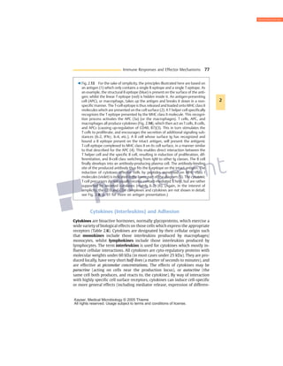
3 Fig. 2.13 For the sake of simplicity, the principles illustrated here are based on
an antigen (1) which only contains a single B epitope and a single T epitope. As
an example, the structural B epitope (blue) is present on the surface of the anti-
gen; whilst the linear T epitope (red) is hidden inside it. An antigen-presenting
cell (APC), or macrophage, takes up the antigen and breaks it down in a non-
specific manner. The T-cell epitope is thus released and loaded onto MHC class II
molecules which are presented on the cell surface (2). AT helper cell specifically
recognizes the T epitope presented by the MHC class II molecule. This recogni-
tion process activates the APC (3a) (or the macrophages). T cells, APC, and
macrophages all produce cytokines (Fig. 2.14), which then act onTcells, B cells,
and APCs (causing up-regulation of CD40, B7)(3). This in turn stimulates the
T cells to proliferate, and encourages the secretion of additional signaling sub-
stances (IL-2, IFNc, IL-4, etc.). A B cell whose surface Ig has recognized and
bound a B epitope present on the intact antigen, will present the antigenic
T cell epitope complexed to MHC class II on its cell surface, in a manner similar
to that described for the APC (4). This enables direct interaction between the
T helper cell and the specific B cell, resulting in induction of proliferation, dif-
ferentiation, and B-cell class switching from IgM to other Ig classes. The B cell
finally develops into an antibody-producing plasma cell. The antibody-binding
site of the produced antibody thus fits the B epitope on the intact antigen. The
induction of cytotoxic effector cells by peptides presented on MHC class I
molecules (violet) is indicated in the lower part of the diagram (5). The cytotoxic
T cell precursors do not usually receive contact-mediated T help, but are rather
supported by secreted cytokines (mainly IL-2) (6). (Again, in the interest of
simplicity, the CD3 and CD4 complexes and cytokines are not shown in detail;
see Fig. 2.8, p. 61 for more on antigen presentation.)
2
Kayser, Medical Microbiology © 2005 Thieme
All rights reserved. Usage subject to terms and conditions of license.
- 2. tiation molecules and regulation of cell surface molecule expression). The
functions of cytokines are usually pleiotropic, in that they display a number
of effects of the same, or of a different, nature on one or more cell types. Below
is a summary of cytokine functions:
& Promotion of inflammation: IL-1, IL-6, TNFa, chemokines (e.g., IL-8).
& Inhibition of inflammation: IL-10, TGFb.
& Promotion of hematopoiesis: GM-CSF, IL-3, G-CSF, M-CSF, IL-5, IL-7.
& Activating B cells: CD40L, IL-6, IL-3, IL-4.
& Activating T cells: IL-2, IL-4, IL-10, IL-13, IL-15.
& Anti-infectious: IFNa, IFNb, IFNc, TNFa.
& Anti-proliferative: IFNa, IFNb, TNFa, TGFb.
78 2 Basic Principles of Immunology
CD4+
T Helper Cell Subpopulations
and APCs during Immune Responses
NK
cells
IFN-γ
IL-12
TH2
TH1
TH
0
APC
Viruses
Bacteria
Viruses
Parasites
Allergens
Mast cells
Basophils
IL-4
–
Macrophages
Eosinophils
B cells
B cells
IFN-γ
IL-2
IL-4
IL-5
IL-10
IL-13
IgM
IgG2a
Delayed type
hypersensitivity
(DTH)
Defenses against
intracellular
microorganisms
IgG1
IgE
+
+
Fig. 2.14 TH1 and TH2 cells are derived from a TH0 cell, and undergo differentia-
tion in the presence of help derived from cytokines, DC, macrophages, and other
cell types. TH1 cells are activated by IL-12 and IFNc and inhibited by IL-4; whilst for
TH2 cells the reverse is true. Viruses and bacteria (particularly intracellular bacter-
ia) can induce a TH1 response by activating natural killer cells. In contrast, allergens
and parasites induce a TH2 response via the release of IL-4. However, the strong in-
vitro differentiation of CD4+ Tcells intoTH1–TH2 subsets is likely to be less sharply
defined in vivo.
2
Kayser, Medical Microbiology © 2005 Thieme
All rights reserved. Usage subject to terms and conditions of license.
- 3. Immune Responses and Effector Mechanisms 79
Antiviral Protection by T Cells
Perforin
T cellT cell
Perforin
Noncytopathic
virus
Cytopathic
virus
Perforin mouse:
uncontrolled
viral proliferation
Control mouse:
perforin lyses
infected cells
T cell
B cell
IL IFN
Neutralizing
antibody
Diffusion,
protection of
many other cells
Lysis caused by virus
Fig. 2.15 Certain viruses destroy the infected host cells (right), others do not
(left). Cytotoxic T cells can destroy freshly infected cells by direct contact (with
the help of perforin), thus inhibiting viral replication (middle). Whether the result
of this lysis is clinically desirable depends on the balance between protection from
viral proliferation, and the damage caused by immunologically mediated cell de-
struction. In perforin knockout mice (perforino/o
), T cells are unable to produce
perforin and therefore do not destroy the infected host cells. Replication of
non-cytopathic viruses thus continues unabated in these mice. Soluble anti-viral
interleukins (especially IFNc and TNFa), and neutralizing antibodies, combat cyto-
pathic viruses (which replicate comparatively rapidly) more efficiently than do cy-
tolytic Tcells; this is because interleukin and antibody molecules can readily diffuse
through tissues and reach a greater number of cells, more rapidly, than can killer T
cells.
2
Kayser, Medical Microbiology © 2005 Thieme
All rights reserved. Usage subject to terms and conditions of license.
- 4. 80 2 Basic Principles of Immunology
Table 2.6 The Most Important Immunological Cytokines and Costimulators
plus Their Receptors and Functions
Cytokines/costimula-
tors/chemokines Receptor
Cytokines/cytokine receptors
produced by Functions
Interleukins
IL-1 CD121 (a)b Macrophages
Endothelial cells
Hypothalamic fever,
NK cell activation,
T and B stimulation
IL-2 (T-cell growth
factor)
CD25 (a)
CD122 (b), cc
T cells T-cell proliferation
IL-3 (multicolony
stimulating factor)
CD123, bc T cells, B cells,
thymic epithelial
cells
Synergistic effect in
hematopoiesis
IL-4 (BCGF-1, BSF-1)
(B-cell growth factor,
B-cell stimulating factor)
CD124, cc T cells, mast cells B-cell activation, switch
to IgE
IL-5 (BCGF-2) CD125, bc T cells, mast cells Growth and differentia-
tion of eosinophilis
IL-6 (interferon/IFNb2,
BSF-2, BCDF)
CD126,
CDw130
T cells,
macrophages
Growth and differentia-
tion of T and B cells,
acute-phase immune
response
IL-7 CDw127, cc Bone marrow
stroma
Growth of pre-B and
pre-T cells
IL-10 T cells Macrophages, reduction
of TH1 cytokines
IL-9 IL-9R, cc T cells Effect on mast cells
IL-10 T helper cells
(especially mouse
TH2), macro-
phages,
Epstein-Barr virus
Efficient inhibitor for
macrophage functions,
inhibits inflammatory
reactions
IL-11 IL-11R,
CDw130
Stromal
fibroblasts
Synergistic effect with
IL-3 and IL-4 in hemato-
poiesis
2
All rights reserved. Usage subject to terms and conditions of license
Kayser, Medical Microbiology © 2005 Thieme
All rights reserved. Usage subject to terms and conditions of license.
- 5. Immune Responses and Effector Mechanisms 81
Table 2.6 Continued: The Most Important Immunological Cytokines. . .
Cytokines/costimula-
tors/chemokines Receptor
Cytokines/cytokine receptors
produced by Functions
IL-12 B cells,
macrophages
Activates natural killer
cells,induces differentia-
tion of CD4+
T cells into
TH1-like cells, encour-
ages IFNc production
IL-13 IL-13R, cc T cells Growth and differentia-
tion of B cells, inhibits
production of inflamma-
tory cytokines by means
of macrophages
IL-15 IL-15R, cc T cells, placenta,
muscle cells
IL-2-like, mainly
intestinal effects
GM-CSF (granulocyte
macrophage colony
stimulating factor)
CDw116, bc Macrophages,
T cells
Stimulates growth and
differentiation of the
myelomonocytic lineage
LIF (leukemia
inhibitory factor)
LIFR, CDw130 Bone marrow
stroma, fibroblasts
Maintains embryonal
stem cells; like IL-6, IL-11
Interferons (IFN)
IFNc CD119 T cells, natural
killer cells
Activation of macro-
phages, enhances MHC
expression, antiviral
IFNa CD118 Leukocytes Antiviral, enhances MHC
class I expression
IFNb CD118 Fibroblasts Antiviral, enhances MHC
class I expression
Immunoglobulin superfamily
B7.1 (CD80) CD28
(promoter);
CTLA-4
(inhibitor)
Antigen-
presenting cells
Costimulation of T cell
responses
B7.2 (CD86) CD28;
CTLA-4
Antigen-
presenting cells
Costimulation of T cell
responses
2
All rights reserved. Usage subject to terms and conditions of license
Kayser, Medical Microbiology © 2005 Thieme
All rights reserved. Usage subject to terms and conditions of license.
- 6. 82 2 Basic Principles of Immunology
Table 2.6 Continued: The Most Important Immunological Cytokines. . .
Cytokines/costimula-
tors/chemokines Receptor
Cytokines/cytokine receptors
produced by Functions
TNF (tumor necrosis factor) family
TNFa (cachexin) p55, p75,
CD120a,
CD120b
Macrophages,
natural killer cells
Local inflammations,
endothelial activation
TNFb (lymphotoxin,
LT, LTa)
p55, p75,
CD120a,
CD120b
T cells, B cells Endothelial activation,
organization of second-
ary lymphoid tissues
LTb T cells, B cells Organization of second-
ary lymphoid tissues
CD40 ligand (CD40-L) CD40 T cells, mast cells B-cell activation, class
switching
Fas ligand CD95 (Fas) T cells Apoptosis, Ca2+
-inde-
pendent cytotoxicity
Chemokines
IL-8 (prototype)
CXCL8
CXCR1,
CXCR2
Activated endo-
thelium, activated
fibroblasts
Attraction of neutro-
phils, degranulation of
neutrophils
MCP-1 (monocyte
chemoattractant
protein)
CCL2
CCR2 Activated endo-
thelium, tissue
macrophages,
synovial cells
Inflammation
MIP-1a (macrophage
inflammatory protein)
CCL3
CCR5, CCR1 T cells, activated
Mf
Proinflammatory
HIVa receptor
MIP-1b
CCL4
CCR5 T cells, activated
Mf
Proinflammatory
HIVa receptor
RANTES (regulated on
activation, normal
T cell expressed and
secreted)
CCL5
CCR5, CCR1,
CCR3
T cells, blood
platelets
Inhibits cellular entry
by M-trophic HIV,
proinflammatory
IP-10 (interferon
gamma-inducible
protein)
CXCL10
CXCR3 Inflamed tissue
due to effects of
IFNc
Proinflammatory
2
All rights reserved. Usage subject to terms and conditions of license
Kayser, Medical Microbiology © 2005 Thieme
All rights reserved. Usage subject to terms and conditions of license.
- 7. Immune Responses and Effector Mechanisms 83
Table 2.6 Continued: The Most Important Immunological Cytokines. . .
Cytokines/costimula-
tors/chemokines Receptor
Cytokines/cytokine receptors
produced by Functions
Chemokines
MIG (monokine
induced by interferon
gamma)
CXCL11
CXCR3 Inflamed tissue,
due to effects of
IFNc
Proinflammatory
Eotaxin
CCL22
CCR3 Endothelium,
epithelial cells
Buildup of infiltrate in
allergic diseases, e.g.,
asthma
MDC (macrophage-
derived chemokine)
CCR4 T-cell zone DCs,
activated B cells,
monocytes
Supports T-B cell colla-
boration during humoral
immune responses
Fractalkine
CXCL1
CX3CR1 Intestinal epithe-
lium, endothelium
Endothelial cells activa-
tion of thrombocytes
Constitutive chemokines
LARC (liver and
activation-regulated
chemokine) MIP-3a
CCR6 Intestinal epithelia,
Peyer’s patches
Participation in mucosal
immune responses
SLC (secondary
lymphoid organ
chemokine)
CCR7 High endothelial
lymph nodes,
T-cell zone
Facilitates entry of naive
T cells, contact between
T cells and DCs
TECK (thymus-
expressed chemokine)
CCR9 Thymic and
intestinal epithelia
Presumed role in T-cell
selection
SDF-1a (stromal
cell-derived factor)
CXCR4
(also known
as fusin)
Stromal cells of
bone marrow
Involved in hemato-
poiesis, inhibits cellular
entry by T-trophic HIV
BCA-1 (B-cell
attractant)
CXCR5 Follicular DCs (?) Contact between TH
and B cells,
and between TH and
follicular DCs
Others:
TGFb (transforming
growth factor b)
Many cells,
including mono-
cytes and T cells
Inhibits cell growth,
inhibits macrophages
and production of IL-1
and TNFa, represents a
switching factor for IgA
2
All rights reserved. Usage subject to terms and conditions of license
Kayser, Medical Microbiology © 2005 Thieme
All rights reserved. Usage subject to terms and conditions of license.