2. Topics to be covered
1. Introduction
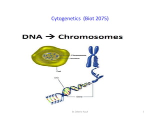
1. Introduction
Definition of Cytogenetics
History of Cytogenetics
Chromosome Theory of Inheritance
2. Organization and structure of genome: genome size variation and c-value paradox
3. Chromosome characterization based on morphology, karyotyping and banding patterns
Chromosome Morphology: the Centromere
chromosome Classification based on the number of centromeres: Acentric, Monocentric &
Dicentric
chromosome Classification Based on Centromere Position: metacentric, submetacentric,
subtelocentric and telocentric.
subtelocentric and telocentric.
shapes of Specialized techniques to visualize chromosomes Chromosomes during
metaphase
Chromosome Classification Based On Size and Other Attributes: size of chromosomes,
satellite chromosomes, NOR, euchromatin and heterochromatin, telomere, variation in
chromosome number and structure.
4. Chromosomal abberations/abnormalities
5. Molecular cytogenetics: Fluorescent in situ Hybridisation (FISH) and Comparative
Genomic Hybridisation (CGH)
6. Cancer cytogenetics
7. Chromosome evolution
2
Dr. Zekeria Yusuf
3. 1. Introduction
Definition of Cytogenetics
• Cytogenetics is a science concerned with the structure,
number, function, & movement of chromosomes and the
numerous variations of these properties as
• they relate to the transmission, recombination and expression
of the genes.
of the genes.
• Cytogenetics was developed from two originally separate
sciences – cytology and genetics. To fully understand the
development of cytogenetics as a discipline, one has to look
into its history.
3
Dr. Zekeria Yusuf
5. History of cytogenetics
• It was the Swiss botanist Nageli who first described thread-like
structures in the nuclei of plant cells in the 1840s, and what he called
“transitory cytoblasts” are now known as chromosomes.
• Later, in 1888, Waldeyer coined the term “chromosome” after staining
techniques had been developed to make them more discernible
(chromos = Greek for colour; soma = Greek for body).
(chromos = Greek for colour; soma = Greek for body).
• Cytogenetics is the study of the structure and properties of
chromosomes, their behaviour during somatic cell division during growth
and development (mitosis), & germ cell division during reproduction
(meiosis), as well as their influence on phenotype. Cytogenetics also
includes the study of factors that cause chromosomal changes
5
Dr. Zekeria Yusuf
6. History of cytogenetics
• Initially, it was difficult to determine the diploid number of
mammalian species because the chromosomes were crowded
in metaphase. In the 1950s, several technical improvements,
such as the addition of colchicines to arrest cells in metaphase
and the use of hypotonic solution to obtain better
chromosome spreads, were made .
• In 1956, the diploid number of chromosomes in man was
established as 46 , and the peripheral leucocyte cell culture .
• Jau-hong Kao et al. (2008) described a chromosome
classification based on the band profile similarity along the
approximate medial axis.
6
Dr. Zekeria Yusuf
7. History of cytogenetics
• Prior to the 1920s, cytological studies were carried out on
biological tissues that were embedded in paraffin, sectioned,
and stained (Wilson, 1925; Darlington, 1937).
• The methods that were in vogue were not sufficiently refined to
allow for the detection of such gross morphological features as
centromeres, secondary constrictions, and satellites of
chromosomes.
• During the 1920s and 1930s innovations were introduced which
facilitated cytological and karyotypic analyses. In 1921, Belling
described a technique for studying meiosis in plant species that
involved the squashing of anthers. This method permitted the
separation of PMCs and facilitated the spreading of their
chromosomes.
7
Dr. Zekeria Yusuf
8. History of cytogenetics
• 1956: Clinical cytogenetics
• 1959: +21 (France), 45,XO (UK)
• 1960: Ph (t(9;22)) in CML
1960s end: Banding techniques: Chr. Identification
• 1977: ISCN (An International System for Human Cytogenetic Nomenclature)
• Walter Flemming – He showed that the chromosomes split longitudinally
during cell division and first applied the name chromatin.
during cell division and first applied the name chromatin.
(ii) Thomas Morgan – He discovered sex linkage working with Drosophila.
(iii) Emil Heitz – He discovered giant chromosomes in the salivary gland cells of
diptherian insects.
Thomas Morgan – He discovered sex linkage working with Drosophila.
8
Dr. Zekeria Yusuf
9. Four overlapping eras of cytogenetics research
• Cytogenetics Era 1 (1910-1970):
• In this era chromosome number of a number of
plant systems became known, structural changes
like interachanges and inversions were studied for
the first time in Stizolobium, Datura & Oentothera,
inversions were studied in maize, and anuploids
developed and cytogenetic maps constructed in
developed and cytogenetic maps constructed in
several crops like maize, wheat, barley, etc.
• Alien addition and substitution lines were also
developed in bread wheat using rye and few
Aegilops/Agropyron species as the source of alien
chromosomes.
9
Dr. Zekeria Yusuf
10. • Cytogenetics Era 2 (1950-1980): In this era, haploid DNA content (C-
value) and composition (unique and repetitive) of nuclear DNA were
determined in a large number of flowering plants using techniques of
cytospectrophotometry and reassociation kinetics.
• This led to recognition of two versions of C-value paradox.
• Firstly, the DNA contents in most eukaryotes were too high for the
number of genes in the corresponding taxa, as estimated on the basis
of known rates of mutations, and
of known rates of mutations, and
• secondly the large-scale variation in DNA contents, could not be
explained with the level of difference in complexity witnessed in these
different organisms. The occurrence of large proportion of repetitive
DNA in each of these eukaryotic genomes partly resolved the C-value
paradox .
10
Dr. Zekeria Yusuf
11. • Cytogenetics Era 3 (1980-Contd.): In this era, starting in
early 1980s, DNA-based molecular markers were
developed and molecular maps constructed in a large
number of animals and plant systems, so that these
molecular markers and the corresponding maps became
an important resource for a variety of research
problems, including their use in diagnostics and plant
breeding.
breeding.
• During this period, another significant development was
the availability of a variety of fluorescence in situ
hybridization (FISH), including multicolour FISH
(McFISH), chromosome orientation FISH (CO-FISH), fibre-
FISH, RNA-FISH, comparative Genomic Hybridization
(CGH) and 3-D FISH
11
Dr. Zekeria Yusuf
12. • Cytogenetics Era 4 (1995-Contd.): This era started
in mid-1990s and gained momentum in the
present century, with two distinct areas of
cytogenetics research;
• first, the whole genome sequencing giving birth
to ‘reverse genetics’, and
• second, the chromatin remodeling giving birth to
• second, the chromatin remodeling giving birth to
the concept of ‘histone code’.
• significant progress has been made during the
last few years to elucidate how the nucleosome
and chromatin structure are modulated for
expression of genes in time and space.
12
Dr. Zekeria Yusuf
38. DNA Content/genome size
• DNA content is defined as the amount of DNA in one copy or in the
haploid chomosomes of an organism. Haploid DNA content is
referred to as the "C-value".
• The DNA content of an organism can be measured by weight or
number of base pairs in a single copy of the entire sequence of DNA
found within cells of that organism.
• DNA content varies greatly among organisms. In general,
• DNA content varies greatly among organisms. In general,
eukaryotes have more DNA content than prokaryotes.
• Among prokaryotes the variation of DNA content or genome size is
small ranging only an order of magnitude, from 0.5 to 5 Mb.
• The genome sizes of eukaryotes, on the other hand, vary >80,000-
fold. Even among animals there is a nearly 3000-fold variation, and
in plants basal genomes sizes vary by a factor of >6000.
38
Dr. Zekeria Yusuf
43. • Gene density in eukaryotic organisms is consistently lower and
more variable than in their prokaryotic counterparts.
• Among eukaryotes, there is a general trend for gene density to
decrease with increasing organism complexity.
• The simple unicellular eukaryote Saccharomyces cerevisiae has
a gene density close to that of prokaryotes (500 genes/Mb).
a gene density close to that of prokaryotes (500 genes/Mb).
• In contrast, the human genome is estimated to have a 50-fold
lower gene density.
43
Dr. Zekeria Yusuf
55. C value
• Definition: The amount of DNA per cell, a quantity termed an
organism’s C value
• Prokaryotic and eukaryotic cells differ dramatically in their C
values
• Each cell of a fruit fly, for example, contains 35 times the amount
of DNA found in a cell of the bacterium E. coli.
• Examination of DNA sequences has revealed that eukaryotic
• Examination of DNA sequences has revealed that eukaryotic
DNA has complexity that is absent from prokaryotic DNA.
• Human cells contain more than 10 times the amount of DNA
found in Drosophila cells, whereas some salamander cells
contain 20 times as much DNA as that of human cells.
• C value paradox - The apparent paradox that there is no
relationship between the size of the genome and the evolutionary
complexity of species.
55
Dr. Zekeria Yusuf
57. C- Value Complexity and C- Value paradox
• Earlier it was believed that DNA-content is correlated
with the complexity of an organism. The idea was that
the more complex the species the more genes it needed
and hence has more C-value.
• How ever the total amount of chromosomal DNA in
different animals and plants does not vary in a consistent
manner with the apparent complexity of the organisms.
• As compared to human (Cvalue 3.3 pg DNA), Amphibians
• As compared to human (Cvalue 3.3 pg DNA), Amphibians
like salamanders (C-value 120 pg DNA), plants like wheat,
broad beans, and garden onions ( C-value 7.0, 14.6, and
16.8 picograms, respectively) are less complex in their
structure and behavior.
• Even in closely related species like the broad bean and
kidney bean c-value varies about three to four times .
57
Dr. Zekeria Yusuf
58. C- Value paradox
• The failure of C values to correspond to phylogenetic complexity is called the
C-value paradox.
• This perplexing variation in genome size occurs mainly because eukaryotic
chromosomes contain variable amounts of DNA with no demonstrable
function, both between genes and within genes in introns.
• This apparently nonfunctional DNA is composed of repetitious DNA
sequences, some of which are never transcribed and most all of which are
likely dispensable.
likely dispensable.
These Repetitious DNA include:
Simple DNA repeats
Moderately repeated DNA
Transposons
Viral retro-transposones
Long interspersed elements
Short interspersed elements
Unclassified spacer DNA
58
Dr. Zekeria Yusuf
59. C- Value paradox
In addition to the non coding DNA sequences several protein
coding genes are present as multiple copies. These include:
Soiltary genes
Duplicated and diverged genes(functional gene families and
non-functional pseudogenes)
Tandem repeated genes encoding rRNA, tRNA & histones
Thus there is no direct correlation between total DNA
content (C-Value) & the number of functional genes, w/c in
turn determines the complexity of an organism’s structure &
functions.
59
Dr. Zekeria Yusuf
74. Human Genes
• About 30,000 genes, not a particularly large
number compared to other species.
• Gene density varies along the chromosomes:
genes are mostly in euchromatin,
genes are mostly in euchromatin,
• Most genes (90-95% probably) code for proteins.
However, there are a significant number of RNA
genes.
74
Dr. Zekeria Yusuf
112. Formed during the diplotene stage in the nuclei of oocytes during the active synthesis of
mRNA molecules for the future use.
It contains a main axis whose chromonemal fibres (DNA molecule) gives out lateral loops
throughout its length.
112
Dr. Zekeria Yusuf
116. Polytene chromosome…
• The nuclei of the salivary gland cells of the larvae of dipterans like
Drosophila have unusually long and wide chromosomes, 100 or 200
times in size of the chromosomes in meiosis and mitosis of the same
species.
• Salivary gland cells do not divide after the glands are formed, yet their
chromosomes replicate several times (a process called endomitosis)
and become exceptionally giant-sized.
• They are discovered by Balbiani (l881) and named by Koller.
• They are discovered by Balbiani (l881) and named by Koller.
• The endomitosis process result in the production of 2X chromosomes,
where X gives the number of multiplication cycle.
• They have alternating dark and light bands. The dark bands are disc-
shaped structures occupying the whole diameter of chromosome.
They contain euchromatin
• The light bands are fibrillar and composed of heterochromatin.
116
Dr. Zekeria Yusuf
117. Holokinetic Chromosome
• In insects of the order Hemiptera & in some monocotyledonous
plants the kinetic activity is distributed over the entire
chromosome.
• The term diffuse centromere bas been used as an alternative.
• In 1966 Flach observed this type of centromere in some
primitive Dicotyledons along with pseudoscorpion & Ascaris.
primitive Dicotyledons along with pseudoscorpion & Ascaris.
117
Dr. Zekeria Yusuf
118. B chromosome
• These are particular kind of chromosome that may or may not
be found in an organism as extra chromosome over and above
the standard diploid or polyploid chromosome complement.
• Also known as accessory or Supernumerary chromosome.
• Many organisms have a special chromosome in addition to the
autosomes which are called B chromosomes.
• Also termed as supernumery or accessory chromosomes or
• Also termed as supernumery or accessory chromosomes or
accessory fragments.
• Smaller than autosomes and the number varies from 0 to 30
/cell.
• In some animals they may be derivatives of sex chromosomes.
• May have negative affects on the cell.
118
Dr. Zekeria Yusuf
121. OCCURRENCE & DISTRIBUTION IN PLANTS
• Darlington,1956: Bs are mainly restricted to diploid species than
polyploid.
• But in some species like Leucanthemum, Agrostis flaccida etc.
Bs were found only in tetraploid species.
• Bs mainly confined to outbreeding species (Moss, 1969).
• Muntzing, 1954 & Moss, 1969 demonstrated that enforced
• Muntzing, 1954 & Moss, 1969 demonstrated that enforced
inbreeding leads to decline in Bs frequency.
121
Dr. Zekeria Yusuf
136. • B chromosome are not essential for normal growth &
development, there effect upon phenotype are
manifold, often pronounced & startling.
• They affect cell size, duration of cell division, protein &
RNA content of cells, distribution of chiasmata &
RNA content of cells, distribution of chiasmata &
chromosome pairing in species hybrid at meiosis.
• Many of the effects of Bs are deleterious to fitness but
there effect on crossing over at meiosis could have
adaptive significance in generating novel & superior
genotype.
136
Dr. Zekeria Yusuf
137. What is so special about chromosomes ?
What is so special about chromosomes ?
1. They are huge:
• One bp = 600 dalton, an average chromosome is 107 bp
• long = 109- 1010 dalton !
• (for comparison a protein of 3x10⁵ is considered very big).
2. They contain a huge amount of nonredundant information (it is
not just a big repetitive polymer but it has a unique sequence).
not just a big repetitive polymer but it has a unique sequence).
• Philosophically - the cell is there to serve, protect and propagate
the chromosomes.
• Practically - the chromosome must be protected at the ends –
telomers and it must have “something” that will enable it to be
moved to daughter cells - centromers
137
Dr. Zekeria Yusuf
138. Chromosome Organization
• Genes located between centromere & telomeres-hundreds to
thousands of genes
Lower eukaryotes (i.e. yeast)
• Genes are relatively small
• Very few introns
Higher eukaryotes (i.e. mammals)
• Genes are long
• Genes are long
• Have many introns
• Non-gene sequences
Repetitive DNA
• Telomere
• Centromere
• Satellite
138
Dr. Zekeria Yusuf
144. What are telomeres?
Our bodies are composed of more than a billion cells.
Cells are continually dying and new cells are continually being
formed Inside the nucleus of a cell, our genes are located on
twisted, double-stranded molecules of DNA called chromosomes.
Unique structures at the end of chromosomes are necessary for
Unique structures at the end of chromosomes are necessary for
chromosomal integrity and overall genomic stability called as
telomeres which protect our genetic data, make it possible for
cells to divide, and hold some secrets to how we grow old and get
cancer.
An entire chromosome has about 150 million base pairs. Each
time a cell divides, an average person loses 30 to 200 base pairs
from the ends of that cell's telomeres.
144
Dr. Zekeria Yusuf
145. What are telomeres?
This is because enzymes that duplicate DNA cannot continue their
duplication all the way to the end of chromosomes. If cells divided
without telomeres, they would lose their ends of chromosomes and
necessary information they contain.
Cells normally can divide only about 50 to 70 times, with telomeres
getting progressively shorter until the cells become senescent, die or
sustain genetic damage that can cause cancer.
sustain genetic damage that can cause cancer.
Example: In human blood cells, the length of telomeres ranges from
8,000 base pairs at birth to 3,000 base pairs as people age and as low
as 1,500 in elderly people.
Telomeres do not shorten with age in tissues such as heart muscle in
which cells do not continually divide.
145
Dr. Zekeria Yusuf
146. Telomere
The major function of telomere is to cap the ends of
chromosomes and protect the chromosomes from RED
mechanism.
As cells divide, telomeres continuously shorten with each
successive cell division.
Telomerase provides the necessary enzymatic activity to
Telomerase provides the necessary enzymatic activity to
restore and maintain the telomere length.
The vast majority of tumour's activate telomerase , and only
few maintain telomeres by ALT mechanism relying on
recombination.
Telomere and telomerase are the attractive targets for
anticancer therapeutics.
146
Dr. Zekeria Yusuf
148. Telomere structure:
• Telomeres are comprised of repeat sequences and bound by
multiple telomeric interacting proteins.
• In mammalian cells, telomere DNA contains double-stranded
tandem repeats of TTAGGG followed by terminal 3¹ G-rich single-
stranded over- hangs.
• Telomere DNA is thought to adopt the T-loop structure, where
the telomere end folds back on itself and the 3’ G strand
the telomere end folds back on itself and the 3’ G strand
overhang invades into the double-stranded DNA(these-called D-
loop).
148
Dr. Zekeria Yusuf
150. Telomeres, a multi protein complex
• Mammalian telomeres have a SIX PROTEIN complex called
“SHELTERIN”.
• TRF1 and TRF2 bind to the TTAGGG sequences in the double
strand telomeric DNA.
• POT1 binds to the sequences in single strand form
• TIN2 and TPP1 proteins keep TRF1, TRF2 and POP1 together.
• TIN2 and TPP1 proteins keep TRF1, TRF2 and POP1 together.
• This six protein complex, SHELTERIN prevents the activation of
the DNA damage response.
• SHELTERIN is required for the recruitment of telomerase.
150
Dr. Zekeria Yusuf
159. What role do telomere play in cancer?
Telomeres were first discovered in cancer cells because, cancer cells
are saturated with an enzyme called telomerase.
Telomerase is the key enzyme for human cells to acquire immortality.
As a cell begins to cancerous, it divides more often and its telomere
becomes very short. If its telomeres get too short, the cell may die,
whereas normal cell is devoid of telomerase activity.
It can escape this fate by becoming cancerous cell by activating
telomerase (or) ALT pathway is activated, resulting in abnormal
telomerase (or) ALT pathway is activated, resulting in abnormal
telomere lengthening & proliferative growth
Telomerase is over expressed in many cancers cells.
When cells lose the function of P53 pathway, they can no longer arrest
cells in G1 an important point in cell cycle for repairing DNA damage
response. Cells without P53 are able to divide with deprotected
telomeres, which cause genomic instability a common feature of
malignant cells.
159
Dr. Zekeria Yusuf
162. What is aging?
• Aging is a degenerative process that is associated with
progressive accumulation of deleterious changes with time,
reduction of physiological function and increase in the
chance of disease and death.
• Some long lived species like human have telomeres that are
much shorter than species like mice, which live only few
years.
• But its evidence shows that telomeres alone, do not reduce
• But its evidence shows that telomeres alone, do not reduce
the life span, but there are some factors which also plays an
important role in aging.
• Cawthons study, found that, when people are divided into 2
groups based on telomere length, the half with longer
telomere lives five years longer than the shorter telomeres.
That suggests lifespan could be increased five years by
increasing the length of telomeres in shorter one.
162
Dr. Zekeria Yusuf
164. • The major cause of aging is ʻʻOxida vestressʻʻand ʻʻGlyca onʻʻ.
• Mitochondrial dysfunction also plays an important role in aging and
age related diseases.
• Protein misfolding can also cause age related disease as we grow old.
Measuring telomerase may be a new way to detect cancer.
• If scientists can learn how to stop telomerase, they might be able to
fight with cancer by making cancer cells age and die.
• If scientists can learn how to stop telomerase, they might be able to
fight with cancer by making cancer cells age and die.
• Some of the drugs are showed positive results by inhibiting
telomerase and associated proteins and finding the way to shortening
of telomere which results in cell death/apoptosis.
• Most of anti-telomerase drugs are still in Clinical phases I and II.
164
Dr. Zekeria Yusuf
165. CHEMICAL STRUCTURE
• Chemically the chromosomes are made of proteins and nucleic
acids.
• PROTEINS It is mainly Protamines, Histones and smaller amount
of acidic proteins.
• NUCLEIC ACIDS It is de-oxy ribose Nucleic Acids (DNA).
• Genes are nothing but the segments of DNA.
• Genes are nothing but the segments of DNA.
165
Dr. Zekeria Yusuf
166. Chemical composition of chromosome
• Deoxyribonuclic acid (DNA): Most essential & stable molecular
constituent of chromosomes. It is made up of deoxyribose
sugar molecule and nucleotides.
• Each chromosome contains a single continuous double –
stranded DNA molecule
• Ribose nuclic acid (RNA): Single stranded structure having
ribose as a sugar molecule
ribose as a sugar molecule
• Histones: are the protein rich in arginine & lysine. They are
aggregated along the DNA strand, Which is coiled around each
• particle to form a complex body known as nucleosomes having
4 histones
• Acidic proteins: are nonhistone proteins & form many enzymes
e.g. DNA polymerase & RNA Polymerase
166
Dr. Zekeria Yusuf
170. STRUCTURE OF EUKARYOTIC CHROMOSOME
• Eukaryotes are diploid-2 sets of
genes
• 2 to 15 times as many genes as
E.coli
• Each chromosome is present in 2
(diploid) or more (polyploid)
copies
copies
• Haploid chromosome complement
contains about 1000mm of
DNA,this is subdivided into 23
chromosome of variable size and
shape
• In which each chromosome contain
15 to 85mm of DNA
170
Dr. Zekeria Yusuf
189. CHROMOSOME CONDENSATION
• Average human cell contains 6.4 billion base
pairs of DNA divided among 46 chromosome
• • Each unreplicated chromosome has a
continous DNA molecule that is about 2m long
continous DNA molecule that is about 2m long
and it is fit into a nucleus of only 10μm in
diameter in a remarkable manner
189
Dr. Zekeria Yusuf
194. Heterochromatin May Provide a Defense Mechanism Against Mobile
DNA Elements
• DNA packaged in heterochromatin often consists of large tandem arrays of
short, repeated sequences that do not code for protein,
• In contrast, euchromatic DNA is rich in genes and other single-copy DNA
sequences. Although this correlation is not absolute (some arrays of
repeated sequences exist in euchromatin and some genes are present in
heterochromatin), this trend suggests that some types of repeated DNA may
be a signal for heterochromatin formation.
• Repeated tandem copies of genes results in silencing of these genes.
• This feature, called repeat-induced gene silencing, may be a mechanism that
• This feature, called repeat-induced gene silencing, may be a mechanism that
cells have for protecting their genomes from being overtaken by mobile
genetic elements.
• These elements can multiply and insert themselves throughout the genome.
• According to this idea, once a cluster of such mobile elements has formed,
the DNA that contains them would be packaged into heterochromatin to
prevent their further proliferation.
• The same mechanism could be responsible for forming the large regions of
heterochromatin that contain large numbers of tandem repeats of a simple
sequence, as occurs around centromeres.
Dr. Zekeria Yusuf 194
224. • Meiosis is a crucial process in the sexual reproduction of
the eukaryotic species, whose purpose is to generate
haploid gametes, which includes two successive divisions
of the nucleus, where the first division is reductional and
the second is equational; the failure of either the first or
the second meiotic division leads to the formation of
restituted nuclei and therefore the formation of 2n
gametes,
gametes,
• however, other possible routes have been proposed, such
as: premeiotic failures, abnormal cytokinesis, post-meiotic
doubling, the ovule's apomeiotic cells, being the irregular
orientations of spindles and abnormal cytokinesis at the
second meiotic division the most accepted nowadays
(Zhang and Kang 2010).
Dr. Zekeria Yusuf 224
225. • C- mitosis: an artificially induced abortive nuclear division in
which the chromosome number is doubled ( as that caused by
exposure of cells to colchicine).
• Mitotic restitution/ mitotic nonreduction- a cell nucleus that
contains a diploid or double number of chromosomes and
contains a diploid or double number of chromosomes and
that results typically from failure of completion of a division in
mitosis.
Dr. Zekeria Yusuf 225
226. • Endomitosis :- This is duplication of chromosomes without
division of nucleus. Endomitosis leads to polyploidy.
• i.e. Increase in number of genome. Colchicine induces
polyploidy in plants. Colchicine is a mitotic poison as it
• arrests the formation of spindle fibres.
• 5. Endoreduplication : Endoreduplication is a modification
• 5. Endoreduplication : Endoreduplication is a modification
of endomitosis. The polytene chromosomes form by
• process of endoreduplication. In endoreduplication, the
chromatids replicate but do not get seperated. This
• process is also known as polyteny.
• * Mustard gas and Ribonucleases are also mitotic poisons.
Dr. Zekeria Yusuf 226
227. AMITOSIS
• * Name 'Amitosis' was given by Remake and detail of amitosis is given by
Flemming. It is most primitive type of cell division. Condensation of chromosomes
not occurs in amitosis. Chromosomes are not visible during division.
• It is a process of division without recognizable chromosomes. Amitosis does not
involve the formation of
• spindle. Division of nucleus is direct. i.e. without sequential changes (prophase,
metaphase, anaphase & telophase).
• In amitosis, division of cytoplasm and nucleus occur simultaneously by the
constriction. In amitosis
• division may be equal or unequal. Amitosis is fastest cell division which may
complete in 20–30 minute.
• division may be equal or unequal. Amitosis is fastest cell division which may
complete in 20–30 minute.
• Amitosis is cell division of prokaryotes. But exceptionaly also occurs in some
eukaryotes.
• eg. yeast–budding occurs by amitosis. In amoeba multiple fission occurs by
amitosis.
• * In Paramecium division of meganucleus.
• * In mammals–growth of foetal membranes (amnion, chorion, allantois, yolk sac)
• * Division of mitochondria and chloroplasts.
Dr. Zekeria Yusuf 227
240. Classification of chrmosome
On basis of :
• Position of centromere
• Numbr of centromere
• According to Denver system
• Depending on function:
• Autosomes: there are 22 pair of autosomes-
• Autosomes: there are 22 pair of autosomes-
responsible for determination of body parts &
their functions
• Sex chromosomes: there is one pair of sex
chromosome in each sex. In male it is XX and in
female it is XY.
240
Dr. Zekeria Yusuf
241. Types of Chromosomes Based on the position of Centromere
1. Metacentric Chromosomes
The two arms are equal in length.
Appears in 'V' shape.
2. Submetacentric Chromosomes
Also called as Heterobrachal.
Chromatids of one side are slightly longer than the other side.
Resemble the letter 'L'.
3. Acrocentric Chromosomes
3. Acrocentric Chromosomes
Centromere is located closer to one end of chromatid.
The small round structure is termed as satellite.
Resemble the lettar 'j'.
4. Telocentric Chromosomes
Also called as monarchial type.
Centromere at the end of chromosomes.
Not seen in human cells.
Resemble the lettar 'i'.
241
Dr. Zekeria Yusuf
243. Chromosomes Diffuse: also known as Holocentric Chromosomes whereby the
entire length of the chromosome acts as the centromere.
243
Dr. Zekeria Yusuf
244. Types of Chromosomes: Autosomes & Allosomes
Autosomes:
chromosomes that are not directly concerned with reproduction
and sex determination are called autosomes.
These are identical in both the two sexes in man.
They have loci occupied by autosomal genes.
The term "autosome” was coined by T.H. Montogomery in 1904.
Allosomes/Heterosome :
Allosomes/Heterosome :
• These chromosomes are directly associated with reproduction
and differ from autosomes in size, form and behaviour.
• Usually there is a single pair of allosomes in mammals termed as
'X" and "Y" chromosomes.
• In bugs of Heteroptera like locusts, the female has two X
chromosomes while the male has one X. The Y chromosome is
absent in these species.
244
Dr. Zekeria Yusuf
245. Homologous and Non Homologous Chromosomes
Homologous chromosomes
• Chromosome pairs of the same length, centromere
position, and staining pattern, with genes for the same
characteristics at corresponding loci.
• The pair (synapse) during meiosis.
• Each pair contains genes for the same biological features,
such as eye color, at the same locations (loci) on the
such as eye color, at the same locations (loci) on the
chromosome.
Non Homologous chromosomes:
• Chromosomes that are not members of the same pair.
• Each chromosome of the pair is obtained from the each
parent in diploids and contains all the gene pool of that
organism.
245
Dr. Zekeria Yusuf
247. Functions of Chromosomes
In charge of all the processes.
“Packaging material” that binds DNA and protein together.
Protein synthesis steps are the responsibility of genes.
Very important roles in the development of an individual.
They are the 'vehicles of heredity'.
DNA provides the genetic information for various cellular functions
essential for survival, growth, development etc.
Chromosomes protect the genetic material (DNA) from being
Chromosomes protect the genetic material (DNA) from being
damaged during cell division.
Essential for the process of cell division and are responsible for the
replication, division and creation of daughter cells.
Centromeres perform an important function in chromosome
movement during cell division.
247
Dr. Zekeria Yusuf
248. Epigenetics?
• Epigenetics means ‘above’ or ‘on top of genetics’
• A study of the changes in gene expression that are
mitotically and/or meiotically heritable and do not involve a
change in the DNA sequence
• Gene functions can be altered by more than just
• change in DNA sequence.
• change in DNA sequence.
• “An Epigenetic trait is a stably heritable phenotype
• resulting from changes in a chromosome without
• alterations in the DNA sequence”
• Gene-regulatory information that is not expressed in DNA
sequences but transmitted from one generation (of cells or
organisms) to the next
• Coined by embryologist C. H. Waddington in 1942
248
Dr. Zekeria Yusuf
249. The fact that non-genetic variations that are obtained during an organism’s life can
be possibly be passed on to that organism’s offspring.
• The epigenome integrates the information encoded in the genome with all the
molecular and chemical cues of cellular, extracellular, and environmental origin.
249
Dr. Zekeria Yusuf
251. • Mechanism of Epigenetic process
• It can be divided into 3 stages
• 1. Epigenator
• 2. Epigenetic Initiator
• 2. Epigenetic Initiator
• 3. Epigenetic Maintainer
251
Dr. Zekeria Yusuf
252. • Epigenator
• Triggers that changes the environment of the cell to create
a
• epigenetic phenotype.
• It can be anything – like nutrition, toxin, radiation,
hormones
hormones
• etc.
• Epigenator signals are transient, they remain in the cell
• environment long enough to trigger the epigenetic process.
• They are not necessary for the subsequent process.
252
Dr. Zekeria Yusuf
253. Epigenetic initiator
• Translates the Epigenator signal to mediate the epigenetic effect
on chromatin.
• Priming of epigenetic initiator by Epigenator –> Initiator identifies
location on a chromosome where epigenetic state is to be
established.
• Initiator could be a DNA-binding protein, a noncoding RNA, or any
other entity that can define the coordinates of the chromatin
other entity that can define the coordinates of the chromatin
structure to be assembled.
• unlike the Epigenator, the Initiator may not dissipate after its
• action, but rather may persist with the Maintainer.
• Initiator will in general be a signal that requires self-reinforcement
• and self-renewal through positive feedback mechanisms.
253
Dr. Zekeria Yusuf
254. • Epigenetic Maintainer
• Signals that sustains the epigenetic chromatin state created by
• initiators.
• Maintainers do not have absolute DNA sequence specificity.
• Consequently, they could operate at any chromosomal location to
• which they are recruited by an Initiator.
• which they are recruited by an Initiator.
• This signals involves many different pathways, including DNA
• methylation, histone modifications, histone variants, nucleosome
• positioning, and others.
254
Dr. Zekeria Yusuf
255. DNA methylation
• Oldest epigenetic
mechanism known
• Addition of methyl
group at cytosine
• residue at CpG
• residue at CpG
dinucleotides.
• These methyl groups
project into the major
groove of DNA and
inhibit transcription.
255
Dr. Zekeria Yusuf
256. DNA methylation…
• Methylation is mostly observed at non-coding regions and interspersed repetitive elements. NOT
seen in CpG islands of active gene.
• The addition of methyl groups is controlled at several different levels in cells and is carried out by a
family of enzymes called DNA methyltransferases (DNMTs). It can be de novo or maintenance,
following DNA replication.
• DNA methylation in mammals mainly occurs on the cytosine nucleotide in a CpG site
• In plants the cytosine can be methylated at CpG, CpHpG, and CpHpH sites, where H represents any
nucleotide
Effects of DNA
Effects of DNA methylation
methylation:
:
1. deactivation of parasitic Transposons
2. Somatic hyper-mutations at Ig locus in B and T cells
3. embryonic development and growth
4. Genomic imprinting
5. X-chromosome inactivation
• Dysregulation in methylation process result in many disorders like ICF (Immunodeficiency,
centromeric instability and facial abnormalities), cancers (deactivation of Tumor suppressor genes)
etc.
256
Dr. Zekeria Yusuf
257. DNA Methylation…
• Role in prokaryotes, as defence mechanism
Escape from the restriction enzymes
Protection from bacteriophages
• In eukaryotes, it controls the mechanism of
transposable elements in the genome
transposable elements in the genome
257
Dr. Zekeria Yusuf
258. Histone modification
• Effects of histone modifications:
• Cis effect – alter inter-nucleosomal contacts and
spacing,
• Trans effect – altered histone-non histone protein
associations
• Pattern of histone modification may provide ON or
OFF epigenetic signature mark
OFF epigenetic signature mark
• Acetylation – association with active chromatin
domain
• Phosphorylation –association with condensed
chromatin which generally fails to support
transcriptional activity.
258
Dr. Zekeria Yusuf
259. Histone modification
• Histone: several small, basic
proteins most commonly found
in association with the DNA in
the chromatin of eukaryotes
• Packaging and ordering the DNA
into structural unit called
nucleosomes
• Histone modifications also
• Histone modifications also
known as epigenetic modifiers
Biological functions of
Biological functions of histone
histone
modifications:
modifications:
In chromatin organization
Gene expression
DNA repair
259
Dr. Zekeria Yusuf
263. Nucleosome positioning, Chromatin remodeling complex and histone
variants
• Sometimes nucleosomes are bound by repressive chromatin associated
factors
• Transcription machinery is not able to gain access to binding site
• It is solved by Chromatin remodeling enzymes.
• Categorized in two families;
1. SNF2H or ISWI – mobilizes nucleosome along the DNA
2. SWI/SNF or Brahma – alter the structure of nucleosome and hence DNA:histone
contacts
contacts
• Additionally there are some ATP dependent “exchanger complexes” replace
core histone with histone variants
• Histones are synthesized and deposited only during S phase
• Replacement with histone variant is independent of cell cycle stage
• Take immediate effect in response to transcriptional activity or stress signals
• Replacement of H3 by H3.3 and H2A by H2A.Z is better studied and they are
correlated with transcriptional activities
• Specific exchanger complexes are observed for histone variants
263
Dr. Zekeria Yusuf
268. EPIGENETIC INHERITANCE
epigenetic marks are erased during two phases of the
life cycle –
• Firstly, just after fertilisation
• Secondly, in the developing primordial germ cells
• Cellular mechanisms may allow for co-transmission
of some epigenetic marks
of some epigenetic marks
• During replication, DNA polymerases working on
the leading and lagging strands are coupled by the
DNA processivity factor proliferating cell nuclear
antigen (PCNA),
• PCNA is implicated in patterning & strand crosstalk
that allows for copy fidelity of epigenetic marks
268
Dr. Zekeria Yusuf
269. Epigenetic inheritance
Epigenetic inheritance
• children who were conceived during a harsh wartime famine
in the Netherlands in the 1940s are at increased risk of
diabetes, heart disease and other conditions — possibly
because of epigenetic alterations to genes involved in these
diseases poor people living in inner cities, where cycles of
drug addiction, neuropsychiatric illness and other problems
often seem to recur in parents and their children.
• laboratory mice trained to fear the smell of acetophenone, a
chemical the scent of which has been compared to those of
• laboratory mice trained to fear the smell of acetophenone, a
chemical the scent of which has been compared to those of
cherries and almonds. He and Dias wafted the scent around a
small chamber, while giving small electric shocks to male
mice. The animals eventually learned to associate the scent
with pain, shuddering in the presence of acetophenone even
without a shock.
• This reaction was passed on to their pups.
269
Dr. Zekeria Yusuf
270. Effect of environmental chemicals on
Effect of environmental chemicals on Epigenetics
Epigenetics
• Cadmium – interact with the methyltransferase DNA binding
domain-interference in enzyme-DNA interaction - reduces
genome methylation
• Arsenic – Detoxification of As is by enzymatic methylation
using SAM-depressed SAM levels - global DNA
hypomethylation
• Nickel - replace magnesium in DNA interactions, enhance
chromatin condensation, and trigger de novo DNA
chromatin condensation, and trigger de novo DNA
methylation - leading to the inactivation of the gene
• Also increases global H3K9 mono- and dimethylation, a/w
increased DNA methylation and long-term gene silencing.
• Chromium - reduce in-vitro H3 phosphorilation and
trimethylation, and acetylation marks in H3 and H4 – a/w
lung cancers
270
Dr. Zekeria Yusuf
271. Effect of nutrition on
Effect of nutrition on Epigenetics
Epigenetics
• Folate, vitamin B-12, methionine, choline (Soymilk, broccoli ),
and betaine (Wheat Bran, Spinach, Sweet Potato, beef etc.)can
affect DNA methylation & histone methylation through altering
1-carbon metabolism.
• Pantothenic acid is a part of CoA to form acetyl-CoA, which is
the source of acetyl group in histone acetylation.
the source of acetyl group in histone acetylation.
• Genistein (soyabean, coffee) and tea catechin affects DNA
methyltransferases (Dnmt)
• Resveratrol (grape, blueberry, raspberry, mulberry), butyrate
(released by gut bacteria), sulforaphane (broccoli), and diallyl
sulfide (garlic and onion) inhibit HDAC and curcumin inhibits
histone acetyltransferases (HAT). 271
Dr. Zekeria Yusuf
273. What is Stress?
• Any external factor that exerts disadvantageous influence
on organisms.
• External environmental conditions imposing biotic and
abiotic stresses during plant growth also are proven to
induce epigenetic changes in plant e.g. pathogen attack,
induce epigenetic changes in plant e.g. pathogen attack,
tissue culture somaclonal variation.
Strategies to minimize stress influence:
• Tolerance, Resistance, Avoidance or Escape
• Physiological alteration in metabolic pathways
• Modification in gene expression pattern
273
Dr. Zekeria Yusuf
275. Implications of epigenetic mechanism in Crop improvement
A. Better understanding on the physiological
mechanisms
• Epigenetic variation can causes heritable
variation
• DNA methylation majorly involve in plant
• DNA methylation majorly involve in plant
defence against herbivorous and pathogens
• Heritable variation in plant growth responses
to jasmonic acid & salicylic acid
275
Dr. Zekeria Yusuf
276. B. Improving Plant Stress Tolerance
• Stress tolerance can be improved by the controlling
transposable elements
• Plant phenotypic variation, improve long-term plant
adaptation to environmental challenges and, thus,
increase productivity
adaptation to environmental challenges and, thus,
increase productivity
C. Evolutionary studies/ epigenetic diversity studies
• Variation of ecologically important plant traits, root
allocation, drought tolerance and nutrient plasticity,
• Rapid evolution based on epigenetic variation alone
should thus be possible
276
Dr. Zekeria Yusuf
277. D. Epigenetic mechanisms, yield, and heterosis
• Hybrids are in general, less methylated than
their parental inbreds
• Heterotic hybrids are less methylated than
• Heterotic hybrids are less methylated than
related nonheterotic hybrids
• Low-yielding inbreds are more methylated
277
Dr. Zekeria Yusuf
278. E. Paramutation
Brink and Coe (1950's) in maize
• Paramutation is the directed, heritable alteration
of the expression of one allele when
heterozygous with another allele
• Only observed with specific alleles
• Only observed with specific alleles
• Newly silenced allele can further silence new
targets
• Paramutation is associated with DNA methylation
changes (trigger and target sequence).
278
Dr. Zekeria Yusuf
282. Chromosome Size
• The size of chromosomes shows a remarkable variation
depending upon the stages of the cell division.
• Longest and thinnest in Interphase.
• Progressive decrease in their length with an increase in
thickness in prophase.
• Most easily observed during metaphase when they are very
• Most easily observed during metaphase when they are very
thick, quite short and well spread in the cell.
• Chromosomes are smallest in anaphase.
• Therefore, chromosomes measurements are generally taken
during mitotic metaphase.
282
Dr. Zekeria Yusuf
285. Karyotype
• Chromosome types (CT) according to Levan et al. (1964).
• arm ratio (r):
• m = metacentric, arm ratio from 1 to 1.7;
• sm = submetacentric, arm ratio from 1.7 to 3;
• st = subtelocentric, arm ratio from 3 to 7;
• a = acrocentric, arm ratio more than 7;
• t = telocentric, only one arm.
• m/sm, sm/st and st/a respectively correspond to the defined borders
• m/sm, sm/st and st/a respectively correspond to the defined borders
of the chromosome types.
• Note that the short arm of an acrocentric chromosome can be very
short and easily be interpreted as satellites or even overlooked.
• The schematic chromosomes given for every chromosome type are
also used in Fig. 4 to visualise the karyotypes of the respective
clades.
285
Dr. Zekeria Yusuf
286. Karyotyping
Karyotyping: is the process of pairing & arranging all the chromosomes in a
standard manner of an individual & providing a hotomicrograph of an individual's
chromosomes
: is a process by which karyotype is obtained.
286
Dr. Zekeria Yusuf
287. Karyotype
• A karyotype is the number and appearance of
chromosomes in the nucleus of a eukaryotic cell.
• It describes the number of chromosomes, and what they
look like under a light microscope.
• Attention is paid to their length, the position of the
centromeres, banding pattern, any differences between the
centromeres, banding pattern, any differences between the
sex chromosomes, and any other physical characteristics.
• The study of whole sets of chromosomes is sometimes
known as karyology.
• Karyotypes can be used for various purposes; such as, to
study chromosomal aberrations, cellular functions,
taxonomic relationships, and to gather information about
past evolutionary events.
287
Dr. Zekeria Yusuf
288. HUMAN CHROMOSOMES
• Normal human somatic cells contain a diploid number of
chromosomes (2n=46), so there are 23 pairs of
chromosomes:
• - 22 pairs are identical in man and women and are called
autosomes;
• - 1 pair consists of different chromosomes in men (XY) and
women (XX) – sex chromosomes.
women (XX) – sex chromosomes.
• The maternally and paternally derived chromosomes
present in a diploid cell that bear equivalent genetic
information, are similar in morphology, and pair during
meiosis are called homologous chromosomes.
• The mature sexual sells – the gametes – contain a haploid
number of chromosomes (n=23); the sperm cells contain
22+X or 22+Y; the egg cells contain 22+X.
288
Dr. Zekeria Yusuf
289. HUMAN CHROMOSOMES
• A display of the metaphase chromosomes of a somatic cell of an
individual to show their number, shape, size and other landmarks
(secondary constriction, satellites, bands) is called karyotype.
• The karyotype is normally shown as a photomicrograph of the
chromosomes arranged in a standard way.
• The process of preparing such a photomicrograph is known as
karyotyping. Individual chromosomes are identified by chromosome
banding and in a formal karyotype; photographs of chromosome pairs
are aligned to provide a visual representation of the organism's
are aligned to provide a visual representation of the organism's
chromosomal constitution.
• A typical metaphase chromosome consists of two sister chromatids
connected by centromere (located in primary constriction).
• Each chromatid contains two arms: short (p) and long (q), separated
by primary constriction.
• The ends of chromatids are called telomeres. Some chromosomes
contain less condensed and less stained fragments called secondary
constriction.
289
Dr. Zekeria Yusuf
290. • There are some morphological criteria based on
chromosome’s size and configuration used for identification of
chromosomes:
• - The chromosomal length. Usually is used absolute length (in
microns) or relative length, calculated using formula:
microns) or relative length, calculated using formula:
The chromosomes may be large, medium and small.
- The position of the centromere, which is characterized by centromere
index, calculated using formula:
290
Dr. Zekeria Yusuf
291. - Presence of satellites. The satellites represent short fragments of heterochromatin at
the end of chromosome, separated by secondary constriction. They are present in
chromosomes 13, 14, 15, 21, 22 (all acrocentric chromosomes, except Y).
- Presence of secondary constrictions. The secondary constrictions represent less
condensed and less stained fragments of chromosomes. They may be present in all
chromosomes, but are usual near the centromere in long arms of chromosomes 1, 9, 16,
19.
291
Dr. Zekeria Yusuf
293. Classification of human chromosomes
• Based on different quantitative criteria (length and centromere index) and
qualitative criteria all human chromosomes are divided in 7 groups, marked A,
B, C, D, E, F, G.
• - Group A (large, metacentric) 1 – 3, the largest chromosomes. Chromosome 1
may contain a secondary constriction (1qh+).
• - Group B (large, submetacentric) – 4, 5.
• - Group C (medium, submetacentric) – 6 – 12 and X. There are 16
chromosomes in women and 15 in men. Chromosome 9 contains a secondary
constriction (9qh+).
• - Group D (medium, acrocentric) – 13 – 15. May contain satellites on short
• - Group D (medium, acrocentric) – 13 – 15. May contain satellites on short
arms.
• - Group E (medium metacentric) – 16 and (small submetacentric) – 17, 18.
Chromosome 16 may contain a secondary constriction (16qh+).
• - Group F (small metacentric) – 19, 20.
• - Group G (small acrocentric) – 21, 22, Y. Chromosome Y may contain a
secondary constriction (Yqh+). There are 4 chromosomes in women (2x 21 +
2x 22) and 5 chromosomes in men (2x 21 + 2x 22 + Y).
•
293
Dr. Zekeria Yusuf
295. Chromosomal formulas
In each chromosome may be distinguished some landmarks:
• - Short arm – p
• - Long arm – q
• - The arms may contain one or some regions separated by secondary
constriction or prominent, large bands.
• The regions are marked with numbers, beginning from the centromere.
Different chromosomes contain diverse number of regions (e.g.
Chromosome 1 contains 3 regions in p-arm and 4 regions in q-arm).
• In each region consists of bands, which are marked with numbers
• In each region consists of bands, which are marked with numbers
in direction from centromere to telomere. The metaphase
chromosomes contain 400 – 500 bands.
• - At the prometaphase stage (short stage between prophase and
metaphase), when the chromosomes are less condensed, the bands
may be divided in some subbands. The prometaphase
chromosomes contain 1800 – 2000 subbands. This stage is used
for high resolution karyotyping.
295
Dr. Zekeria Yusuf
296. • The normal karyotype formulas: the number of chromosomes,
coma, sex chromosomes: 46,XX – for women 46,XY – for men
296
Dr. Zekeria Yusuf
297. • The formulas of karyotypes containing numeric errors: Errors
of sex chromosomes - the number of chromosomes, coma, sex
chromosomes:
• 47,XXX – a woman with an extra X chromosome;
• 47,XXY – a man with an extra X chromosome;
• 47,XXY – a man with an extra X chromosome;
• 47,XYY – a man with an extra Y chromosome;
• 45, X – a woman missing of an X chromosome.
Errors of autosomes - the number of chromosomes, coma, sex
chromosomes, coma, plus (minus) chromosome;
• 47, XX, +13 – a woman with an extra 13 chromosome;
• 47, XY, +21 – a man with an extra 21 chromosome
297
Dr. Zekeria Yusuf
299. • Variations in chromosome number and structure in
persons with normal phenotype
• In women after 60 years old ~ 7% of cells may loose one of
chromosome X and become 45,X In men after 70 years old ~
2% of cells may loose the chromosome Y and become 45,X
• Some chromosomes (1, 9, 16, Y) contain a very long
• Some chromosomes (1, 9, 16, Y) contain a very long
secondary constriction.
• Some times satellites may be observed in chromosomes 17,
18. The bands width (Q, G, C) may differ in chromosomes
from different origin.
• These peculiarities offer possibility to identify the origin of
chromosomes (maternal or paternal)
299
Dr. Zekeria Yusuf
300. Symboles used in describing karyotype: {According to International system of Human
cytogenetic Nomenclature (ISCN) 1985}:
1. A-G: Chromosome groups
2. 1-22: Autosome numbers
3. X, Y: Sex Chromosome
4. p(= petite): Short arm of chromosome
5. Q ('g' = grande): Long arm of chromosome
6. mat: Maternal origin
7. pat: paternal origin
8. t: translocation
9. rob: robertsonian translocation
10. rep: reciprocal
10. rep: reciprocal
11. r: ring chromomsome
12. i: Iso chromosoe
13. del: Deletion
14. dup: Duplication
15. inv: Inversion
16. fra: Fragile site
17. ter: terminal end of chromome
18. + or - : Befor a chromosome: indicate gain or loss of that chromosome. Eg:47, xx+21;
means female with trisomy 21; Down’s syndrome.
After a chromosome: indicate gain or loss of part of that chromosome , e.g. 46, XY, 5p-;
means male with 46 chromosome but having deletion of short arm of chromosome 5 ( cri-
du – chat syndrome). 300
Dr. Zekeria Yusuf
312. Application of karyotype
The benefits of
The benefits of karyotyping
karyotyping are:
are:
It can view the entire genome.
It can visualize individual cells & individual chromosomes.
Reveals structural features of each chromosomes.
Helps in studying chromosome banding pattern.
Helps in the identification of chromosomal aberrations.
Helps in the identification of chromosomal aberrations.
Diagnosis of prenatal genetic defects.
Aids in studying evolutionary changes .
312
Dr. Zekeria Yusuf
313. Application of karyotype
• In general, karyotyping is indicated as firstline testing for:
1. Common aneuploidy assessment, e.g. trisomies 21, 18 or
a sex chromosome aneuploidy.
2. Ambiguous genitalia/indeterminate gender.
3. Delayed puberty/inappropriate secondary sexual
development.
development.
4. Short stature or amenorrhea in females.
5. Isolated clinical features, e.g. cleft lip, heart disease.
6. Chromosome breakage syndromes.
7. Infertility.
313
Dr. Zekeria Yusuf
314. 314
Dr. Zekeria Yusuf
Other limitations of
Other limitations of karyotyping
karyotyping are:
are:
Resolution limited to around 5 Mb.
An actively growing source of cells is required.
It is important to note that classic karyotyping is time consuming, with the
preparation of cells for examination taking several days.
317. • chromosome banding pattern is comprised of
alternating light and dark stripes, or bands, that
appear along its length after being stained with a
dye.
• A unique banding pattern is used to identify each
• A unique banding pattern is used to identify each
chromosome and to diagnose
chromosomal aberrations
chromosome breakage
loss
duplication or inverted segments.
317
Dr. Zekeria Yusuf
318. Chromosome Banding
• Uniformly staining. The chromosomes are stained without special treatment
at the metaphase stage. Usually Giemsa dye or orcein are used for staining.
This method provides information only about the number and morphology of
chromosomes.
• The chromosomes could be grouped on the basis of their relative sizes and
the relative lengths of their two arms, i.e. the positions of their centromeres.
Chromosome banding:
• If chromosomes are treated briefly with protease before staining then each
chromosome has a characteristic-banding pattern.
• Different dyes provide different patterns of banding. The two main banding
• Different dyes provide different patterns of banding. The two main banding
techniques used are Giemsa banding (G banding) and reverse banding (R
banding) which are grossly complementary and enable the detection of some
150200 bands generally agreed upon and officially recognized by the ISCN
(International System for Human Cytogenetic Nomenclature).
• A still higher resolution can be obtained by synchronizing cell division and
blocking the chromosomes in a prometaphase stage, so that the
chromosomes are more elongated and may show up to 600 and even 1000
bands.
318
Dr. Zekeria Yusuf
326. Why Molecular Cytogenetical Techniques
• Molecular cytogenetical techniques are use to detect chromosomal
aberrations.
• There are many chromosomal aberrations are observe in the
population.
In situ hybridization:
• Hybridization refers to the binding or annealing of complementary
DNA or RNA sequences
• Main purpose – detection of specific nucleic acid sequences in
chromosomes.
chromosomes.
• In early studies, radio isotopes were used as labels for nucleic acids,
and detection of hybridized sequence were done with
autoradiography.
• As technology advanced, detection by enzymatic and fluorescent
means become available for quick and safe analysis.
• Uses- Detection of missing,additional chromosomes,chromosome
rearrangements and microdeletions.
326
Dr. Zekeria Yusuf
328. Fluorescence in situ hybridization (FISH)
• is a cytogenetic technique that uses fluorescent probes that
bind to only those parts of the chromosome with a high
degree of sequence complementarity,
• It is used to detect and localize the presence or absence of
specific DNA sequences on chromosomes,
• FISH is often used for finding specific features in DNA for use in
genetic counseling, medicine, and species identification.
328
Dr. Zekeria Yusuf
333. Comparative Genomic Hybridization (CGH)
• Comparative genomic hybridization is a
molecular cytogenetic method for analyzing
copy number variations with the help of
hybridization technique.
hybridization technique.
333
Dr. Zekeria Yusuf
336. Chromosomal mutations:
• Arise spontaneously or can be induced by chemicals or radiation.
• Major contributors to human miscarriage, stillbirths, and genetic disorders.
• ~1/2 of spontaneous abortions result from chromosomal mutations.
• Visible (microscope) mutations occur in 6/1,000 live births.
• ~11% of men with fertility problems and 6% of men with mental deficiencies
• ~11% of men with fertility problems and 6% of men with mental deficiencies
possess chromosomal mutations.
336
Dr. Zekeria Yusuf
338. Variation in chromosome number:
Organism with one complete set of chromosomes is said to be euploid (applies to haploid
and diploid organisms).
Aneuploidy = variation in the number of individual chromosomes (but not the total
number of sets of chromosomes).
Nondisjunction during meiosis I or II (Chapter 12) aneuploidy.
Fig. 12.18
338
Dr. Zekeria Yusuf
339. Variation in chromosome number:
• Aneuploidy not generally well-tolerated in animals; primarily detected after
spontaneous abortion.
• Four main types of aneuploidy:
Nullisomy = loss of one homologous chromosome pair.
Monosomy = loss of a single chromosome.
Trisomy = one extra chromosome.
Trisomy = one extra chromosome.
Tetrasomy = one extra chromosome pair.
• Sex chromosome aneuploidy occurs more often than autosome aneuploidy
(inactivation of X compensates).
• e.g., autosomal trisomy accounts for ~1/2 of fetal deaths.
339
Dr. Zekeria Yusuf
341. Variation in chromosome number:
Down Syndrome (trisomy-21, OMIM-190685):
• Occurs in 1/286 conceptions and 1/699 live births.
• Probability of non-disjunction trisomy-21 occurring varies with age of ovaries
and testes.
• Trisomy-21 also occurs by Robertsonian translocation joins long arm of
chromosome 21 with long arm of chromosome 14 or 15.
• Familial down syndrome arises when carrier parents (heterozygotes) mate
with normal parents.
• 1/2 gametes are inviable.
• 1/3 of live offspring are trisomy-21; 1/3 are carrier heterozygotes, and 1/3 are
normal.
341
Dr. Zekeria Yusuf
343. Relationship between age of mother and risk of trisomy-21:
Age Risk of trisomy-21
16-26 7.7/10,000
27-34 4/10,000
35-39 ~3/1000
40-44 1/100
45-47 ~3/100
343
Dr. Zekeria Yusuf
344. Fig. 16.22
Variation in chromosome number:
Changes in complete sets of
chromosomes:
Monoploidy = one of each chromosome
(no homologous pair)
Polyploidy = more than one pair of each
chromosome.
344
Dr. Zekeria Yusuf
345. Variation in chromosome number:
Monoploidy and polyploidy:
• Result from either (1) meiotic division without cell division or (2) non-disjunction for
all chromosomes.
• Lethal in most animals. Monoploidy is rare in adult diploid species because recessive
lethal mutations are expressed.
• Polyploidy tolerated in plants because of self-fertilization; plays an important role in
plant speciation and diversification.
plant speciation and diversification.
• Two lineages of plants become reproductively isolated following genome
duplication, can lead to instantaneous speciation.
• Examples include
• 15% of angiosperm speciation events
• 31% of ferns
• crops like canola, wheat, cotton
345
Dr. Zekeria Yusuf
388. Chromosomal structural mutations - deletion:
• Begins with a chromosome break.
• Ends at the break point are ‘sticky’, not protected by telomeres.
• Induced by heat, radiation, viruses, chemicals, transposable elements, and
recombination errors.
• No reversion; DNA is missing.
• Cytological effects of large deletions are visible in polytene chromosomes.
Fig. 16.2 388
Dr. Zekeria Yusuf
389. Chromosomal structure mutations - effects of deletions:
• Deletion of one allele of a homozygous wild type normal.
• Deletion of heterozygote normal or mutant (possibly lethal).
• Pseudodominance deletion of the dominant allele of a heterozygote results in
phenotype of recessive allele.
• Deletion of centromere typically results in chromosome loss
(usually lethal; no known living human has a complete autosome deleted).
(usually lethal; no known living human has a complete autosome deleted).
• Human diseases:
• Cri-du-chat syndrome (OMIM-123450)
• Deletion of part of chromosome 5; 1/50,000 births
• Crying babies sound like cats; mental disability
• Prager-Willi syndrome (OMIM-176270)
• Deletion of part of chromosome 15; 1/10,000-25,000
• Weak infants, feeding problems as infants, eat to death by age 5 or 6 if
not treated; mental disability 389
Dr. Zekeria Yusuf
390. Chromosomal structure mutations - duplication:
• Duplication = doubling of chromosome segments.
• Tandem, reverse tandem, and tandem terminal duplications are three types of
chromosome duplications.
• Duplications result in un-paired loops visible cytologically.
Fig. 16.5
390
Dr. Zekeria Yusuf
392. http://en.wikipedia.org/wiki/Gene_duplication
Gene duplication relaxes selective constraints on gene function.
1. Old gene copy can serve original function.
2. New gene copies can serve similar but novel function
(subfunctionalization & neofunctionalization).
3. Genes can also be lost.
http://en.wikipedia.org/wiki/Gene_duplication
392
Dr. Zekeria Yusuf
393. Multi-gene families - result from duplications:
Hemoglobins (Hb)
• Genes for the -chain are clustered on one chromosome, and genes for the -chain
occur on another chromosome.
• Each Hb gene contains multiple ORFs; adults and embyros also use different
hemoglobins genes.
• Adult and embryonic hemoglobins on same chromosomes share similar sequences
• Adult and embryonic hemoglobins on same chromosomes share similar sequences
that arose by duplication.
• and hemoglobins also are similar; gene duplication followed by sequence
divergence and periodic gene conversion.
• Different Hb genes contribute to different isoforms with different biochemical
properties (e.g., fetal vs. adult hemoglobin differ in their affinity for oxygen).
393
Dr. Zekeria Yusuf
394. Linkage map of human hemoglobins
In humans, 8 genes total on 2 different linkage groups:
•-chain: , 1, 2
•-chain: , G, A, ,
In birds, 7 genes total on 2 different linkage groups:
•-chain: , D, A
•-chain: , , H, A
•The -chain genes are ordered in the sequence they are expressed.
394
Dr. Zekeria Yusuf
395. Vijay G. Sankaran and Stuart H. Orkin
Cold Spring Harb Perspect Med 2013; doi: 10.1101/cshperspect.a011643 395
Dr. Zekeria Yusuf
396. Chromosomal structural mutations - inversion:
• Chromosome segment excises and reintegrates in opposite orientation.
• Two types of inversions:
• Pericentric = include the centromere
• Paracentric = do not include the centromere
• Generally do not result in lost DNA.
Fig. 16.7
396
Dr. Zekeria Yusuf
397. Chromosomal structure mutations - inversion:
• Linked genes often are inverted together, so gene order typically remains the same.
• Homozygous: ADCBEFGH no developmental problems
ADCBEFGH
• Heterozygote: ABCDEFGH unequal-crossing
ADCBEFGH
• Gamete formation differs, depending on whether it is a paracentric inversion or a
• Gamete formation differs, depending on whether it is a paracentric inversion or a
pericentric inversion.
397
Dr. Zekeria Yusuf
398. • 1. Paracentric: if the centromere is outside the
inverted segment
• 2. Pericentric: if the centromere is within the inverted
segment
• Ordinarily, no genetic material is gained or lost in an
inversion
• Ordinarily, no genetic material is gained or lost in an
inversion
• Thus an individual, whether homozygous or
heterozygous for the inversion, generally shows no
• phenotypic effect.
• While no genetic material is lost, if breakpoints occur
within genes, can cause mutations.
Dr. Zekeria Yusuf 398
399. Fig. 16.8, Unequal crossing-over w/paracentric inversion:
(inversion does not include the centromere)
Results:
1 normal chromosome
2 deletion chromosomes
(inviable)
1 inversion chromosome
(all genes present; viable)
399
Dr. Zekeria Yusuf
400. Fig. 16.9, Unequal crossing-over w/pericentric inversion:
(inversion includes the centromere)
Results:
1 normal chromosome
2 deletion/duplication
chromosomes
(inviable)
1 inversion chromosome
(all genes present; viable)
(all genes present; viable)
400
Dr. Zekeria Yusuf
401. A
B
A
B
b
a
b
a
A b
Heterozygote
Chromosomal inversions suppress recombination in heterozygotes!
B a
A
B
b
a
A
a
b
B
Non-recombinant Recombinant
Viable Gametes
All genes present
Inviable Gametes
Genes missing
401
Dr. Zekeria Yusuf
402. Chromosomal structural mutations - translocation:
• Change in location of chromosome segment; no DNA is lost or gained. May change
expression = position effect.
• Intrachomosomal
• Interchromosomal
• Reciprocal - segments are exchanged.
• Non-reciprocal - no two-way exchange.
• Several human tumors are associated with chromosome translocations;
myelogenous leukemia (OMIM-151410) and Burkitt lymphoma (OMIM-113970).
myelogenous leukemia (OMIM-151410) and Burkitt lymphoma (OMIM-113970).
Fig. 16.10
402
Dr. Zekeria Yusuf
403. How translocation affects the products of meiotic segregation:
Gamete formation differs for homozygotes and heterozygotes:
Homozygotes: translocations lead to altered gene linkage.
• If duplications/deletions are unbalanced, offspring may be inviable.
• Homozygous reciprocal translocations “normal” gametes.
Heterozygotes: must pair normal chromosomes (N) with translocated chromosomes (T);
heterozygotes are “semi-sterile”.
heterozygotes are “semi-sterile”.
Segregation occurs in three different ways with results very similar to end product
of inversions:
50% gametes viable & 50% gametes inviable
403
Dr. Zekeria Yusuf
404. Fig. 16.11, Meiosis in translocation heterozygotes with no cross-over.
404
Dr. Zekeria Yusuf
416. Wheat Genome
Wheat Genome
• Why…?
• Wheat – Important cereal crop
• Food- 30% of the world population, Rich in nutrients
• Challenges : Increasing population, Climatic changes
• Need for increasing the productivity
• Explore the genome content to understand molecular basis
• Explore the genome content to understand molecular basis
for Agronomic traits – accelerate them
416
Dr. Zekeria Yusuf
417. Advances in wheat genome
Advances in wheat genome
1. A chromosome-based draft sequence of the hexaploid bread
wheat (Triticum aestivum) genome
2. Structural and functional partitioning of bread wheat
chromosome 3B
3. Ancient hybridizations among the ancestral genomes of bread
3. Ancient hybridizations among the ancestral genomes of bread
wheat
4. Genome interplay in the grain transcriptome of hexaploid bread
wheat
417
Dr. Zekeria Yusuf
428. Genome…
Recent polyploidy: revealed by cytogenetics and
hybridization
Recent rearrangements or duplications: revealed by
molecular cytogenetics
Ancient, evolutionary polyploidy: revealed by
sequencing
Understanding polyploidy is important for speciation,
Understanding polyploidy is important for speciation,
evolution and breeding
Different sequence classes evolve at different rates and
many are saltatory rather than clocks
Consequences and applications???
428
Dr. Zekeria Yusuf