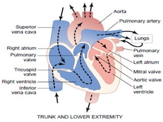
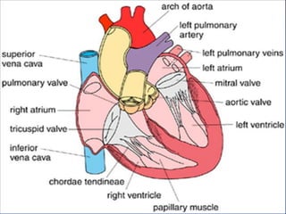
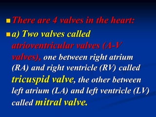
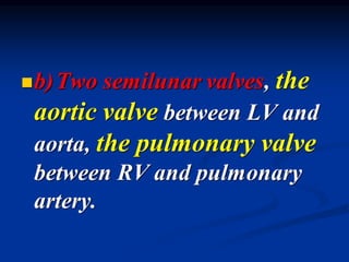
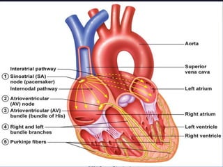
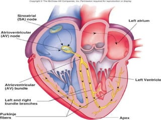
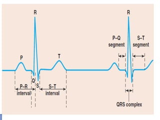
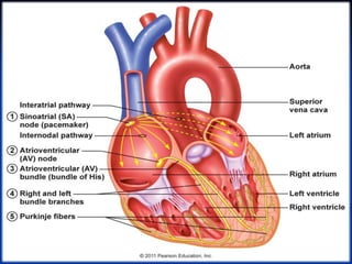
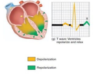
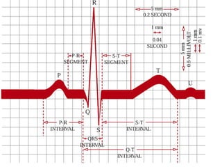
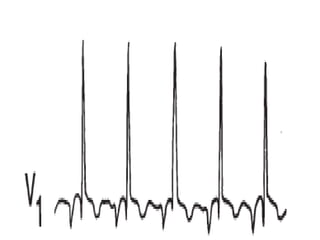
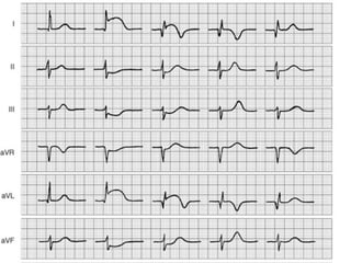
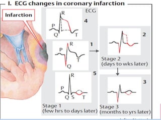
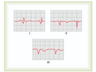
SlideShare a Scribd company logo
1 of 172
Download to read offline
Cardiovascular system
(CVS)
◼ The blood circulates in a closed system
called the cardiovascular system (CVS),
which consists of the heart and blood
vessels.
◼ The heart is a muscular organ divided
into right and left sides. Each side is
formed of two chambers (atrium and
ventricle).
◼ The wall of the heart is formed of
cardiac muscle (myocardium).
◼ The atrial myocardium is thinner
that of the ventricles.
◼ The atria act mainly as blood
reservoirs, while the ventricles act
mainly as pumping chambers.
◼There are 4 valves in the heart:
◼a) Two valves called
atrioventricular valves (A-V
valves), one between right atrium
(RA) and right ventricle (RV) called
tricuspid valve, the other between
left atrium (LA) and left ventricle (LV)
called mitral valve.
◼b)Two semilunar valves, the
aortic valve between LV and
aorta, the pulmonary valve
between RV and pulmonary
artery.
◼Division of circulation:
◼ 1- The systemic (greater) circulation:
◼ This starts from the left ventricle→
aorta → large arteries→ small
arteries→ arterioles→ capillaries→
venules→ veins→ superior and inferior
venae cavae→ right atrium.
◼2-The pulmonary (lesser)
circulation:
◼It starts from the right
ventricle→ pulmonary artery→
lungs→ pulmonary capillaries→
4 pulmonary veins → left atrium.
◼3-The special circulations:
◼These include the circulations
at specific sites e.g. the
capillary, venous, lymph,
coronary and cerebral
circulations.
◼Characteristic of the different
blood vessels:
◼ 1- The aorta, pulmonary artery and
large arteries are elastic vessels.
◼ 2- The medium- size and small
arteries are muscular low- resistance
vessels.
◼3- The arterioles are muscular
high- resistance vessels.
◼4- The capillaries are exchange
vessels.
◼5- The veins and pulmonary
vessels are volume reservoir.
◼Functions of the heart:
◼a)Function of the ventricles:
The only function of ventricles is
pumping of the blood into large
artery (LV→ aorta and RV →
pulmonary).
◼Function of the atria:
◼ 1- They are the entry- ways to the
ventricles.
◼ 2- Booster pump action.
◼ 2- The atrial walls contain stretch
receptors.
◼ 3- The atrial cells secrete the atrial
natriuretic peptide (ANP).
◼The cardiac muscle fibers
◼ There 3 types of cardiac muscle fibers:
◼ 1- The contractile cardiac muscle
fibers (99 %).
◼ These form the atrial and ventricular walls.
◼ They contain actin and myosin filaments.
◼ They perform the mechanical work of
pumping.
◼2-The nodal conducting
fibers.
◼They are modified cardiac
muscle fibers present in both
the sinoatrial and
atrioventricular nodes.
◼They contain more sarcoplasm
and very little myofibrils.
◼The nodal muscle fibers are
smaller than the contractile
fibers and are poor in gap
junctions.
◼ 3- The purkinje conducting fibers.
◼ They are modified cardiac muscle
fibers present in the His purkinje
system.
◼ They contain more sarcoplasm and
very little myofibrils.
◼ The fibers are larger than the
contractile fibers and are rich in gap
junctions
◼The nodal and the purkinje
conducting fibers form the
conduction system of the heart.
◼They do not contract, but are
specialized for initiation and
conduction of the impulses.
◼Nerve supply to the heart:
◼The heart receives
autonomic nerve supply
(sympathetic and
parasympathetic).
◼The fuel of the heart
◼ The heart is characterized by its ability to
oxidize several types of fuel substances.
◼ The major substances for the heart during
rest are the free fatty acids, glucose
and lactate.
◼ During muscular exercise the main
substrate becomes the lactate.
◼The cardiac conduction system:
This consists of the following:
◼1- The nodal system: this includes 2
nodes present in the right atrium
◼ a- The sinoatrial node (SAN).
◼ b- The atrioventricular node (AVN).
◼2-The internodal system: This
consists of 3 bundles called
the anterior, middle and
posterior internodal bundles
found in the right atrial wall.
◼3- The purkinje system:
This consists of 3 components:
◼ a- The atrioventricular bundle
(bundle of His).
◼ b- The right and left bundle
branches.
◼ c- The purkinje fibers.
The arterial blood
pressure
◼ This is the lateral pressure exerted
by the blood on the arterial walls.
◼It is generated by:
◼ 1- Pumping of the blood by the
ventricle into the aorta.
◼ 2- Presence of the peripheral
resistance to blood flow.
◼The systolic B.P:
◼Normally averages 120 mmHg
(range 90 -140 mmHg) and is
produced by ejection of blood
into the aorta during left
ventricular systole.
◼The diastolic B.P:
◼ Normally averages 80 mmHg (range 60 -
90 mmHg) and is produced as result of
the elastic recoil of the aorta during
ventricular diastole.
◼ The arterial B.P is often reported as
the systolic over the diastolic
pressure (e.g. 120/80).
◼The function of the arterial
blood pressure:
◼ 1- It maintains tissue perfusion
(blood flow) throughout different
tissues.
◼ 2- It produces the capillary
hydrostatic pressure.
◼3-The diastolic B.P performs
the following functions:
◼ a) It maintains blood flow to the tissues
during ventricular diastole.
◼ b) It is essential for normal coronary
blood flow.
◼ c) It prevents blood stasis in the arteries
during ventricular diastole.
◼Physiological factors that
affect the arterial B.P:
◼ 1- Body region.
◼ 2- Age.
◼ 3- Sex.
◼ 4- Body built.
◼ 5- Diurnal variation.
◼6- Meals.
◼7- Exercise.
◼8- Emotion.
◼9- Sexual intercourse.
◼10-Sleep.
◼11-Temperature.
◼Factors that determine and
maintain A.B.P:
◼1-Cardiac output (CO):
◼ It is the amount of blood pumped by
each ventricle per minute.
◼2-Peripheral resistance (PR):
it is determined by 3 factors:
◼ a) The diameter of the vessels.
◼ b) Blood viscosity.
◼ c) The length of the vessels.
◼3- Elasticity of the aorta and
large arteries.
◼4- Blood volume and circulatory
capacity.
Regulation of the A.B.P.:
A) Short- term mechanisms:
◼ These are potent mechanisms that
maintain survival. They act within a
few seconds and their action lasts for
several hours. They are mostly
nervous reflexes and include the
following:
◼(1)Arterial baroreflexes
(pressure buffer system):
◼ The receptors of these reflexes are located in
the carotid sinus and aortic arch.
◼ Arterial B.P. increases →stimulation of the
CIC and VDC and inhibition of the
VCC this results in reduction of the arterial
B.P by decreasing:
◼ (a) The cardiac pumping power
and CO (through causing
bradycardia)
◼ (b) The peripheral resistance
(through producing V.D. in both
the arterioles and venules)
◼ (c) Catecholamine secretion from
the adrenal medullae.
◼(2) Arterial chemoreflexes:
◼ The receptors of these reflexes are
located in the carotid and aortic
bodies and they are stimulated
when the arterial B.P. is reduced
below 60 mmHg (mainly as a result
of local ischaemia and hypoxia).
◼Arterial B.P. decreases
→stimulation of the VCC and
inhibition of the CIC and
VDC this results in elevation
of the arterial B.P by
increasing:
◼ (a) The cardiac pumping power and CO
(through causing tachycardia)
◼ (b) The peripheral resistance (through
producing V.C. in both the arterioles
and venules)
◼ (c) Catecholamine secretion from the
adrenal medullae.
◼3- The CNS ischaemic response:
◼Reduction of ABP below 60 mmHg
→ brain ischaemia → local
hypoxia → stimulation of the VCC
→ generalizes VC → ↑ ABP and
maintains the cerebral blood flow.
◼4-The abdominal compression
reflex:
◼ When the VCC is stimulated →
contraction of abdominal muscles → ↑
intra-abdominal pressure →
compresses the abdominal veins → ↑
venous return → ↑ cardiac output → ↑
ABP.
◼B) intermediate- term
mechanisms:
◼ They act within a few minutes and
their action lasts for several days.
◼ During this time, the nervous (rapid)
mechanisms usually fatigue and
become less effective. They include
the following:
◼1-Capillary fluid shift
mechanism:
◼Increases in the blood volume →
↑ capillary hydrostatic pressure →
↑ fluid filtration into tissue spaces
→ ↓ blood volume → ↓ ABP.
◼2- Stress relaxation mechanism:
◼↑ ABP → stretches the arteries
and increases the tension in their
walls → after sometime, the
arteries relax and tension in
their decreases → ↓ ABP.
◼ 3- Renin - angiotensin mechanism:
◼ ↓ ABP → renal ischaemia →
stimulates secretion of renin →
convert the angiotensinogen to
angiotensin I which converted to
angiotensin II by angiotensin-
converting enzyme → VC effect → ↑
ABP.
◼4-Right atrial mechanism:
◼ Increases in the blood volume →
stimulates the volume receptors in the
right atrium → following effects:
◼ a) Generalized VD.
◼ b) Reflex inhibition of secretion of
antidiuretic hormone.
◼ c) Secretion of atrial natriuretic
peptide (ANP).
◼C) long- term mechanisms:
◼ These mechanisms control the ABP by
adjusting the body fluids and blood
volume through modifying the
excretion of water and salt by the
kidneys. This occurs by:
◼ 1- Glomerular filtration.
◼ 2- Secretion of the aldosterone
hormone.
◼ ↓ ABP → ↓ glomerular filtration →
↓ renal excretion of water and salt
→ ↑ body fluid and blood volume
→ ↑ ABP.
◼ At the same time, renin is secreted
and angiotensin II is formed → VC
and stimulates aldosterone
secretion.
The electrocardiogram
(ECG)
◼The electrocardiogram is a record of
the electrical activity of the heart from
the surface of the body.
◼ It is produced by the sum of all action
potentials occurring in the cardiac
muscle fibers with each cardiac cycle.
◼The ECG is useful for the
diagnosis of:
◼ 1- Atrial and ventricular hypertrophy.
◼ 2- Myocardial infarction and ischaemia.
◼ 3- Arrhythmias.
◼ 4- Disturbances of electrolyte metabolism.
◼ 5- Drugs effect (digitalis).
◼ 6- Pericarditis.
◼Recording points on the body
surface:
◼There are nine standard points on
the body surface from which the
ECG should be recorded.
◼Six points are on the chest wall and the
other three points are at the limbs.
◼Chest points:
◼V1: at the right 4 intercostal
space near the sternum.
◼V2: at the left 4 intercostal
space near the sternum.
◼V3: midway between V2 and V4.
◼V4: at the 5 left intercostal space at
the midclavicular line.
◼V5: at the 5 left intercostal space
at the anterior axillary line.
◼V6: at the 5 left intercostal space
at the midaxillary line.
◼Limb points:
◼VL: at the junction of the left
arm with the trunk.
◼VR: at the junction of the right
arm with the trunk.
◼VF: at the junction of the left
lower limb with the trunk.
◼The ECG leads:
◼There are 3 types of leads:
◼Unipolar leads: (chest leads and
limb leads)
◼Bipolar leads: (LI, LII and LIII)
◼Augmented leads: (aVL, aVR and
aVF)
Normal ECG
◼ The ECG consists of 5 main waves
called P, Q, R, S, T and sometimes
U wave.
◼ The Q, R and S waves form a
complex called the QRS complex.
◼ The termination of the QRS
complex at the isoelectric line is
called the J point.
◼P wave
◼ This is a small blunt wave that is
produced as a result of atrial
depolarization.
◼ Its amplitude averages 0.1(up to
0.25) mV while its duration averages
0.08 (up to o.11) seconds.
◼It is +ve in the leads that face
the LV.
◼The first part of the wave is
due to RA activation while
its terminal part is due to LA
activation.
◼ Abnormalities of the P wave
◼ In the LA hypertrophy → P mitrale
(broad and notched) and their duration
increases.
◼ In the RA hypertrophy → P pulmonale (tall
and peaked) with less normal duration.
◼ In AV nodal rhythm → inverted P wave.
◼ In atrial fibrillation → disappear of P wave.
◼QRS complex
◼ This is produced as a result of
ventricular depolarization and its
duration is 0.06 - 0.1 second.
◼ The Q wave is due to depolarization
of interventricular septum and it is
normally –ve in the leads facing the
LV.
◼ The R wave is due to depolarization
of the apex and ventricular wall and
it is +ve in the leads facing the LV.
◼ The S wave is due to depolarization
of the posterobasal part of the LV
and the pulmonary conus and it is -ve
in the leads facing the LV.
◼Abnormalities of the QRS
complex:
◼ These occur in cases of:
◼ 1- Ventricular hypertrophy.
◼ 2- Myocardial infarction.
◼ 3- Bundle branch block.
◼ 4- Electrolyte disturbances.
◼T wave
◼ This is a large blunt wave that is
produced as a result of ventricular
repolarization.
◼ Its amplitude averages 0.2 (up to 0.4)
mV, while its duration averages 0.2 (up
to 0.25) second.
◼ It is normally +ve in the leads facing the
LV.
◼Abnormalities of the T wave
◼The T wave becomes inverted in
cases of:
◼ 1- Myocardial ischaemia.
◼ 2- Ventricular hypertrophy.
◼ 3- Bundle branch block.
◼ 4- Digitalis over dosage.
◼The T wave amplitude
increases in cases of:
◼1-Sympathetic over activity.
◼2-Muscular exercise.
◼3-Hyperkalemia.
◼U wave
◼This is a small +ve wave that
sometimes follows the T wave.
◼It is due to slow repolarization of
either the papillary muscles or
the purkinje network.
◼ECG segments:
◼An ECG segment is part of
the ECG record on the
isoelectric line between 2
waves.
◼P-R segment:
◼ It is the segment of the ECG from the
end of P wave to the start of the QRS
complex.
◼ Normal duration is 0.04- 0.13 second.
◼ During this segment, the atria are
completely depolarized but the
ventricles are completely polarized.
◼S-T segment:
◼ It is the segment of the ECG from
the end of S wave to the start of the
T wave.
◼ Normal duration is 0.12 second.
◼ During this segment, all ventricular
muscle fibers are completely
depolarized.
◼T-P segment:
◼ It is the segment of the ECG from
the end of T wave to the start of the
P wave of the next cycle.
◼ Normal duration is 0.25 second.
◼ During this segment, all atrial and
ventricular muscle fibers are
polarized.
◼ECG intervals
◼An ECG interval is part of
the ECG record that
includes a segment and
one or more waves.
◼P-R interval:
◼ It is the interval on the ECG from the
start of P wave to the start of R wave.
◼ Normal duration is 0.12 – 0.21
second.
◼ It is taken as index for the duration of
the AV nodal delay.
◼It is prolonged in:
◼ 1- First degree of heart block.
◼ 2- Increased vagal tone.
◼It is shortened in:
◼ 1- A-V nodal rhythm.
◼ 2- Sympathetic over activity.
◼ 3- Wolff-Parkinson-White syndrome
(WPW).
◼Q-T interval
◼ It is the interval on the ECG from the
start of Q wave to the end of T wave.
◼ Normal duration is 0.36 – 0.42 second
and it is called the electrical systole of
the heart.
◼ It is taken as index for the duration of
both the ventricular action potential and
refractory period.
◼T-Q interval
◼It is the interval on the ECG from
the end of T wave to the onset of
the next Q wave.
◼Normal duration is 0.4 second
and it is called the electrical
diastole of the heart.
◼It is shortened before atrial
and ventricular extrasystoles
but is prolonged mainly after
ventricular extrasystoles due
to compensatory pause.
◼Axis deviation
◼This occurs if the electric axis of
the heart is beyond the normal
range (between -30 and +110)
and it may be to right or to the
left.
◼Right axis deviation: this occurs in:
◼ 1- Tall thin subjects (normally).
◼ 2- Right ventricular hypertrophy.
◼ 3- Right bundle branch block.
◼ On in ECG, there are deep –ve waves
(S waves) in lead I and high +ve waves
(R waves) in lead III.
◼Left axis deviation: this occurs in:
◼ 1- Short subjects and pregnant women
(normally).
◼ 2- Left ventricular hypertrophy.
◼ 3- Left bundle branch block.
◼ On in ECG, there are high +ve waves
(R waves) in lead I and deep –ve waves
(S waves) in lead III.
◼ECG manifestation of
ventricular hypertrophy
◼1- Right ventricular hypertrophy.
◼2- Left ventricular hypertrophy.
◼ ECG characteristics of coronary
insufficiency:
◼ According to severity, coronary
insufficiency is associated with either:
◼ 1-Ischaemia: (inverted symmetric T wave)
◼ 2- Injury: (S-T segment elevation or
depression).
◼ 3- Necrosis (infarction): deep
(pathological) Q wave.
◼When a coronary artery is
occluded:
◼ 1- Necrosis usually occurs in the centre of
the affected area (deep Q wave).
◼ 2- This is surrounded by area of injury (S-
T segment shift).
◼ 3- This is surrounded by an ischaemic
area (inversion of T wave).
Heart rate
◼The cardiovascular centres:
◼These are collections of neurons
located in the medulla oblongata
which regulate the functions of
the CVS:
◼ 1- The cardioinhibitory centre
(CIC).
◼ 2- The vasomotor centre (VMC):
this includes 2 component:
◼ a- Vasoconstrictor centre (VCC or
pressor area).
◼ b- Vasodilator centre (VDC or
depressor area).
◼ Physiological variation of heart rate:
◼ 1- Age.
◼ 2- Sex.
◼ 3- Time of the day (circadian rhythm).
◼ 4- Rest and sleep.
◼ 5- Physical training.
◼ 6- Posture.
◼Regulation of the heart rate
◼The heart rate refers to the
ventricular rate of beating /
minute.
◼Normally, it averages 75 beats
/minute (60 – 100 b/minute).
◼A heart rate higher than 100
beats/min is called
tachycardia.
◼A heart rate lower than 60
beats/min is called
bradycardia.
◼The frequency of discharge of
the S-A node is regulated by:
◼ 1- Nervous factor.
◼ 2- Chemical factors.
◼ 3- Certain factors that directly affected
the SA node activity.
◼Nervous regulation of the H.R
◼ The activity of these centres is affected by:
◼a-Supraspinal centres:
◼ 1- The cerebral cortex: the HR is
affected by emotions and conditioned
reflexes.
◼ 2- The hypothalamus and limbic
system: these structures are concerned
with emotional reactions.
◼3-The respiratory centre:
◼Respiratory sinus arrhythmia:
this term refers to increase of HR
during inspiration and its
decrease during expiration.
◼The tachycardia that occurs
during inspiration is due to:
◼ a- Irradiation of impulses from the
inspiratory centre which excite the
VCC.
◼ b- Lung inflation through a
pulmonary stretch reflex.
◼ c- Bainbridge reflex or effect.
◼b-Reflexes initiated from the
CVS:
◼1-Marey,s reflex or law
◼ ↑of ABP → ↓ HR.
◼ ↓of ABP → ↑ HR.
◼Effects of stimulation of arterial
baroreceptors:
◼ a- Stimulation of both the CIC and
VDC and inhibition of the VCC.
◼ b- Inhibition of respiratory centre
→ apnea.
◼ c- Inhibition of secretion of the
ADH.
◼ d- Inhibition of the brain activity
and muscle tone.
◼2- Bainbridge reflex
(atrial stretch reflex):
◼An increase in the right
atrial pressure leads to heart
acceleration.
◼ Increase in RA pressure →
Stimulation of the type A atrial
receptors → stimulate VCC → heart
acceleration.
◼ Atrial distension → Stimulation of
the type B atrial receptors → heart
slowing.
◼Effects of rise of the right atrial
pressure:
◼ a- Tachycardia (Bainbridge reflex).
◼ b- Tachypnea (Harrison,s reflex).
◼ c- Generalized VD and decrease of the
ABP.
◼ d- Coronary VD (anrep,s reflex).
◼ e- Inhibition of secretion of ADH.
◼ f- Stimulation of secretion of ANP.
◼3- The ventricular stretch reflex:
◼ Distension of the LV leads to
bradycardia and hypotension.
◼ Stimulation of certain baroreceptors
located near the coronary vessels →
stimulation of CIC and VDC and
inhibition of the VCC.
◼4- The Bezold- Jarisch reflex
(coronary chemoreflex):
◼ Injection of certain substances
(serotonin) into the coronary
arteries that supply the LV leads to
apnea followed by rapid breathing,
hypotension and bradycardia.
◼c- Reflexes initiated from
extravascular structures:
◼1- From the lungs
◼ a- The pulmonary stretch reflex:
◼ Lung inflation leads to tachycardia.
◼ Stimulation of baroreceptors located in
bronchial walls → stimulation of VCC →
tachycardia.
◼ b- The pulmonary chemoreflex.
◼2- From skeletal muscles (Alam-
Smirk reflex):
◼ Voluntary contraction of the skeletal
muscles leads to tachycardia and
tachypnea.
◼3- From the skin and viscera:
◼ Cold and mild pain → tachycardia.
◼ Severe pain → bradycardia.
◼4-From the eye (oculo- cardiac
reflex):
◼Applying pressure to the eyeball
results in reflex decrease of the
heart rate.
◼5-From the trigger zones
(trigger zone reflexes):
◼Signals from certain sensitive
areas in the body as the trigger
area (larynx, testes and
epigastric region) → slowing of
the heart.
◼2- Chemical regulation of the heart
rate
◼ A-Effects of changes in blood gases
◼1- Hypoxia:
◼ A moderate O2 lack increases the heart
rate by 3 mechanisms:
◼ a- Direct mechanism.
◼ b- Central mechanism.
◼ c- Reflex mechanism
◼Severe hypoxia → ↓ HR
due to:
◼- Inhibition of S-A node
activity.
◼- Paralysis of CVS center in
medulla oblongata.
◼2-Hypercapnia and acidosis
◼ Moderate hypercapnia and acidosis
→ ↑ HR by 3 mechanisms:
◼ a- Inhibition of the CIC.
◼ b- Stimulation of the VCC
(peripheral chemoreceptors).
◼ c- Stimulation of the VCC (central
chemoreceptors).
◼Severe hypercapnia → ↓ HR
due to:
◼- Inhibition of S-A node activity.
◼- Paralysis of CVS center in
medulla oblongata.
◼B- Effects of hormones ,
drugs and chemical:
◼ 1- Adrenaline. 2-Noradrenaline.
◼ 3- Thyroxin. 4-Atropine.
◼ 5- Histamine.
◼ 6- Bile salts.
◼ 7- Autonomic drugs.
◼3- Factors that directly
affect the S-A node activity:
◼1-Physical factors.
◼2-Mechanical factors.
◼3-Chemical factors.
The cardiac output
◼Cardiac output is the volume of
blood pumped by each ventricle per
minute.
◼ It equals the stroke volume x heart rate.
◼ In normal adult male during rest, it is
about 5.5 L/ min.
◼ Stroke volume is the volume of blood
pumped by each ventricle per beat.
◼Factors that affect the end
diastolic volume:
◼ 1- Right atrial pressure (RAP).
◼ 2- Blood volume:
Hypovolaemia → ↓ MCP → ↓ VR
and EDV.
◼3- Venous tone:
Venoconstriction → ↑MCP → ↑
VR and EDV.
◼4- Intrapericardial pressure:
↑ intrapericradial pressure → ↓
EDV.
◼5- Atrial fibrillation:
AF → weakness of atrial contraction
→ ↓ EDV.
◼6- Intrathoracic pressure:
↑ of its negativity → help ventricular
relaxation and ↑VR → ↑ EDV.
◼7- Skeletal muscle contraction.
◼8- Posture.
◼9- Ventricular stiffness and
compliance.
◼10- Atrial systole.
◼Factors that affect the cardiac
output:
◼The cardiac output is affected
by changes in either HR or
the stroke volume.
◼ Effects of changes in the HR on the
CO:
◼ If the HR decreases below 70 beats /
minute.
◼ If the HR decreases below 50 beats /
minute.
◼ If the HR increases from 70 to 180 beats /
minute.
◼ If the HR increases above 180 beats /
minute.
◼Factors that affect the stroke
volume:
◼1- The cardiac pumping power
(maximal CO that can be pumped
per minute) : it is affected by the
following factors:
◼A- The preload (EDV):
This is an intrinsic autoregulatory
mechanism that controls the power
of cardiac contractility by Starling's
law.
◼The resting cardiac pumping
power is about 10 L/min.
◼B- Sympathetic nerve supply:
The resting cardiac sympathetic
tone increases the cardiac
pumping power to 13-15 L/min.
◼Maximal sympathetic stimulation
increases it to about 25 L/min.
◼C-The afterload: an increase in
the afterload reduces the cardiac
pumping power.
◼D- Ventricular hypertrophy: in
athletes, the cardiac pumping
power increase up to 35 L/min.
◼2-The venous return (VR): The
volume of the venous return is
affected by the following factors:
◼ A-The mean circulatory pressure
(MCP): this the main pressure in
systemic circulation.
◼ Normally, it is about 7 mmHg and it
affects venous return directly.
◼B-The right atrial pressure:
It is closely related to the central
venous pressure (CVP) and VR
is inversely proportional to it.
◼C- The resistance to the VR
(RVR):
◼This occurs mostly in the
veins and normally, it is about
1.4 mmHg/ liter of blood flow
and it affects the VR inversely.
◼Regulation of the cardiac
output:
◼1- Extrinsic regulation
◼This regulates both the stroke
volume and heart rate by effect
of:
◼a-The autonomic nerves to
the heart:
◼1-Sympathetic stimulation:
◼ Sympathetic stimulation increases
the cardiac output by increasing the
stroke volume and heart rate
because sympathetic stimulation
leads to:
◼ ↑ the rhythmicity of SA node
(+ve chronotropic effect).
◼ ↑ the force of cardiac muscle
contraction (+ve inotropic
effect).
◼ ↑ the coronary blood flow.
◼2- Parasympathetic stimulation:
◼ Parasympathetic stimulation decreases the
cardiac output by:
◼ ↓ the rhythmicity of SA node (-ve
chronotropic effect).
◼ ↓ the force of cardiac muscle contraction
(-ve inotropic effect).
◼ ↓ the coronary blood flow.
◼b-Hormones and drugs:
◼1- Glucagon hormone: increase
the cardiac output by increasing the
force of cardiac muscle contration.
◼2- Catecholamines: increase the
cardiac output by increasing SV and
HR.
◼3- Acetylcholine: decrease the
cardiac output by decreasing the HR.
◼4- Thyroxin: increase the cardiac
output by increasing the HR.
◼5- Xanthines: such as caffeine:
increase the cardiac output by
increasing the force of contraction.
◼6- Digitalis: increase the cardiac
output by increasing the force of
contraction.
◼7- Hypoxia, hypercapnia,
acidosis and myocardial
ischemia: decrease the cardiac
output by decreasing the HR and
cardiac contractility.
◼2-Intrinsic regulation:
◼This regulates the stroke
volume only and it includes
heterometric and homeometric
autoregulation.
◼a-Heterometric autoregulation:
◼ This regulation of the stroke volume
through changing the initial length of the
muscle fibers.
◼ Filling of the ventricle → ventricular
expansion → ↑ in EDV and length of
muscle fibers → ↑ the force of contraction
→ ↑ stroke volume.
◼B- Homeometric autoregulation:
◼ This regulation of the stroke volume
without change in the length of the
muscle fibers.
◼ A prolonged increase in EDV is
followed by its decrease to its normal
level.
◼ESV decreases less than its
normal level by more forceful
ventricular contraction.
◼The stroke volume remains
elevated.
◼Physiological variations of
the cardiac output:
◼1-Sleep.
◼2-Posture.
◼3-Meals.
◼4-Temperature.
◼5-Pregnancy.
◼6-Emotions.
◼7-Muscular exercise.
CVS Physiology mecical physiology le .pdf

More Related Content

Similar to CVS Physiology mecical physiology le .pdf

Cardiovascularsystem 110221045748-phpapp02
Cardiovascularsystem 110221045748-phpapp02Cardiovascularsystem 110221045748-phpapp02
Cardiovascularsystem 110221045748-phpapp02
Tracy Phillips
 
Cardiac anatomy and physiology
Cardiac anatomy and physiologyCardiac anatomy and physiology
Cardiac anatomy and physiology
MitchellChapman5
 
Cardiac A&P Review
Cardiac A&P ReviewCardiac A&P Review
Cardiac A&P Review
TeleClinEd
 

Similar to CVS Physiology mecical physiology le .pdf (20)

coronarycirculation-
coronarycirculation-coronarycirculation-
coronarycirculation-
 
coronarycirculation-pptx
coronarycirculation-pptxcoronarycirculation-pptx
coronarycirculation-pptx
 
Venous circulation
Venous circulationVenous circulation
Venous circulation
 
coronary circulation -1305080032111-phpappa02 pdf
coronary circulation -1305080032111-phpappa02 pdfcoronary circulation -1305080032111-phpappa02 pdf
coronary circulation -1305080032111-phpappa02 pdf
 
Coronary circulation
Coronary circulationCoronary circulation
Coronary circulation
 
Cardiovascular System Part 1
Cardiovascular System Part 1Cardiovascular System Part 1
Cardiovascular System Part 1
 
Physical assessment
Physical assessmentPhysical assessment
Physical assessment
 
Cardiovascularsystem 110221045748-phpapp02
Cardiovascularsystem 110221045748-phpapp02Cardiovascularsystem 110221045748-phpapp02
Cardiovascularsystem 110221045748-phpapp02
 
Arterial blood pressure
Arterial blood pressureArterial blood pressure
Arterial blood pressure
 
Cardiac anatomy and physiology
Cardiac anatomy and physiologyCardiac anatomy and physiology
Cardiac anatomy and physiology
 
Cardiovascular system.pptx
 Cardiovascular system.pptx Cardiovascular system.pptx
Cardiovascular system.pptx
 
Coronary circulation
Coronary circulationCoronary circulation
Coronary circulation
 
Cardiac A&P Review
Cardiac A&P ReviewCardiac A&P Review
Cardiac A&P Review
 
Cardiovascular system
Cardiovascular systemCardiovascular system
Cardiovascular system
 
Echo class 2_05262015
Echo class 2_05262015Echo class 2_05262015
Echo class 2_05262015
 
Ppt cvs phsiology a small review for anaesthetist
Ppt cvs phsiology a small review for anaesthetistPpt cvs phsiology a small review for anaesthetist
Ppt cvs phsiology a small review for anaesthetist
 
4-lecture cardiovascular Pharmacy 2024.pptx
4-lecture cardiovascular Pharmacy 2024.pptx4-lecture cardiovascular Pharmacy 2024.pptx
4-lecture cardiovascular Pharmacy 2024.pptx
 
Cardio 1
Cardio 1Cardio 1
Cardio 1
 
7. CARDIOVASCULAR PHYSIOLOGY.pdf bsc nursing
7. CARDIOVASCULAR  PHYSIOLOGY.pdf bsc nursing7. CARDIOVASCULAR  PHYSIOLOGY.pdf bsc nursing
7. CARDIOVASCULAR PHYSIOLOGY.pdf bsc nursing
 
cardiac output,
cardiac output, cardiac output,
cardiac output,
 

More from MosaHasen

7- HssssssssssssssssssTN-2020ثثثثثث.pptx
7- HssssssssssssssssssTN-2020ثثثثثث.pptx7- HssssssssssssssssssTN-2020ثثثثثث.pptx
7- HssssssssssssssssssTN-2020ثثثثثث.pptx
MosaHasen
 
Vital_signs-BcccccccccccccvvvvvvvvP-1.ppt
Vital_signs-BcccccccccccccvvvvvvvvP-1.pptVital_signs-BcccccccccccccvvvvvvvvP-1.ppt
Vital_signs-BcccccccccccccvvvvvvvvP-1.ppt
MosaHasen
 
Lecture 4-Valvularcccccccc Heart Diseases.ppt
Lecture 4-Valvularcccccccc Heart Diseases.pptLecture 4-Valvularcccccccc Heart Diseases.ppt
Lecture 4-Valvularcccccccc Heart Diseases.ppt
MosaHasen
 
Vital_signdddddddddddddddddddds-BP-1.ppt
Vital_signdddddddddddddddddddds-BP-1.pptVital_signdddddddddddddddddddds-BP-1.ppt
Vital_signdddddddddddddddddddds-BP-1.ppt
MosaHasen
 
Lecture 4-Valvuddddlar Heart Diseases.ppt
Lecture 4-Valvuddddlar Heart Diseases.pptLecture 4-Valvuddddlar Heart Diseases.ppt
Lecture 4-Valvuddddlar Heart Diseases.ppt
MosaHasen
 
autonoddddddddddddddddddddddddmic 1 .ppt
autonoddddddddddddddddddddddddmic 1 .pptautonoddddddddddddddddddddddddmic 1 .ppt
autonoddddddddddddddddddddddddmic 1 .ppt
MosaHasen
 
autdddddddddddddddddddddddddonomic 1 .ppt
autdddddddddddddddddddddddddonomic 1 .pptautdddddddddddddddddddddddddonomic 1 .ppt
autdddddddddddddddddddddddddonomic 1 .ppt
MosaHasen
 
Vital_sfffffffffffffffffffffigns-BP-1.ppt
Vital_sfffffffffffffffffffffigns-BP-1.pptVital_sfffffffffffffffffffffigns-BP-1.ppt
Vital_sfffffffffffffffffffffigns-BP-1.ppt
MosaHasen
 
Lecture 4-Valvulfffar Heart Diseases.ppt
Lecture 4-Valvulfffar Heart Diseases.pptLecture 4-Valvulfffar Heart Diseases.ppt
Lecture 4-Valvulfffar Heart Diseases.ppt
MosaHasen
 
Lecture 4-Valvular Heddddart Diseases.ppt
Lecture 4-Valvular Heddddart Diseases.pptLecture 4-Valvular Heddddart Diseases.ppt
Lecture 4-Valvular Heddddart Diseases.ppt
MosaHasen
 
Vital_signs-BPdddddddddddddddddddd-1.ppt
Vital_signs-BPdddddddddddddddddddd-1.pptVital_signs-BPdddddddddddddddddddd-1.ppt
Vital_signs-BPdddddddddddddddddddd-1.ppt
MosaHasen
 
autonomddddddddddddddddddddddddic 1 .ppt
autonomddddddddddddddddddddddddic 1 .pptautonomddddddddddddddddddddddddic 1 .ppt
autonomddddddddddddddddddddddddic 1 .ppt
MosaHasen
 
Lecture 4-Valvular Hikkeart Diseases.ppt
Lecture 4-Valvular Hikkeart Diseases.pptLecture 4-Valvular Hikkeart Diseases.ppt
Lecture 4-Valvular Hikkeart Diseases.ppt
MosaHasen
 
Lecture 4-Vajjjlvular Heart Diseases.ppt
Lecture 4-Vajjjlvular Heart Diseases.pptLecture 4-Vajjjlvular Heart Diseases.ppt
Lecture 4-Vajjjlvular Heart Diseases.ppt
MosaHasen
 
Traudddddddddddddddddddddddddddddma .ppt
Traudddddddddddddddddddddddddddddma .pptTraudddddddddddddddddddddddddddddma .ppt
Traudddddddddddddddddddddddddddddma .ppt
MosaHasen
 
Traumaaaaaaaaaaaaaaaaaaaaaaaaaaaaaa .ppt
Traumaaaaaaaaaaaaaaaaaaaaaaaaaaaaaa .pptTraumaaaaaaaaaaaaaaaaaaaaaaaaaaaaaa .ppt
Traumaaaaaaaaaaaaaaaaaaaaaaaaaaaaaa .ppt
MosaHasen
 
parhhhhhhhhhhhhhhhhhhhhhhhhhhathyriod.ppt
parhhhhhhhhhhhhhhhhhhhhhhhhhhathyriod.pptparhhhhhhhhhhhhhhhhhhhhhhhhhhathyriod.ppt
parhhhhhhhhhhhhhhhhhhhhhhhhhhathyriod.ppt
MosaHasen
 
Vital_signs-BddddddddddddddddddddP-1.ppt
Vital_signs-BddddddddddddddddddddP-1.pptVital_signs-BddddddddddddddddddddP-1.ppt
Vital_signs-BddddddddddddddddddddP-1.ppt
MosaHasen
 
hydrdddddddddddddddddddddddddddddo..pptx
hydrdddddddddddddddddddddddddddddo..pptxhydrdddddddddddddddddddddddddddddo..pptx
hydrdddddddddddddddddddddddddddddo..pptx
MosaHasen
 
autonomicfdfdddddddddddddddddddddf 1 .ppt
autonomicfdfdddddddddddddddddddddf 1 .pptautonomicfdfdddddddddddddddddddddf 1 .ppt
autonomicfdfdddddddddddddddddddddf 1 .ppt
MosaHasen
 

More from MosaHasen (20)

7- HssssssssssssssssssTN-2020ثثثثثث.pptx
7- HssssssssssssssssssTN-2020ثثثثثث.pptx7- HssssssssssssssssssTN-2020ثثثثثث.pptx
7- HssssssssssssssssssTN-2020ثثثثثث.pptx
 
Vital_signs-BcccccccccccccvvvvvvvvP-1.ppt
Vital_signs-BcccccccccccccvvvvvvvvP-1.pptVital_signs-BcccccccccccccvvvvvvvvP-1.ppt
Vital_signs-BcccccccccccccvvvvvvvvP-1.ppt
 
Lecture 4-Valvularcccccccc Heart Diseases.ppt
Lecture 4-Valvularcccccccc Heart Diseases.pptLecture 4-Valvularcccccccc Heart Diseases.ppt
Lecture 4-Valvularcccccccc Heart Diseases.ppt
 
Vital_signdddddddddddddddddddds-BP-1.ppt
Vital_signdddddddddddddddddddds-BP-1.pptVital_signdddddddddddddddddddds-BP-1.ppt
Vital_signdddddddddddddddddddds-BP-1.ppt
 
Lecture 4-Valvuddddlar Heart Diseases.ppt
Lecture 4-Valvuddddlar Heart Diseases.pptLecture 4-Valvuddddlar Heart Diseases.ppt
Lecture 4-Valvuddddlar Heart Diseases.ppt
 
autonoddddddddddddddddddddddddmic 1 .ppt
autonoddddddddddddddddddddddddmic 1 .pptautonoddddddddddddddddddddddddmic 1 .ppt
autonoddddddddddddddddddddddddmic 1 .ppt
 
autdddddddddddddddddddddddddonomic 1 .ppt
autdddddddddddddddddddddddddonomic 1 .pptautdddddddddddddddddddddddddonomic 1 .ppt
autdddddddddddddddddddddddddonomic 1 .ppt
 
Vital_sfffffffffffffffffffffigns-BP-1.ppt
Vital_sfffffffffffffffffffffigns-BP-1.pptVital_sfffffffffffffffffffffigns-BP-1.ppt
Vital_sfffffffffffffffffffffigns-BP-1.ppt
 
Lecture 4-Valvulfffar Heart Diseases.ppt
Lecture 4-Valvulfffar Heart Diseases.pptLecture 4-Valvulfffar Heart Diseases.ppt
Lecture 4-Valvulfffar Heart Diseases.ppt
 
Lecture 4-Valvular Heddddart Diseases.ppt
Lecture 4-Valvular Heddddart Diseases.pptLecture 4-Valvular Heddddart Diseases.ppt
Lecture 4-Valvular Heddddart Diseases.ppt
 
Vital_signs-BPdddddddddddddddddddd-1.ppt
Vital_signs-BPdddddddddddddddddddd-1.pptVital_signs-BPdddddddddddddddddddd-1.ppt
Vital_signs-BPdddddddddddddddddddd-1.ppt
 
autonomddddddddddddddddddddddddic 1 .ppt
autonomddddddddddddddddddddddddic 1 .pptautonomddddddddddddddddddddddddic 1 .ppt
autonomddddddddddddddddddddddddic 1 .ppt
 
Lecture 4-Valvular Hikkeart Diseases.ppt
Lecture 4-Valvular Hikkeart Diseases.pptLecture 4-Valvular Hikkeart Diseases.ppt
Lecture 4-Valvular Hikkeart Diseases.ppt
 
Lecture 4-Vajjjlvular Heart Diseases.ppt
Lecture 4-Vajjjlvular Heart Diseases.pptLecture 4-Vajjjlvular Heart Diseases.ppt
Lecture 4-Vajjjlvular Heart Diseases.ppt
 
Traudddddddddddddddddddddddddddddma .ppt
Traudddddddddddddddddddddddddddddma .pptTraudddddddddddddddddddddddddddddma .ppt
Traudddddddddddddddddddddddddddddma .ppt
 
Traumaaaaaaaaaaaaaaaaaaaaaaaaaaaaaa .ppt
Traumaaaaaaaaaaaaaaaaaaaaaaaaaaaaaa .pptTraumaaaaaaaaaaaaaaaaaaaaaaaaaaaaaa .ppt
Traumaaaaaaaaaaaaaaaaaaaaaaaaaaaaaa .ppt
 
parhhhhhhhhhhhhhhhhhhhhhhhhhhathyriod.ppt
parhhhhhhhhhhhhhhhhhhhhhhhhhhathyriod.pptparhhhhhhhhhhhhhhhhhhhhhhhhhhathyriod.ppt
parhhhhhhhhhhhhhhhhhhhhhhhhhhathyriod.ppt
 
Vital_signs-BddddddddddddddddddddP-1.ppt
Vital_signs-BddddddddddddddddddddP-1.pptVital_signs-BddddddddddddddddddddP-1.ppt
Vital_signs-BddddddddddddddddddddP-1.ppt
 
hydrdddddddddddddddddddddddddddddo..pptx
hydrdddddddddddddddddddddddddddddo..pptxhydrdddddddddddddddddddddddddddddo..pptx
hydrdddddddddddddddddddddddddddddo..pptx
 
autonomicfdfdddddddddddddddddddddf 1 .ppt
autonomicfdfdddddddddddddddddddddf 1 .pptautonomicfdfdddddddddddddddddddddf 1 .ppt
autonomicfdfdddddddddddddddddddddf 1 .ppt
 

Recently uploaded

Call Girls Aurangabad Just Call 8250077686 Top Class Call Girl Service Available
Call Girls Aurangabad Just Call 8250077686 Top Class Call Girl Service AvailableCall Girls Aurangabad Just Call 8250077686 Top Class Call Girl Service Available
Call Girls Aurangabad Just Call 8250077686 Top Class Call Girl Service Available
Dipal Arora
 

Recently uploaded (20)

Call Girls Service Jaipur {9521753030} ❤️VVIP RIDDHI Call Girl in Jaipur Raja...
Call Girls Service Jaipur {9521753030} ❤️VVIP RIDDHI Call Girl in Jaipur Raja...Call Girls Service Jaipur {9521753030} ❤️VVIP RIDDHI Call Girl in Jaipur Raja...
Call Girls Service Jaipur {9521753030} ❤️VVIP RIDDHI Call Girl in Jaipur Raja...
 
Premium Call Girls Cottonpet Whatsapp 7001035870 Independent Escort Service
Premium Call Girls Cottonpet Whatsapp 7001035870 Independent Escort ServicePremium Call Girls Cottonpet Whatsapp 7001035870 Independent Escort Service
Premium Call Girls Cottonpet Whatsapp 7001035870 Independent Escort Service
 
Call Girls Jabalpur Just Call 8250077686 Top Class Call Girl Service Available
Call Girls Jabalpur Just Call 8250077686 Top Class Call Girl Service AvailableCall Girls Jabalpur Just Call 8250077686 Top Class Call Girl Service Available
Call Girls Jabalpur Just Call 8250077686 Top Class Call Girl Service Available
 
O898O367676 Call Girls In Ahmedabad Escort Service Available 24×7 In Ahmedabad
O898O367676 Call Girls In Ahmedabad Escort Service Available 24×7 In AhmedabadO898O367676 Call Girls In Ahmedabad Escort Service Available 24×7 In Ahmedabad
O898O367676 Call Girls In Ahmedabad Escort Service Available 24×7 In Ahmedabad
 
(👑VVIP ISHAAN ) Russian Call Girls Service Navi Mumbai🖕9920874524🖕Independent...
(👑VVIP ISHAAN ) Russian Call Girls Service Navi Mumbai🖕9920874524🖕Independent...(👑VVIP ISHAAN ) Russian Call Girls Service Navi Mumbai🖕9920874524🖕Independent...
(👑VVIP ISHAAN ) Russian Call Girls Service Navi Mumbai🖕9920874524🖕Independent...
 
Top Rated Hyderabad Call Girls Erragadda ⟟ 9332606886 ⟟ Call Me For Genuine ...
Top Rated  Hyderabad Call Girls Erragadda ⟟ 9332606886 ⟟ Call Me For Genuine ...Top Rated  Hyderabad Call Girls Erragadda ⟟ 9332606886 ⟟ Call Me For Genuine ...
Top Rated Hyderabad Call Girls Erragadda ⟟ 9332606886 ⟟ Call Me For Genuine ...
 
Call Girls Siliguri Just Call 8250077686 Top Class Call Girl Service Available
Call Girls Siliguri Just Call 8250077686 Top Class Call Girl Service AvailableCall Girls Siliguri Just Call 8250077686 Top Class Call Girl Service Available
Call Girls Siliguri Just Call 8250077686 Top Class Call Girl Service Available
 
Call Girls Ooty Just Call 8250077686 Top Class Call Girl Service Available
Call Girls Ooty Just Call 8250077686 Top Class Call Girl Service AvailableCall Girls Ooty Just Call 8250077686 Top Class Call Girl Service Available
Call Girls Ooty Just Call 8250077686 Top Class Call Girl Service Available
 
Night 7k to 12k Navi Mumbai Call Girl Photo 👉 BOOK NOW 9833363713 👈 ♀️ night ...
Night 7k to 12k Navi Mumbai Call Girl Photo 👉 BOOK NOW 9833363713 👈 ♀️ night ...Night 7k to 12k Navi Mumbai Call Girl Photo 👉 BOOK NOW 9833363713 👈 ♀️ night ...
Night 7k to 12k Navi Mumbai Call Girl Photo 👉 BOOK NOW 9833363713 👈 ♀️ night ...
 
Call Girls Aurangabad Just Call 8250077686 Top Class Call Girl Service Available
Call Girls Aurangabad Just Call 8250077686 Top Class Call Girl Service AvailableCall Girls Aurangabad Just Call 8250077686 Top Class Call Girl Service Available
Call Girls Aurangabad Just Call 8250077686 Top Class Call Girl Service Available
 
Call Girls Dehradun Just Call 9907093804 Top Class Call Girl Service Available
Call Girls Dehradun Just Call 9907093804 Top Class Call Girl Service AvailableCall Girls Dehradun Just Call 9907093804 Top Class Call Girl Service Available
Call Girls Dehradun Just Call 9907093804 Top Class Call Girl Service Available
 
Call Girls Bangalore Just Call 8250077686 Top Class Call Girl Service Available
Call Girls Bangalore Just Call 8250077686 Top Class Call Girl Service AvailableCall Girls Bangalore Just Call 8250077686 Top Class Call Girl Service Available
Call Girls Bangalore Just Call 8250077686 Top Class Call Girl Service Available
 
♛VVIP Hyderabad Call Girls Chintalkunta🖕7001035870🖕Riya Kappor Top Call Girl ...
♛VVIP Hyderabad Call Girls Chintalkunta🖕7001035870🖕Riya Kappor Top Call Girl ...♛VVIP Hyderabad Call Girls Chintalkunta🖕7001035870🖕Riya Kappor Top Call Girl ...
♛VVIP Hyderabad Call Girls Chintalkunta🖕7001035870🖕Riya Kappor Top Call Girl ...
 
Top Rated Bangalore Call Girls Richmond Circle ⟟ 9332606886 ⟟ Call Me For Ge...
Top Rated Bangalore Call Girls Richmond Circle ⟟  9332606886 ⟟ Call Me For Ge...Top Rated Bangalore Call Girls Richmond Circle ⟟  9332606886 ⟟ Call Me For Ge...
Top Rated Bangalore Call Girls Richmond Circle ⟟ 9332606886 ⟟ Call Me For Ge...
 
Call Girls Agra Just Call 8250077686 Top Class Call Girl Service Available
Call Girls Agra Just Call 8250077686 Top Class Call Girl Service AvailableCall Girls Agra Just Call 8250077686 Top Class Call Girl Service Available
Call Girls Agra Just Call 8250077686 Top Class Call Girl Service Available
 
Call Girls Bareilly Just Call 8250077686 Top Class Call Girl Service Available
Call Girls Bareilly Just Call 8250077686 Top Class Call Girl Service AvailableCall Girls Bareilly Just Call 8250077686 Top Class Call Girl Service Available
Call Girls Bareilly Just Call 8250077686 Top Class Call Girl Service Available
 
Premium Call Girls In Jaipur {8445551418} ❤️VVIP SEEMA Call Girl in Jaipur Ra...
Premium Call Girls In Jaipur {8445551418} ❤️VVIP SEEMA Call Girl in Jaipur Ra...Premium Call Girls In Jaipur {8445551418} ❤️VVIP SEEMA Call Girl in Jaipur Ra...
Premium Call Girls In Jaipur {8445551418} ❤️VVIP SEEMA Call Girl in Jaipur Ra...
 
All Time Service Available Call Girls Marine Drive 📳 9820252231 For 18+ VIP C...
All Time Service Available Call Girls Marine Drive 📳 9820252231 For 18+ VIP C...All Time Service Available Call Girls Marine Drive 📳 9820252231 For 18+ VIP C...
All Time Service Available Call Girls Marine Drive 📳 9820252231 For 18+ VIP C...
 
Top Quality Call Girl Service Kalyanpur 6378878445 Available Call Girls Any Time
Top Quality Call Girl Service Kalyanpur 6378878445 Available Call Girls Any TimeTop Quality Call Girl Service Kalyanpur 6378878445 Available Call Girls Any Time
Top Quality Call Girl Service Kalyanpur 6378878445 Available Call Girls Any Time
 
Call Girls Gwalior Just Call 8617370543 Top Class Call Girl Service Available
Call Girls Gwalior Just Call 8617370543 Top Class Call Girl Service AvailableCall Girls Gwalior Just Call 8617370543 Top Class Call Girl Service Available
Call Girls Gwalior Just Call 8617370543 Top Class Call Girl Service Available
 

CVS Physiology mecical physiology le .pdf

  • 1.
  • 3. ◼ The blood circulates in a closed system called the cardiovascular system (CVS), which consists of the heart and blood vessels. ◼ The heart is a muscular organ divided into right and left sides. Each side is formed of two chambers (atrium and ventricle).
  • 4. ◼ The wall of the heart is formed of cardiac muscle (myocardium). ◼ The atrial myocardium is thinner that of the ventricles. ◼ The atria act mainly as blood reservoirs, while the ventricles act mainly as pumping chambers.
  • 5.
  • 6.
  • 7. ◼There are 4 valves in the heart: ◼a) Two valves called atrioventricular valves (A-V valves), one between right atrium (RA) and right ventricle (RV) called tricuspid valve, the other between left atrium (LA) and left ventricle (LV) called mitral valve.
  • 8. ◼b)Two semilunar valves, the aortic valve between LV and aorta, the pulmonary valve between RV and pulmonary artery.
  • 9. ◼Division of circulation: ◼ 1- The systemic (greater) circulation: ◼ This starts from the left ventricle→ aorta → large arteries→ small arteries→ arterioles→ capillaries→ venules→ veins→ superior and inferior venae cavae→ right atrium.
  • 10. ◼2-The pulmonary (lesser) circulation: ◼It starts from the right ventricle→ pulmonary artery→ lungs→ pulmonary capillaries→ 4 pulmonary veins → left atrium.
  • 11. ◼3-The special circulations: ◼These include the circulations at specific sites e.g. the capillary, venous, lymph, coronary and cerebral circulations.
  • 12. ◼Characteristic of the different blood vessels: ◼ 1- The aorta, pulmonary artery and large arteries are elastic vessels. ◼ 2- The medium- size and small arteries are muscular low- resistance vessels.
  • 13. ◼3- The arterioles are muscular high- resistance vessels. ◼4- The capillaries are exchange vessels. ◼5- The veins and pulmonary vessels are volume reservoir.
  • 14. ◼Functions of the heart: ◼a)Function of the ventricles: The only function of ventricles is pumping of the blood into large artery (LV→ aorta and RV → pulmonary).
  • 15. ◼Function of the atria: ◼ 1- They are the entry- ways to the ventricles. ◼ 2- Booster pump action. ◼ 2- The atrial walls contain stretch receptors. ◼ 3- The atrial cells secrete the atrial natriuretic peptide (ANP).
  • 16. ◼The cardiac muscle fibers ◼ There 3 types of cardiac muscle fibers: ◼ 1- The contractile cardiac muscle fibers (99 %). ◼ These form the atrial and ventricular walls. ◼ They contain actin and myosin filaments. ◼ They perform the mechanical work of pumping.
  • 17. ◼2-The nodal conducting fibers. ◼They are modified cardiac muscle fibers present in both the sinoatrial and atrioventricular nodes.
  • 18. ◼They contain more sarcoplasm and very little myofibrils. ◼The nodal muscle fibers are smaller than the contractile fibers and are poor in gap junctions.
  • 19. ◼ 3- The purkinje conducting fibers. ◼ They are modified cardiac muscle fibers present in the His purkinje system. ◼ They contain more sarcoplasm and very little myofibrils. ◼ The fibers are larger than the contractile fibers and are rich in gap junctions
  • 20. ◼The nodal and the purkinje conducting fibers form the conduction system of the heart. ◼They do not contract, but are specialized for initiation and conduction of the impulses.
  • 21. ◼Nerve supply to the heart: ◼The heart receives autonomic nerve supply (sympathetic and parasympathetic).
  • 22. ◼The fuel of the heart ◼ The heart is characterized by its ability to oxidize several types of fuel substances. ◼ The major substances for the heart during rest are the free fatty acids, glucose and lactate. ◼ During muscular exercise the main substrate becomes the lactate.
  • 23. ◼The cardiac conduction system: This consists of the following: ◼1- The nodal system: this includes 2 nodes present in the right atrium ◼ a- The sinoatrial node (SAN). ◼ b- The atrioventricular node (AVN).
  • 24.
  • 25.
  • 26. ◼2-The internodal system: This consists of 3 bundles called the anterior, middle and posterior internodal bundles found in the right atrial wall.
  • 27. ◼3- The purkinje system: This consists of 3 components: ◼ a- The atrioventricular bundle (bundle of His). ◼ b- The right and left bundle branches. ◼ c- The purkinje fibers.
  • 28.
  • 30. ◼ This is the lateral pressure exerted by the blood on the arterial walls. ◼It is generated by: ◼ 1- Pumping of the blood by the ventricle into the aorta. ◼ 2- Presence of the peripheral resistance to blood flow.
  • 31. ◼The systolic B.P: ◼Normally averages 120 mmHg (range 90 -140 mmHg) and is produced by ejection of blood into the aorta during left ventricular systole.
  • 32. ◼The diastolic B.P: ◼ Normally averages 80 mmHg (range 60 - 90 mmHg) and is produced as result of the elastic recoil of the aorta during ventricular diastole. ◼ The arterial B.P is often reported as the systolic over the diastolic pressure (e.g. 120/80).
  • 33. ◼The function of the arterial blood pressure: ◼ 1- It maintains tissue perfusion (blood flow) throughout different tissues. ◼ 2- It produces the capillary hydrostatic pressure.
  • 34. ◼3-The diastolic B.P performs the following functions: ◼ a) It maintains blood flow to the tissues during ventricular diastole. ◼ b) It is essential for normal coronary blood flow. ◼ c) It prevents blood stasis in the arteries during ventricular diastole.
  • 35. ◼Physiological factors that affect the arterial B.P: ◼ 1- Body region. ◼ 2- Age. ◼ 3- Sex. ◼ 4- Body built. ◼ 5- Diurnal variation.
  • 36. ◼6- Meals. ◼7- Exercise. ◼8- Emotion. ◼9- Sexual intercourse. ◼10-Sleep. ◼11-Temperature.
  • 37. ◼Factors that determine and maintain A.B.P: ◼1-Cardiac output (CO): ◼ It is the amount of blood pumped by each ventricle per minute. ◼2-Peripheral resistance (PR): it is determined by 3 factors:
  • 38. ◼ a) The diameter of the vessels. ◼ b) Blood viscosity. ◼ c) The length of the vessels. ◼3- Elasticity of the aorta and large arteries. ◼4- Blood volume and circulatory capacity.
  • 39. Regulation of the A.B.P.:
  • 40.
  • 41. A) Short- term mechanisms: ◼ These are potent mechanisms that maintain survival. They act within a few seconds and their action lasts for several hours. They are mostly nervous reflexes and include the following:
  • 42. ◼(1)Arterial baroreflexes (pressure buffer system): ◼ The receptors of these reflexes are located in the carotid sinus and aortic arch. ◼ Arterial B.P. increases →stimulation of the CIC and VDC and inhibition of the VCC this results in reduction of the arterial B.P by decreasing:
  • 43.
  • 44. ◼ (a) The cardiac pumping power and CO (through causing bradycardia) ◼ (b) The peripheral resistance (through producing V.D. in both the arterioles and venules) ◼ (c) Catecholamine secretion from the adrenal medullae.
  • 45. ◼(2) Arterial chemoreflexes: ◼ The receptors of these reflexes are located in the carotid and aortic bodies and they are stimulated when the arterial B.P. is reduced below 60 mmHg (mainly as a result of local ischaemia and hypoxia).
  • 46. ◼Arterial B.P. decreases →stimulation of the VCC and inhibition of the CIC and VDC this results in elevation of the arterial B.P by increasing:
  • 47. ◼ (a) The cardiac pumping power and CO (through causing tachycardia) ◼ (b) The peripheral resistance (through producing V.C. in both the arterioles and venules) ◼ (c) Catecholamine secretion from the adrenal medullae.
  • 48. ◼3- The CNS ischaemic response: ◼Reduction of ABP below 60 mmHg → brain ischaemia → local hypoxia → stimulation of the VCC → generalizes VC → ↑ ABP and maintains the cerebral blood flow.
  • 49. ◼4-The abdominal compression reflex: ◼ When the VCC is stimulated → contraction of abdominal muscles → ↑ intra-abdominal pressure → compresses the abdominal veins → ↑ venous return → ↑ cardiac output → ↑ ABP.
  • 50. ◼B) intermediate- term mechanisms: ◼ They act within a few minutes and their action lasts for several days. ◼ During this time, the nervous (rapid) mechanisms usually fatigue and become less effective. They include the following:
  • 51. ◼1-Capillary fluid shift mechanism: ◼Increases in the blood volume → ↑ capillary hydrostatic pressure → ↑ fluid filtration into tissue spaces → ↓ blood volume → ↓ ABP.
  • 52. ◼2- Stress relaxation mechanism: ◼↑ ABP → stretches the arteries and increases the tension in their walls → after sometime, the arteries relax and tension in their decreases → ↓ ABP.
  • 53. ◼ 3- Renin - angiotensin mechanism: ◼ ↓ ABP → renal ischaemia → stimulates secretion of renin → convert the angiotensinogen to angiotensin I which converted to angiotensin II by angiotensin- converting enzyme → VC effect → ↑ ABP.
  • 54. ◼4-Right atrial mechanism: ◼ Increases in the blood volume → stimulates the volume receptors in the right atrium → following effects: ◼ a) Generalized VD. ◼ b) Reflex inhibition of secretion of antidiuretic hormone. ◼ c) Secretion of atrial natriuretic peptide (ANP).
  • 55. ◼C) long- term mechanisms: ◼ These mechanisms control the ABP by adjusting the body fluids and blood volume through modifying the excretion of water and salt by the kidneys. This occurs by: ◼ 1- Glomerular filtration. ◼ 2- Secretion of the aldosterone hormone.
  • 56. ◼ ↓ ABP → ↓ glomerular filtration → ↓ renal excretion of water and salt → ↑ body fluid and blood volume → ↑ ABP. ◼ At the same time, renin is secreted and angiotensin II is formed → VC and stimulates aldosterone secretion.
  • 57.
  • 59. ◼The electrocardiogram is a record of the electrical activity of the heart from the surface of the body. ◼ It is produced by the sum of all action potentials occurring in the cardiac muscle fibers with each cardiac cycle.
  • 60. ◼The ECG is useful for the diagnosis of: ◼ 1- Atrial and ventricular hypertrophy. ◼ 2- Myocardial infarction and ischaemia. ◼ 3- Arrhythmias. ◼ 4- Disturbances of electrolyte metabolism. ◼ 5- Drugs effect (digitalis). ◼ 6- Pericarditis.
  • 61. ◼Recording points on the body surface: ◼There are nine standard points on the body surface from which the ECG should be recorded. ◼Six points are on the chest wall and the other three points are at the limbs.
  • 62. ◼Chest points: ◼V1: at the right 4 intercostal space near the sternum. ◼V2: at the left 4 intercostal space near the sternum. ◼V3: midway between V2 and V4.
  • 63. ◼V4: at the 5 left intercostal space at the midclavicular line. ◼V5: at the 5 left intercostal space at the anterior axillary line. ◼V6: at the 5 left intercostal space at the midaxillary line.
  • 64.
  • 65.
  • 66. ◼Limb points: ◼VL: at the junction of the left arm with the trunk. ◼VR: at the junction of the right arm with the trunk. ◼VF: at the junction of the left lower limb with the trunk.
  • 67. ◼The ECG leads: ◼There are 3 types of leads: ◼Unipolar leads: (chest leads and limb leads) ◼Bipolar leads: (LI, LII and LIII) ◼Augmented leads: (aVL, aVR and aVF)
  • 68.
  • 69.
  • 70.
  • 72.
  • 73.
  • 74. ◼ The ECG consists of 5 main waves called P, Q, R, S, T and sometimes U wave. ◼ The Q, R and S waves form a complex called the QRS complex. ◼ The termination of the QRS complex at the isoelectric line is called the J point.
  • 75.
  • 76.
  • 77. ◼P wave ◼ This is a small blunt wave that is produced as a result of atrial depolarization. ◼ Its amplitude averages 0.1(up to 0.25) mV while its duration averages 0.08 (up to o.11) seconds.
  • 78. ◼It is +ve in the leads that face the LV. ◼The first part of the wave is due to RA activation while its terminal part is due to LA activation.
  • 79. ◼ Abnormalities of the P wave ◼ In the LA hypertrophy → P mitrale (broad and notched) and their duration increases. ◼ In the RA hypertrophy → P pulmonale (tall and peaked) with less normal duration. ◼ In AV nodal rhythm → inverted P wave. ◼ In atrial fibrillation → disappear of P wave.
  • 80. ◼QRS complex ◼ This is produced as a result of ventricular depolarization and its duration is 0.06 - 0.1 second. ◼ The Q wave is due to depolarization of interventricular septum and it is normally –ve in the leads facing the LV.
  • 81. ◼ The R wave is due to depolarization of the apex and ventricular wall and it is +ve in the leads facing the LV. ◼ The S wave is due to depolarization of the posterobasal part of the LV and the pulmonary conus and it is -ve in the leads facing the LV.
  • 82. ◼Abnormalities of the QRS complex: ◼ These occur in cases of: ◼ 1- Ventricular hypertrophy. ◼ 2- Myocardial infarction. ◼ 3- Bundle branch block. ◼ 4- Electrolyte disturbances.
  • 83. ◼T wave ◼ This is a large blunt wave that is produced as a result of ventricular repolarization. ◼ Its amplitude averages 0.2 (up to 0.4) mV, while its duration averages 0.2 (up to 0.25) second. ◼ It is normally +ve in the leads facing the LV.
  • 84. ◼Abnormalities of the T wave ◼The T wave becomes inverted in cases of: ◼ 1- Myocardial ischaemia. ◼ 2- Ventricular hypertrophy. ◼ 3- Bundle branch block. ◼ 4- Digitalis over dosage.
  • 85. ◼The T wave amplitude increases in cases of: ◼1-Sympathetic over activity. ◼2-Muscular exercise. ◼3-Hyperkalemia.
  • 86.
  • 87. ◼U wave ◼This is a small +ve wave that sometimes follows the T wave. ◼It is due to slow repolarization of either the papillary muscles or the purkinje network.
  • 88. ◼ECG segments: ◼An ECG segment is part of the ECG record on the isoelectric line between 2 waves.
  • 89.
  • 90. ◼P-R segment: ◼ It is the segment of the ECG from the end of P wave to the start of the QRS complex. ◼ Normal duration is 0.04- 0.13 second. ◼ During this segment, the atria are completely depolarized but the ventricles are completely polarized.
  • 91. ◼S-T segment: ◼ It is the segment of the ECG from the end of S wave to the start of the T wave. ◼ Normal duration is 0.12 second. ◼ During this segment, all ventricular muscle fibers are completely depolarized.
  • 92. ◼T-P segment: ◼ It is the segment of the ECG from the end of T wave to the start of the P wave of the next cycle. ◼ Normal duration is 0.25 second. ◼ During this segment, all atrial and ventricular muscle fibers are polarized.
  • 93. ◼ECG intervals ◼An ECG interval is part of the ECG record that includes a segment and one or more waves.
  • 94. ◼P-R interval: ◼ It is the interval on the ECG from the start of P wave to the start of R wave. ◼ Normal duration is 0.12 – 0.21 second. ◼ It is taken as index for the duration of the AV nodal delay.
  • 95. ◼It is prolonged in: ◼ 1- First degree of heart block. ◼ 2- Increased vagal tone. ◼It is shortened in: ◼ 1- A-V nodal rhythm. ◼ 2- Sympathetic over activity. ◼ 3- Wolff-Parkinson-White syndrome (WPW).
  • 96. ◼Q-T interval ◼ It is the interval on the ECG from the start of Q wave to the end of T wave. ◼ Normal duration is 0.36 – 0.42 second and it is called the electrical systole of the heart. ◼ It is taken as index for the duration of both the ventricular action potential and refractory period.
  • 97. ◼T-Q interval ◼It is the interval on the ECG from the end of T wave to the onset of the next Q wave. ◼Normal duration is 0.4 second and it is called the electrical diastole of the heart.
  • 98. ◼It is shortened before atrial and ventricular extrasystoles but is prolonged mainly after ventricular extrasystoles due to compensatory pause.
  • 99. ◼Axis deviation ◼This occurs if the electric axis of the heart is beyond the normal range (between -30 and +110) and it may be to right or to the left.
  • 100. ◼Right axis deviation: this occurs in: ◼ 1- Tall thin subjects (normally). ◼ 2- Right ventricular hypertrophy. ◼ 3- Right bundle branch block. ◼ On in ECG, there are deep –ve waves (S waves) in lead I and high +ve waves (R waves) in lead III.
  • 101.
  • 102.
  • 103. ◼Left axis deviation: this occurs in: ◼ 1- Short subjects and pregnant women (normally). ◼ 2- Left ventricular hypertrophy. ◼ 3- Left bundle branch block. ◼ On in ECG, there are high +ve waves (R waves) in lead I and deep –ve waves (S waves) in lead III.
  • 104.
  • 105.
  • 106. ◼ECG manifestation of ventricular hypertrophy ◼1- Right ventricular hypertrophy. ◼2- Left ventricular hypertrophy.
  • 107.
  • 108.
  • 109. ◼ ECG characteristics of coronary insufficiency: ◼ According to severity, coronary insufficiency is associated with either: ◼ 1-Ischaemia: (inverted symmetric T wave) ◼ 2- Injury: (S-T segment elevation or depression). ◼ 3- Necrosis (infarction): deep (pathological) Q wave.
  • 110. ◼When a coronary artery is occluded: ◼ 1- Necrosis usually occurs in the centre of the affected area (deep Q wave). ◼ 2- This is surrounded by area of injury (S- T segment shift). ◼ 3- This is surrounded by an ischaemic area (inversion of T wave).
  • 111.
  • 112.
  • 113.
  • 114.
  • 115.
  • 117. ◼The cardiovascular centres: ◼These are collections of neurons located in the medulla oblongata which regulate the functions of the CVS:
  • 118. ◼ 1- The cardioinhibitory centre (CIC). ◼ 2- The vasomotor centre (VMC): this includes 2 component: ◼ a- Vasoconstrictor centre (VCC or pressor area). ◼ b- Vasodilator centre (VDC or depressor area).
  • 119. ◼ Physiological variation of heart rate: ◼ 1- Age. ◼ 2- Sex. ◼ 3- Time of the day (circadian rhythm). ◼ 4- Rest and sleep. ◼ 5- Physical training. ◼ 6- Posture.
  • 120. ◼Regulation of the heart rate ◼The heart rate refers to the ventricular rate of beating / minute. ◼Normally, it averages 75 beats /minute (60 – 100 b/minute).
  • 121. ◼A heart rate higher than 100 beats/min is called tachycardia. ◼A heart rate lower than 60 beats/min is called bradycardia.
  • 122. ◼The frequency of discharge of the S-A node is regulated by: ◼ 1- Nervous factor. ◼ 2- Chemical factors. ◼ 3- Certain factors that directly affected the SA node activity.
  • 123. ◼Nervous regulation of the H.R ◼ The activity of these centres is affected by: ◼a-Supraspinal centres: ◼ 1- The cerebral cortex: the HR is affected by emotions and conditioned reflexes. ◼ 2- The hypothalamus and limbic system: these structures are concerned with emotional reactions.
  • 124. ◼3-The respiratory centre: ◼Respiratory sinus arrhythmia: this term refers to increase of HR during inspiration and its decrease during expiration. ◼The tachycardia that occurs during inspiration is due to:
  • 125. ◼ a- Irradiation of impulses from the inspiratory centre which excite the VCC. ◼ b- Lung inflation through a pulmonary stretch reflex. ◼ c- Bainbridge reflex or effect.
  • 126. ◼b-Reflexes initiated from the CVS: ◼1-Marey,s reflex or law ◼ ↑of ABP → ↓ HR. ◼ ↓of ABP → ↑ HR. ◼Effects of stimulation of arterial baroreceptors:
  • 127. ◼ a- Stimulation of both the CIC and VDC and inhibition of the VCC. ◼ b- Inhibition of respiratory centre → apnea. ◼ c- Inhibition of secretion of the ADH. ◼ d- Inhibition of the brain activity and muscle tone.
  • 128. ◼2- Bainbridge reflex (atrial stretch reflex): ◼An increase in the right atrial pressure leads to heart acceleration.
  • 129. ◼ Increase in RA pressure → Stimulation of the type A atrial receptors → stimulate VCC → heart acceleration. ◼ Atrial distension → Stimulation of the type B atrial receptors → heart slowing.
  • 130. ◼Effects of rise of the right atrial pressure: ◼ a- Tachycardia (Bainbridge reflex). ◼ b- Tachypnea (Harrison,s reflex). ◼ c- Generalized VD and decrease of the ABP. ◼ d- Coronary VD (anrep,s reflex). ◼ e- Inhibition of secretion of ADH. ◼ f- Stimulation of secretion of ANP.
  • 131. ◼3- The ventricular stretch reflex: ◼ Distension of the LV leads to bradycardia and hypotension. ◼ Stimulation of certain baroreceptors located near the coronary vessels → stimulation of CIC and VDC and inhibition of the VCC.
  • 132. ◼4- The Bezold- Jarisch reflex (coronary chemoreflex): ◼ Injection of certain substances (serotonin) into the coronary arteries that supply the LV leads to apnea followed by rapid breathing, hypotension and bradycardia.
  • 133. ◼c- Reflexes initiated from extravascular structures: ◼1- From the lungs ◼ a- The pulmonary stretch reflex: ◼ Lung inflation leads to tachycardia. ◼ Stimulation of baroreceptors located in bronchial walls → stimulation of VCC → tachycardia. ◼ b- The pulmonary chemoreflex.
  • 134. ◼2- From skeletal muscles (Alam- Smirk reflex): ◼ Voluntary contraction of the skeletal muscles leads to tachycardia and tachypnea. ◼3- From the skin and viscera: ◼ Cold and mild pain → tachycardia. ◼ Severe pain → bradycardia.
  • 135. ◼4-From the eye (oculo- cardiac reflex): ◼Applying pressure to the eyeball results in reflex decrease of the heart rate.
  • 136. ◼5-From the trigger zones (trigger zone reflexes): ◼Signals from certain sensitive areas in the body as the trigger area (larynx, testes and epigastric region) → slowing of the heart.
  • 137. ◼2- Chemical regulation of the heart rate ◼ A-Effects of changes in blood gases ◼1- Hypoxia: ◼ A moderate O2 lack increases the heart rate by 3 mechanisms: ◼ a- Direct mechanism. ◼ b- Central mechanism. ◼ c- Reflex mechanism
  • 138. ◼Severe hypoxia → ↓ HR due to: ◼- Inhibition of S-A node activity. ◼- Paralysis of CVS center in medulla oblongata.
  • 139. ◼2-Hypercapnia and acidosis ◼ Moderate hypercapnia and acidosis → ↑ HR by 3 mechanisms: ◼ a- Inhibition of the CIC. ◼ b- Stimulation of the VCC (peripheral chemoreceptors). ◼ c- Stimulation of the VCC (central chemoreceptors).
  • 140. ◼Severe hypercapnia → ↓ HR due to: ◼- Inhibition of S-A node activity. ◼- Paralysis of CVS center in medulla oblongata.
  • 141. ◼B- Effects of hormones , drugs and chemical: ◼ 1- Adrenaline. 2-Noradrenaline. ◼ 3- Thyroxin. 4-Atropine. ◼ 5- Histamine. ◼ 6- Bile salts. ◼ 7- Autonomic drugs.
  • 142. ◼3- Factors that directly affect the S-A node activity: ◼1-Physical factors. ◼2-Mechanical factors. ◼3-Chemical factors.
  • 143.
  • 145. ◼Cardiac output is the volume of blood pumped by each ventricle per minute. ◼ It equals the stroke volume x heart rate. ◼ In normal adult male during rest, it is about 5.5 L/ min. ◼ Stroke volume is the volume of blood pumped by each ventricle per beat.
  • 146. ◼Factors that affect the end diastolic volume: ◼ 1- Right atrial pressure (RAP). ◼ 2- Blood volume: Hypovolaemia → ↓ MCP → ↓ VR and EDV.
  • 147. ◼3- Venous tone: Venoconstriction → ↑MCP → ↑ VR and EDV. ◼4- Intrapericardial pressure: ↑ intrapericradial pressure → ↓ EDV.
  • 148. ◼5- Atrial fibrillation: AF → weakness of atrial contraction → ↓ EDV. ◼6- Intrathoracic pressure: ↑ of its negativity → help ventricular relaxation and ↑VR → ↑ EDV.
  • 149. ◼7- Skeletal muscle contraction. ◼8- Posture. ◼9- Ventricular stiffness and compliance. ◼10- Atrial systole.
  • 150. ◼Factors that affect the cardiac output: ◼The cardiac output is affected by changes in either HR or the stroke volume.
  • 151. ◼ Effects of changes in the HR on the CO: ◼ If the HR decreases below 70 beats / minute. ◼ If the HR decreases below 50 beats / minute. ◼ If the HR increases from 70 to 180 beats / minute. ◼ If the HR increases above 180 beats / minute.
  • 152. ◼Factors that affect the stroke volume: ◼1- The cardiac pumping power (maximal CO that can be pumped per minute) : it is affected by the following factors:
  • 153. ◼A- The preload (EDV): This is an intrinsic autoregulatory mechanism that controls the power of cardiac contractility by Starling's law. ◼The resting cardiac pumping power is about 10 L/min.
  • 154. ◼B- Sympathetic nerve supply: The resting cardiac sympathetic tone increases the cardiac pumping power to 13-15 L/min. ◼Maximal sympathetic stimulation increases it to about 25 L/min.
  • 155. ◼C-The afterload: an increase in the afterload reduces the cardiac pumping power. ◼D- Ventricular hypertrophy: in athletes, the cardiac pumping power increase up to 35 L/min.
  • 156. ◼2-The venous return (VR): The volume of the venous return is affected by the following factors: ◼ A-The mean circulatory pressure (MCP): this the main pressure in systemic circulation. ◼ Normally, it is about 7 mmHg and it affects venous return directly.
  • 157. ◼B-The right atrial pressure: It is closely related to the central venous pressure (CVP) and VR is inversely proportional to it.
  • 158. ◼C- The resistance to the VR (RVR): ◼This occurs mostly in the veins and normally, it is about 1.4 mmHg/ liter of blood flow and it affects the VR inversely.
  • 159. ◼Regulation of the cardiac output: ◼1- Extrinsic regulation ◼This regulates both the stroke volume and heart rate by effect of:
  • 160. ◼a-The autonomic nerves to the heart: ◼1-Sympathetic stimulation: ◼ Sympathetic stimulation increases the cardiac output by increasing the stroke volume and heart rate because sympathetic stimulation leads to:
  • 161. ◼ ↑ the rhythmicity of SA node (+ve chronotropic effect). ◼ ↑ the force of cardiac muscle contraction (+ve inotropic effect). ◼ ↑ the coronary blood flow.
  • 162. ◼2- Parasympathetic stimulation: ◼ Parasympathetic stimulation decreases the cardiac output by: ◼ ↓ the rhythmicity of SA node (-ve chronotropic effect). ◼ ↓ the force of cardiac muscle contraction (-ve inotropic effect). ◼ ↓ the coronary blood flow.
  • 163. ◼b-Hormones and drugs: ◼1- Glucagon hormone: increase the cardiac output by increasing the force of cardiac muscle contration. ◼2- Catecholamines: increase the cardiac output by increasing SV and HR.
  • 164. ◼3- Acetylcholine: decrease the cardiac output by decreasing the HR. ◼4- Thyroxin: increase the cardiac output by increasing the HR. ◼5- Xanthines: such as caffeine: increase the cardiac output by increasing the force of contraction.
  • 165. ◼6- Digitalis: increase the cardiac output by increasing the force of contraction. ◼7- Hypoxia, hypercapnia, acidosis and myocardial ischemia: decrease the cardiac output by decreasing the HR and cardiac contractility.
  • 166. ◼2-Intrinsic regulation: ◼This regulates the stroke volume only and it includes heterometric and homeometric autoregulation.
  • 167. ◼a-Heterometric autoregulation: ◼ This regulation of the stroke volume through changing the initial length of the muscle fibers. ◼ Filling of the ventricle → ventricular expansion → ↑ in EDV and length of muscle fibers → ↑ the force of contraction → ↑ stroke volume.
  • 168. ◼B- Homeometric autoregulation: ◼ This regulation of the stroke volume without change in the length of the muscle fibers. ◼ A prolonged increase in EDV is followed by its decrease to its normal level.
  • 169. ◼ESV decreases less than its normal level by more forceful ventricular contraction. ◼The stroke volume remains elevated.
  • 170. ◼Physiological variations of the cardiac output: ◼1-Sleep. ◼2-Posture. ◼3-Meals.