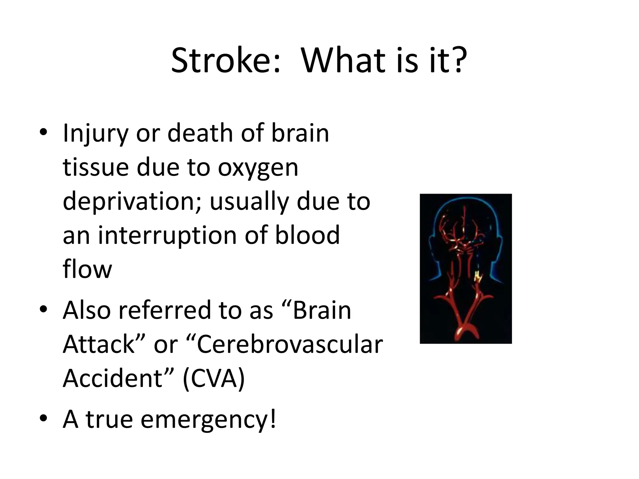
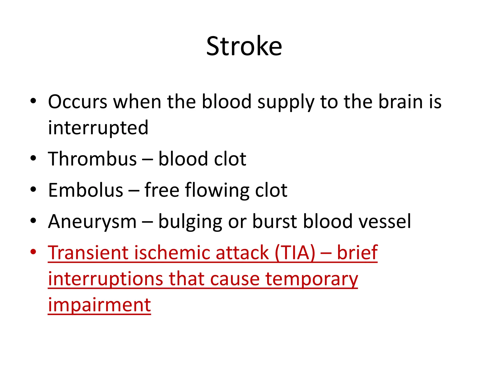
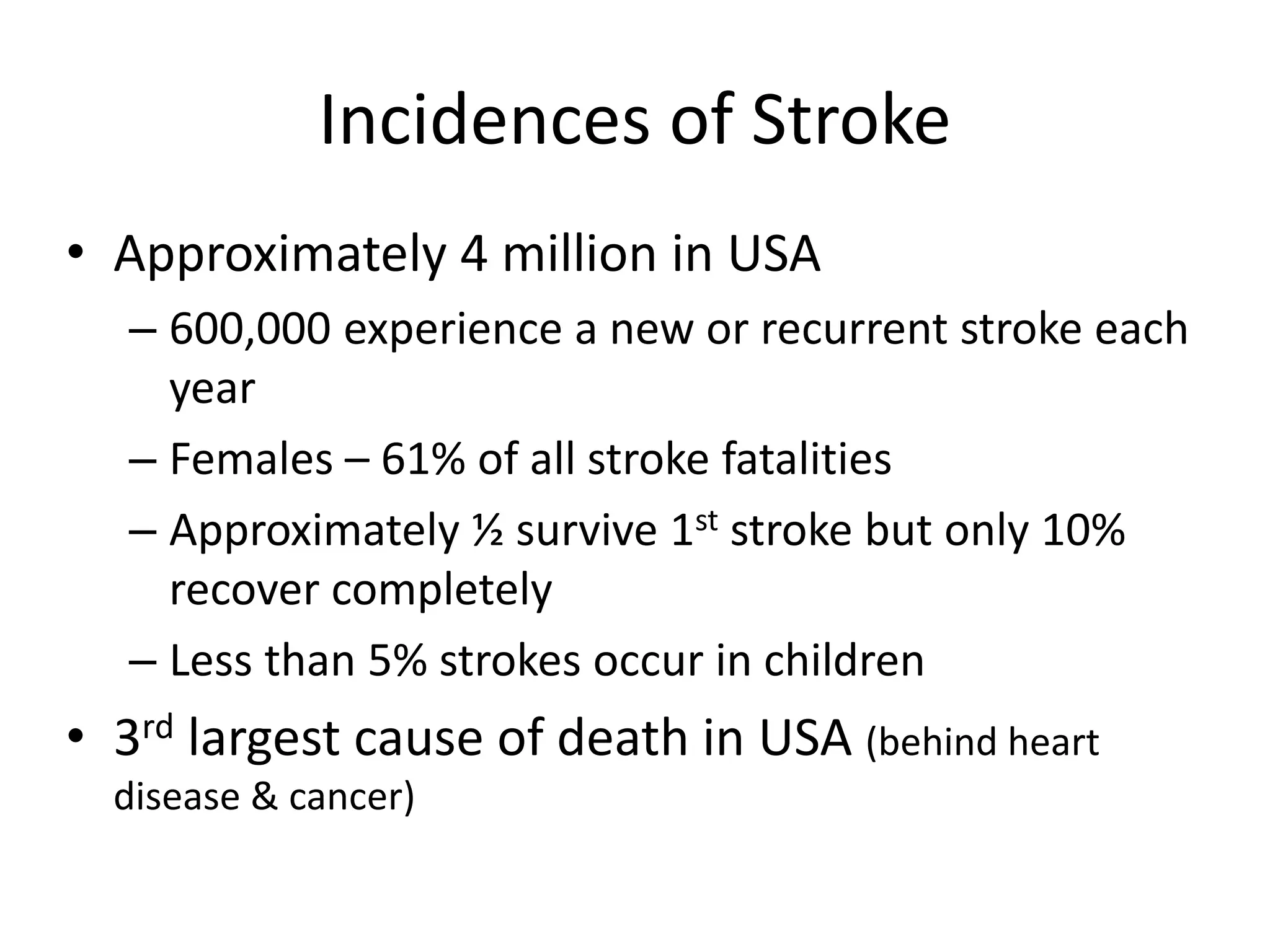
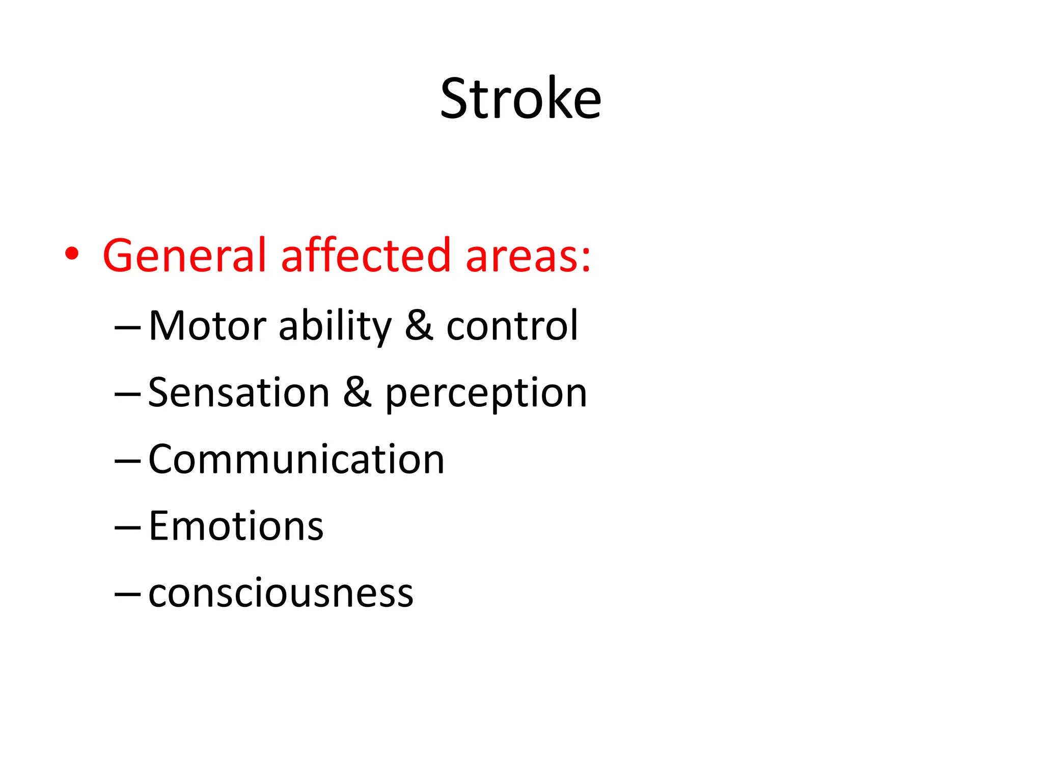
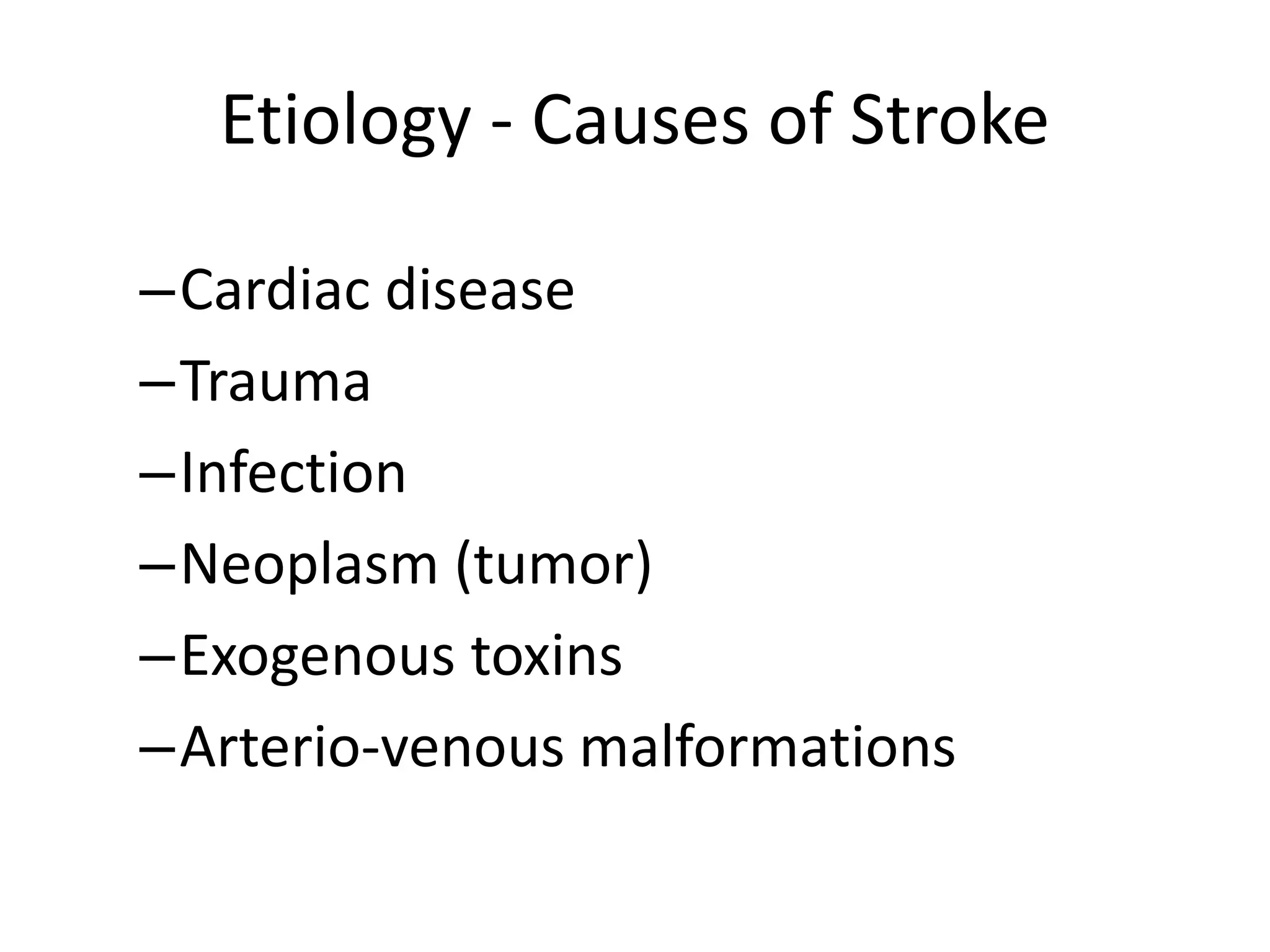
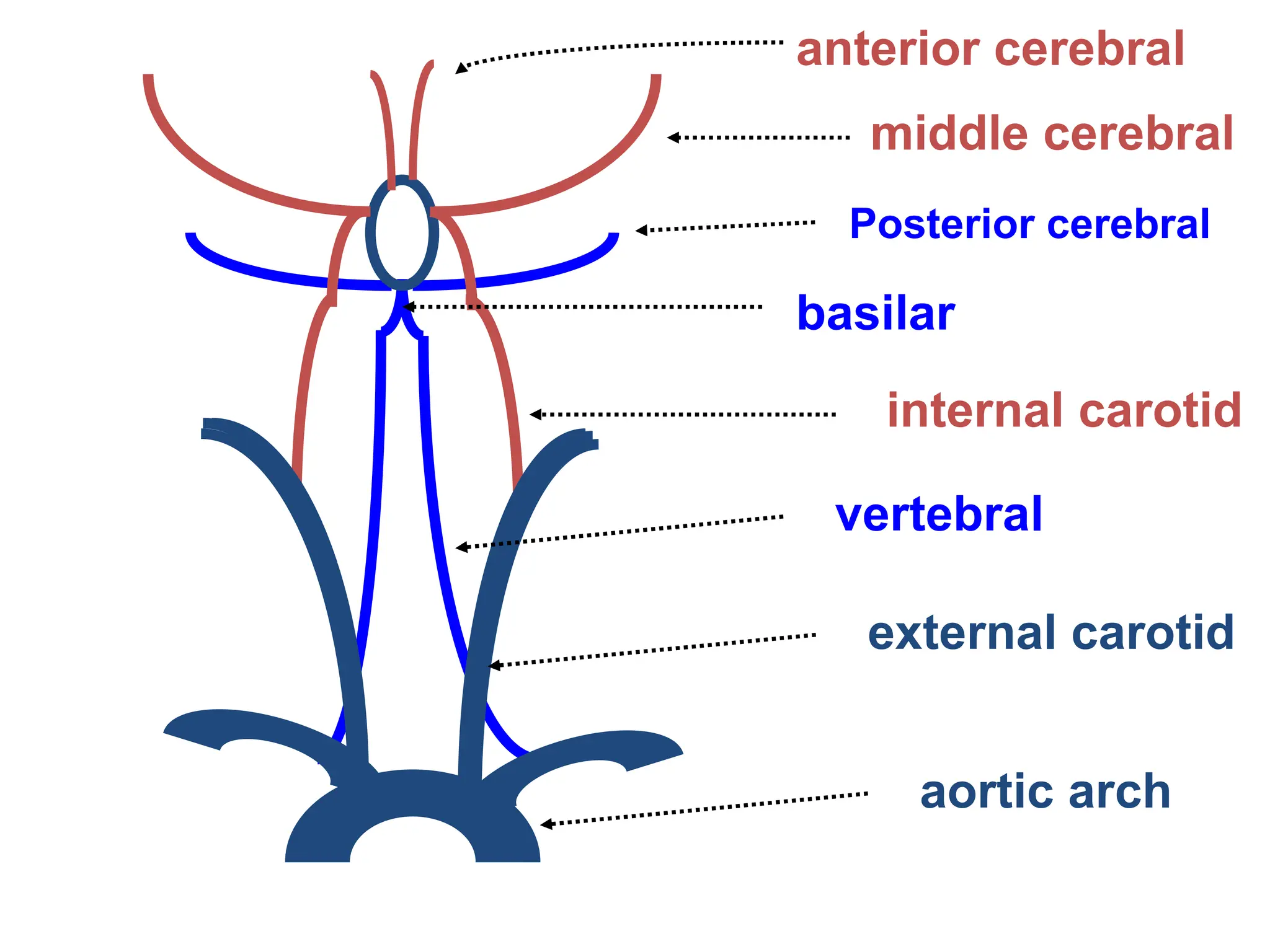
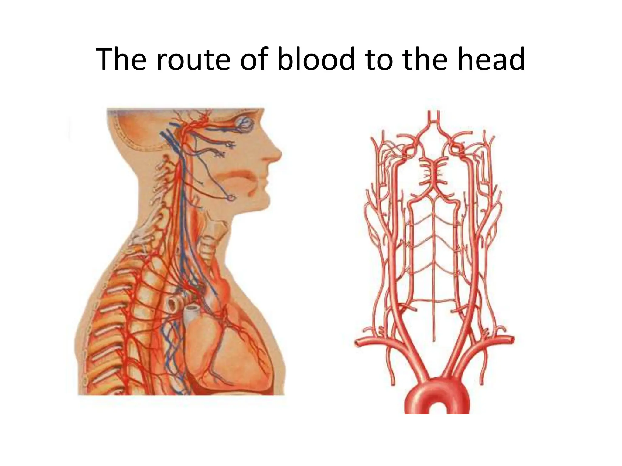
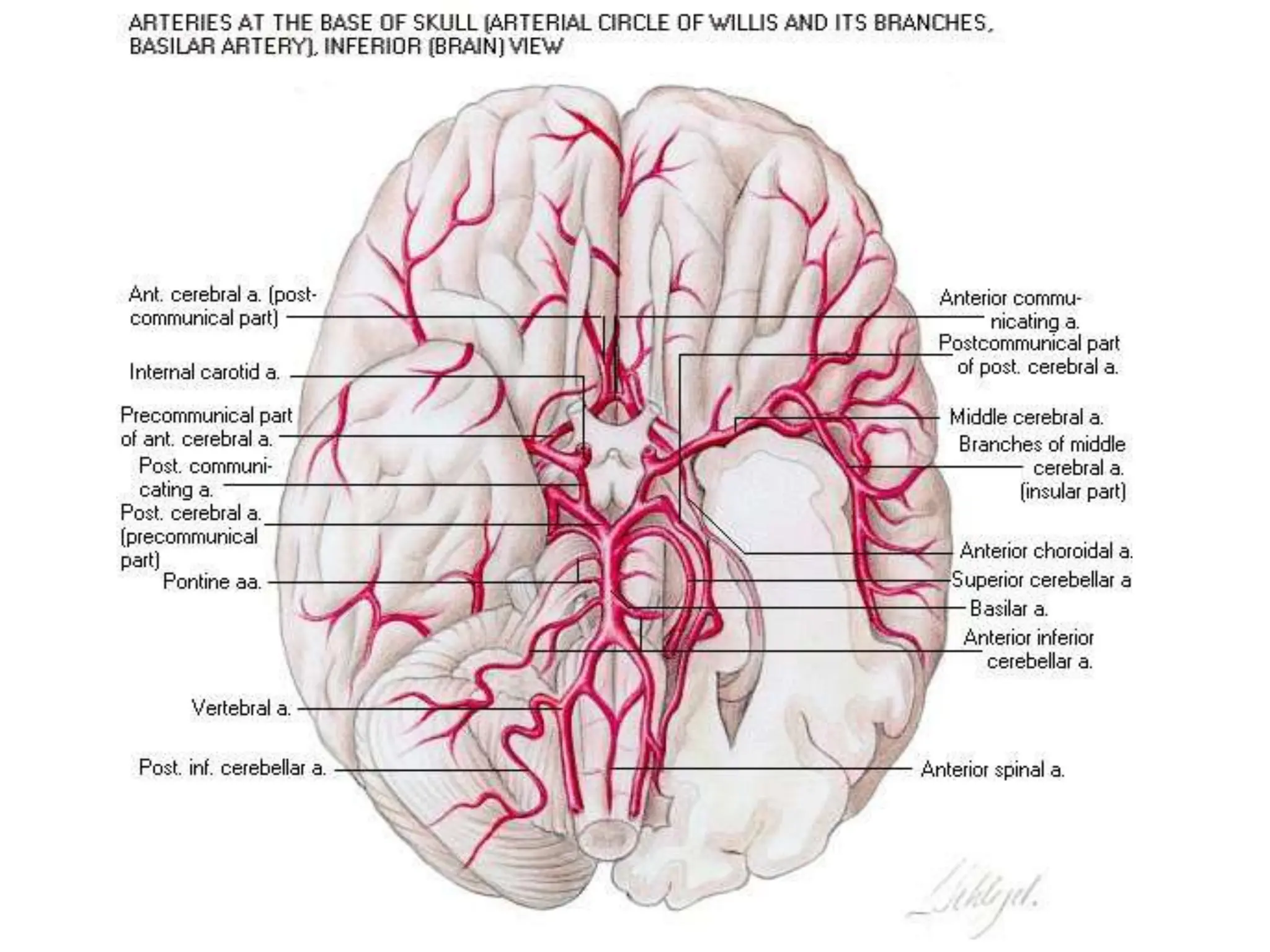
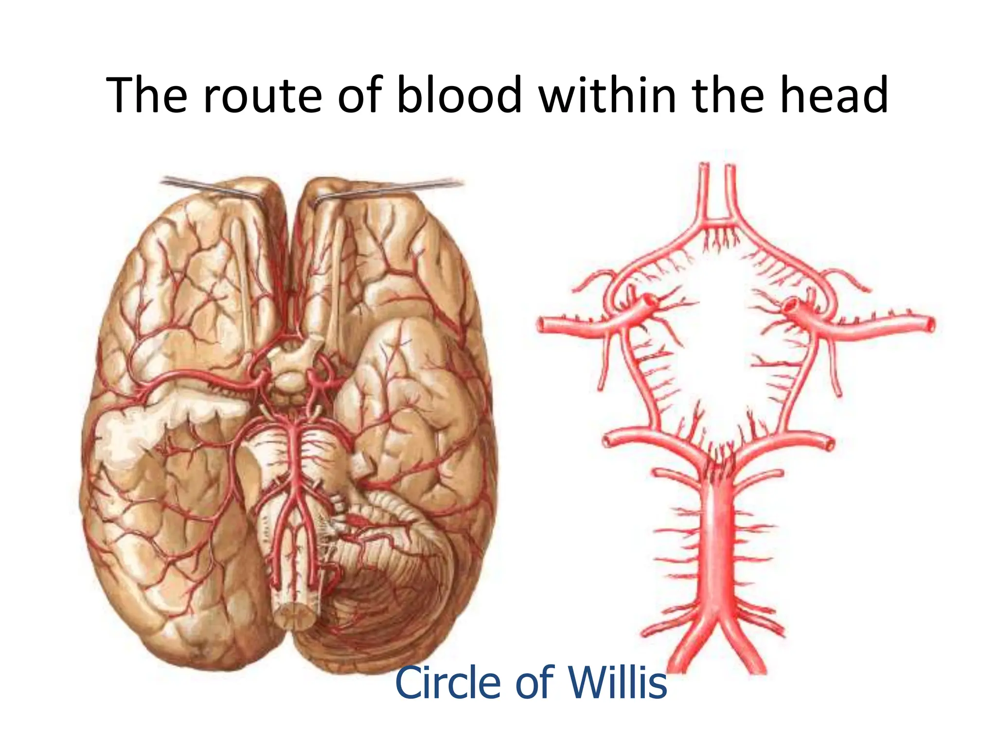
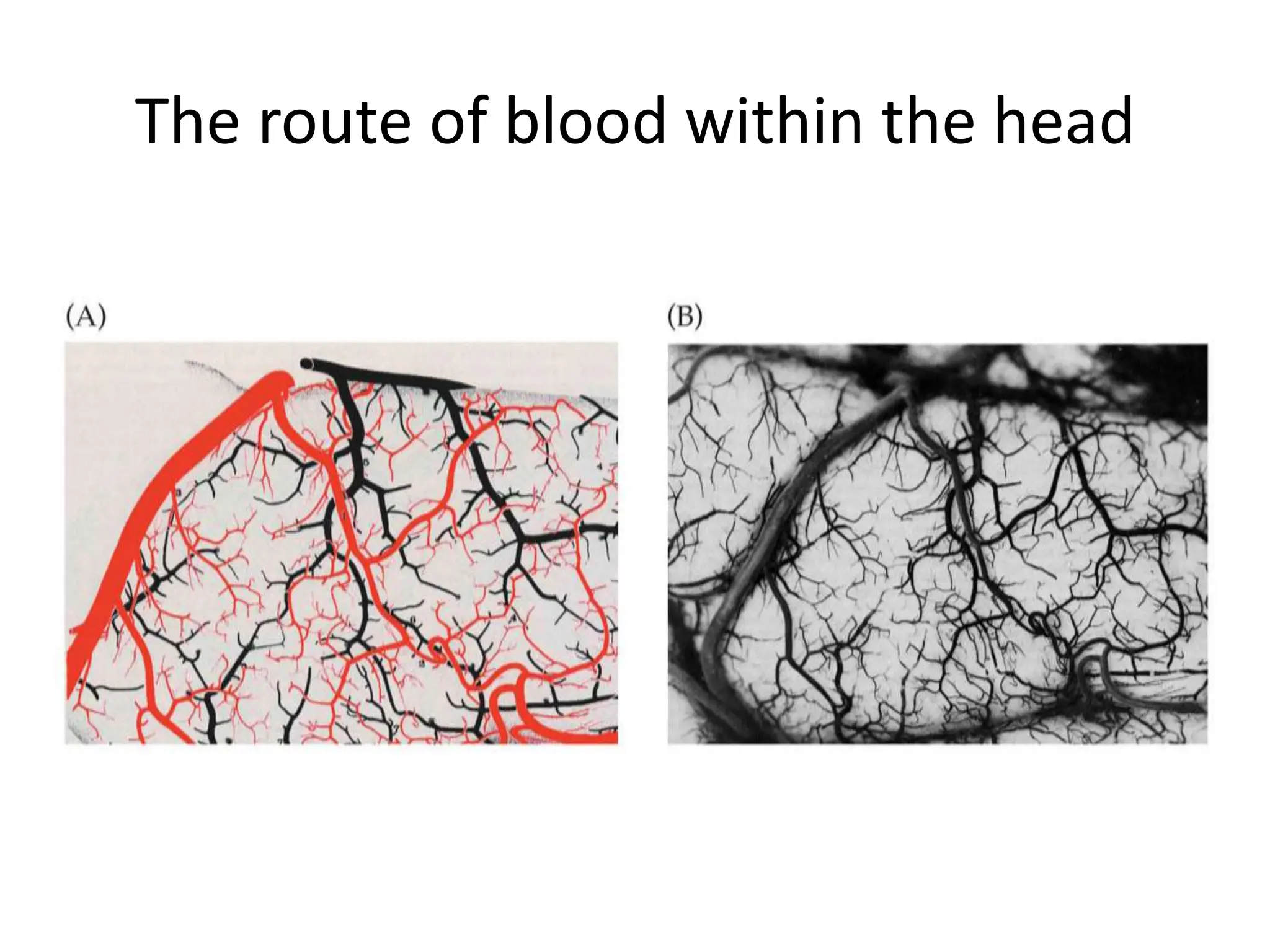
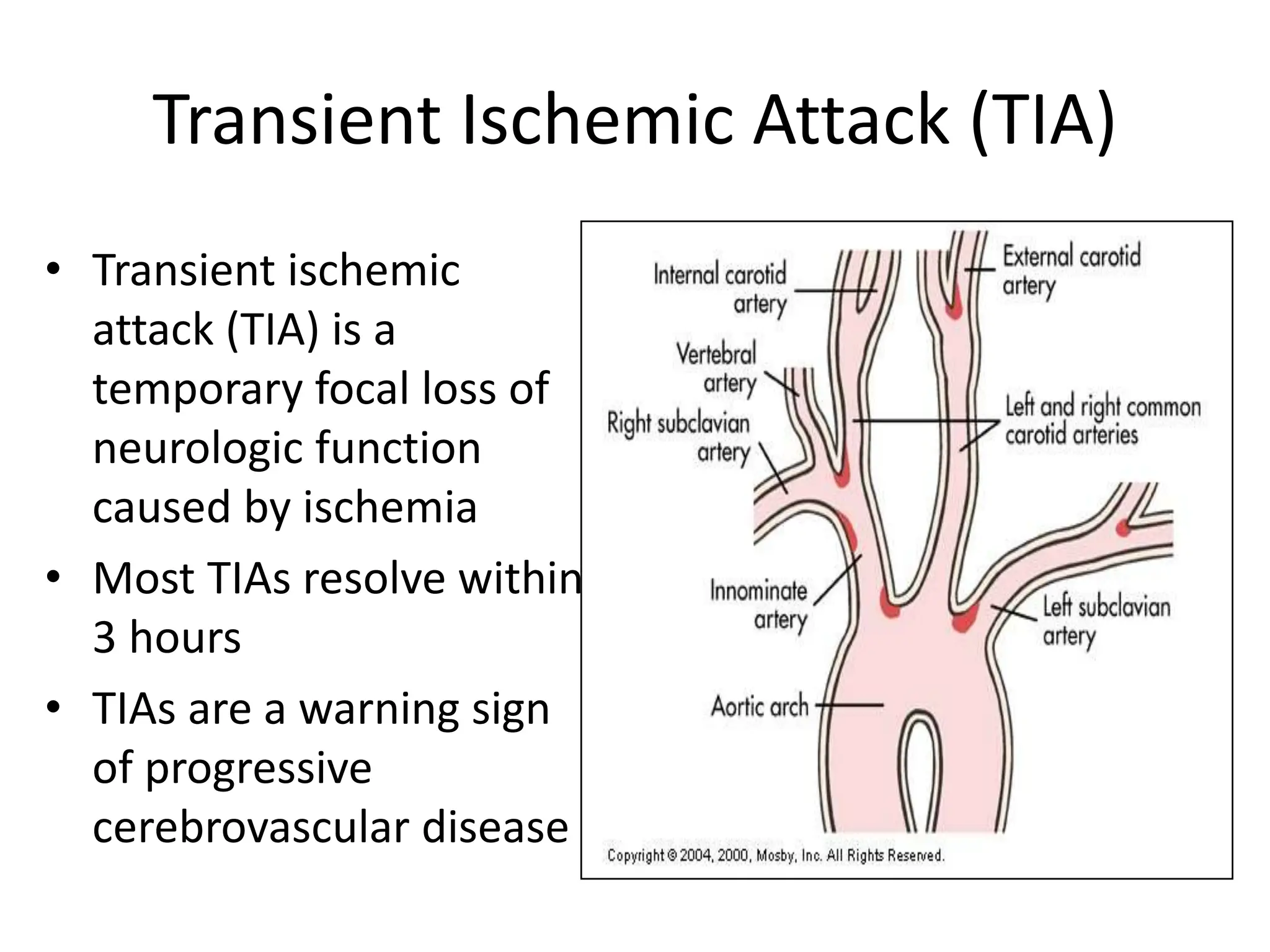
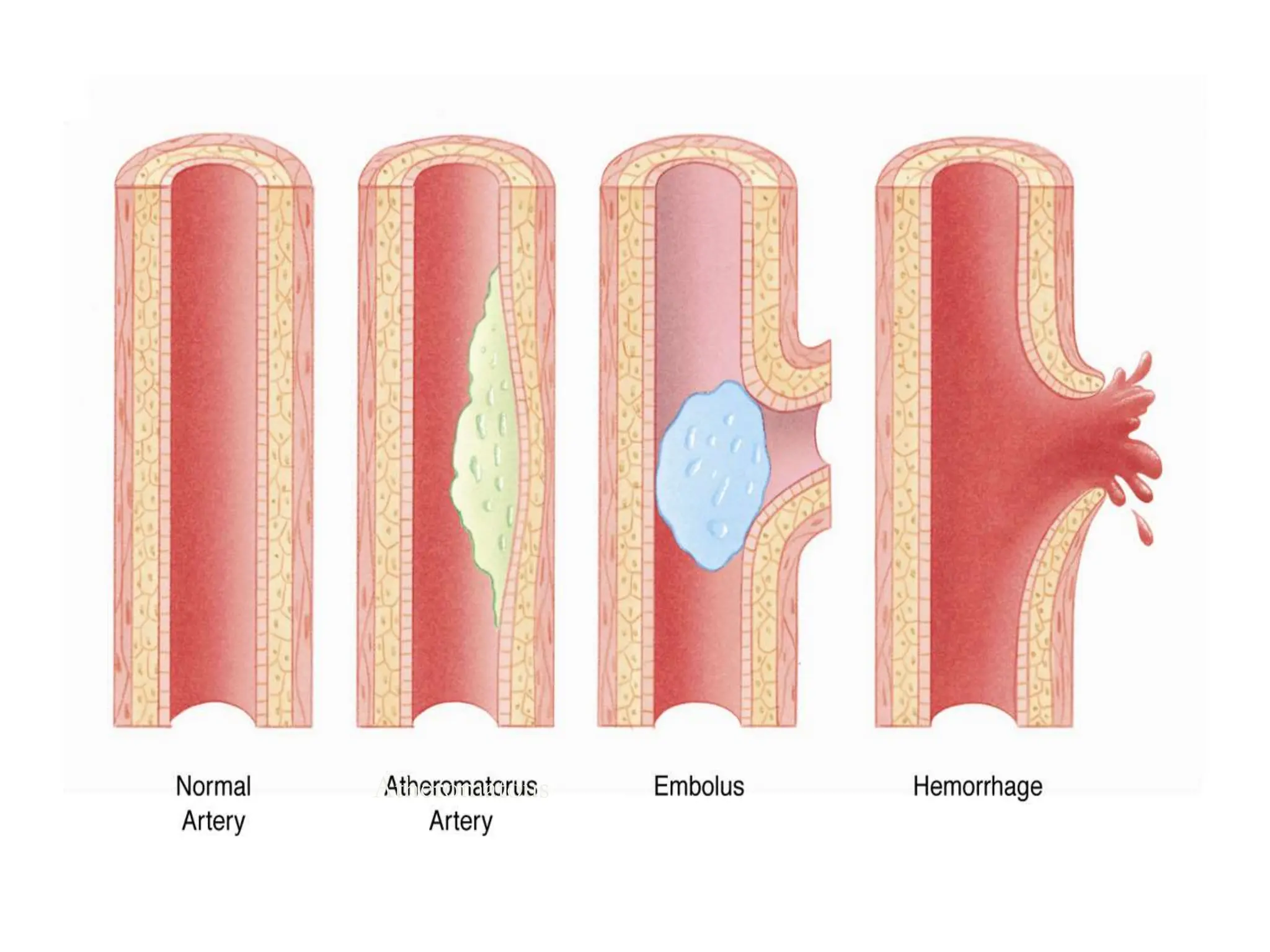
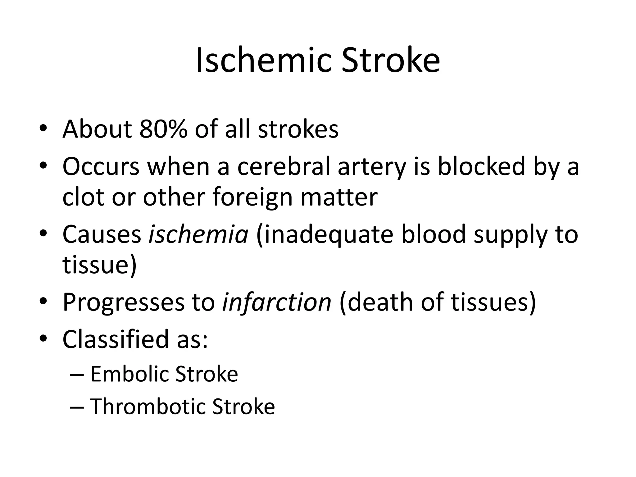
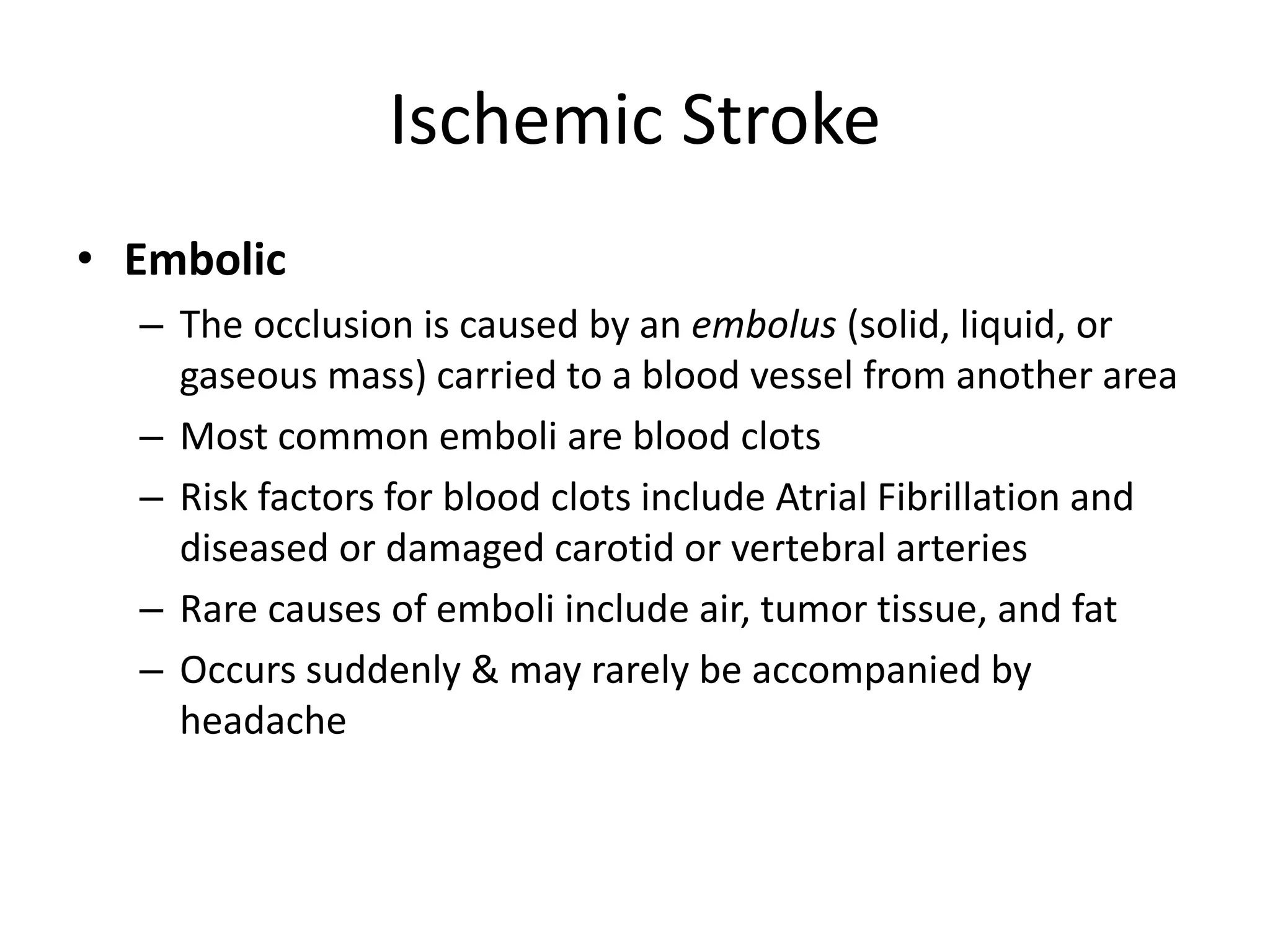
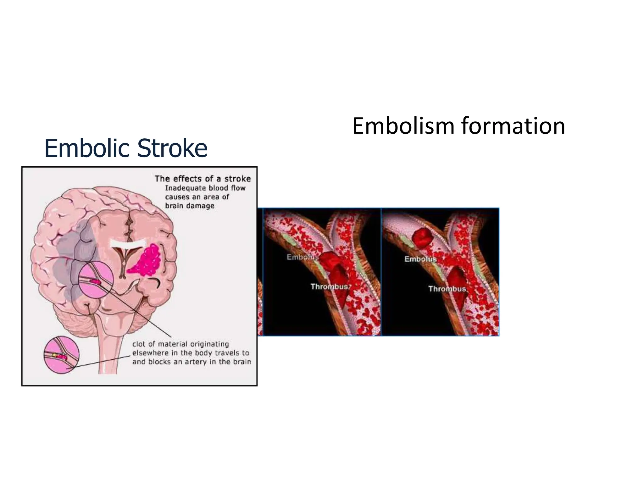
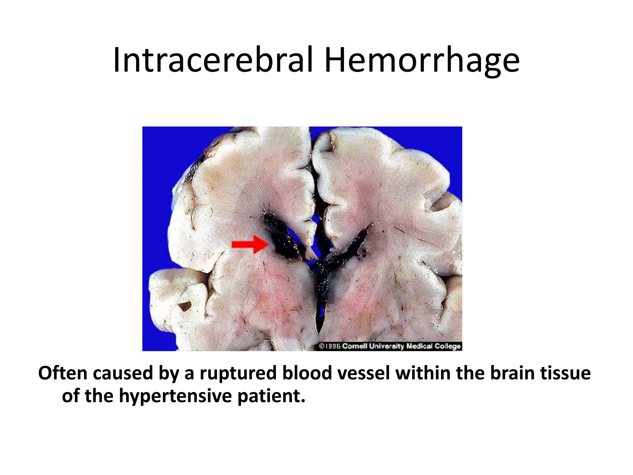
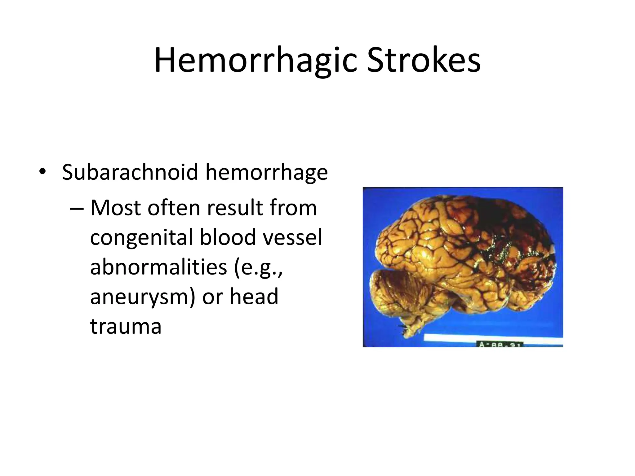
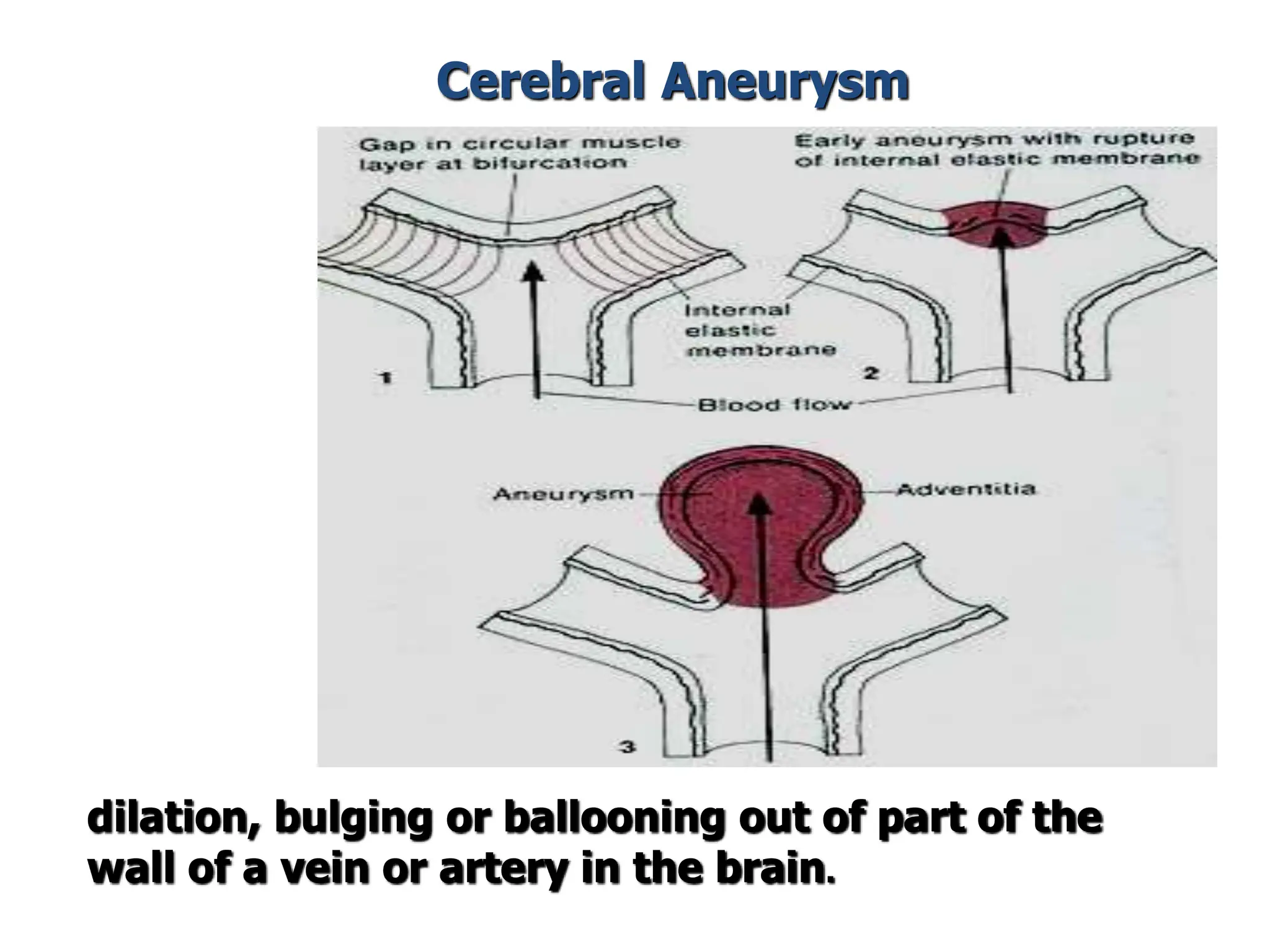
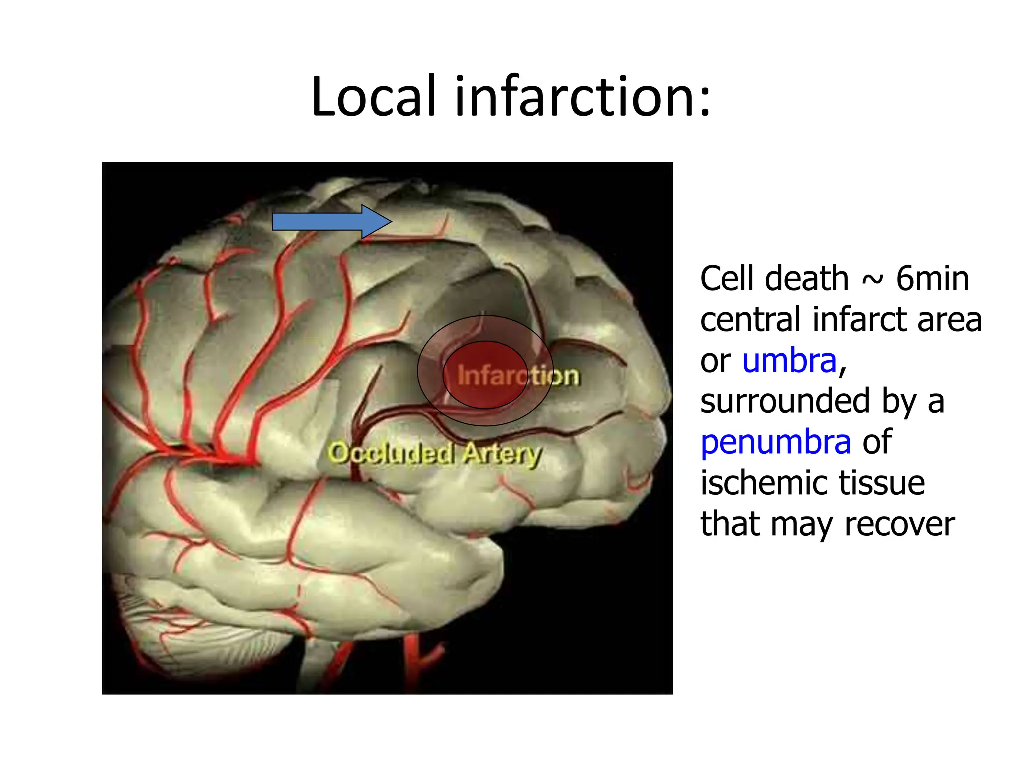
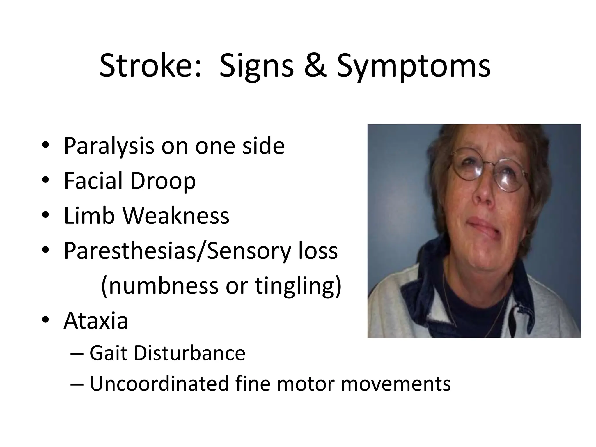
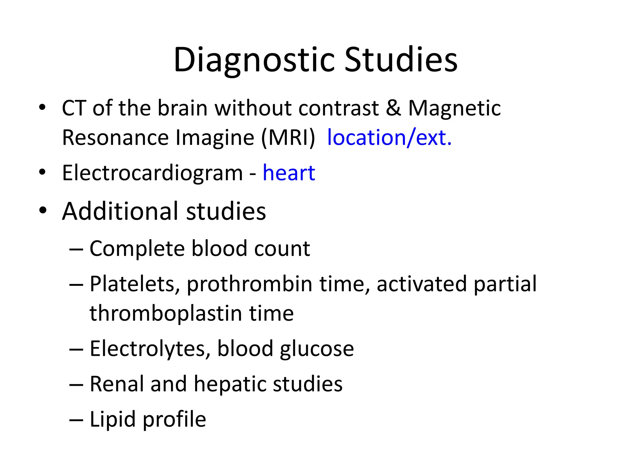
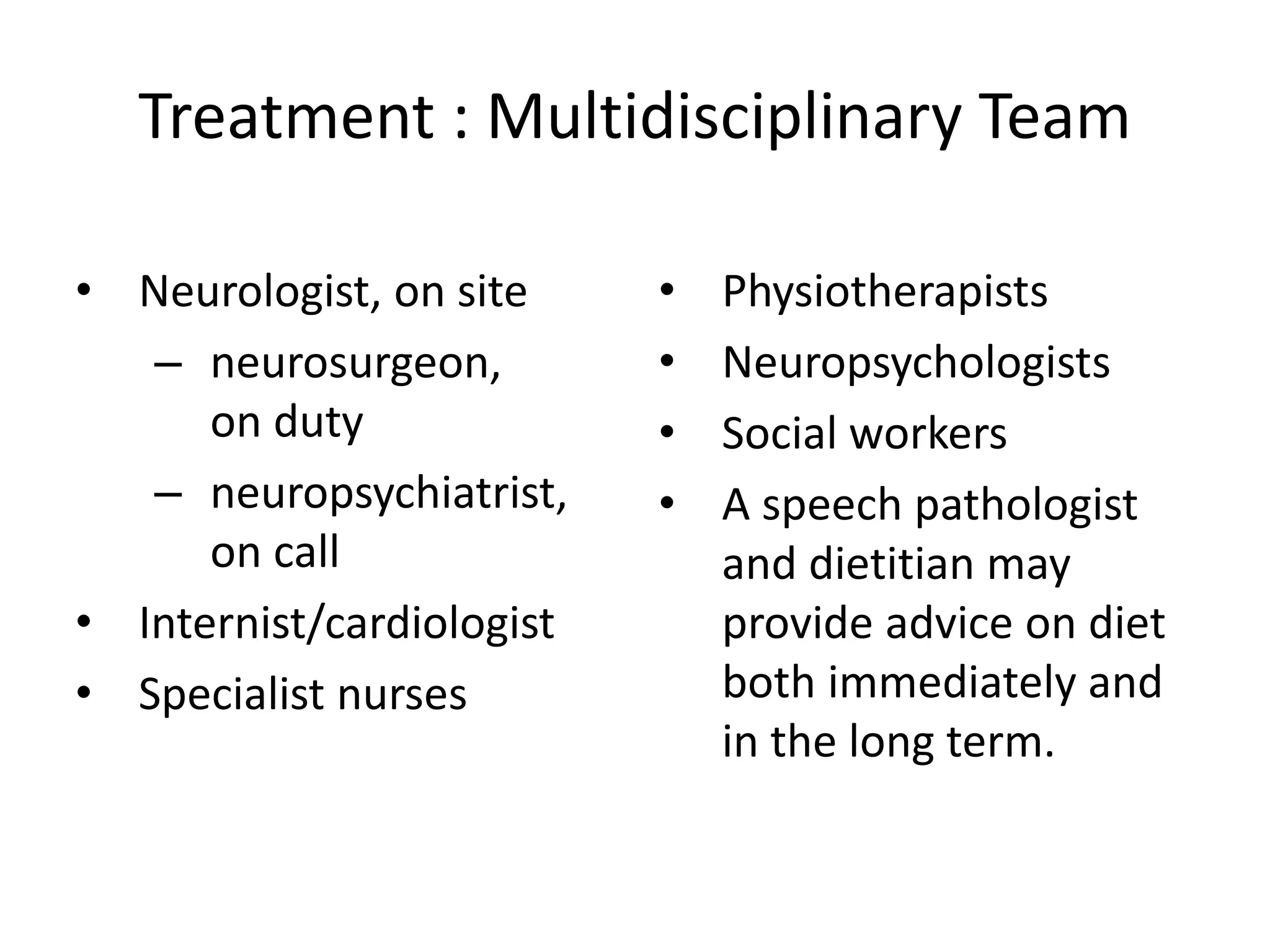
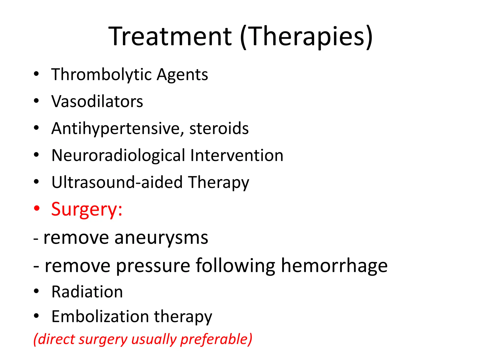
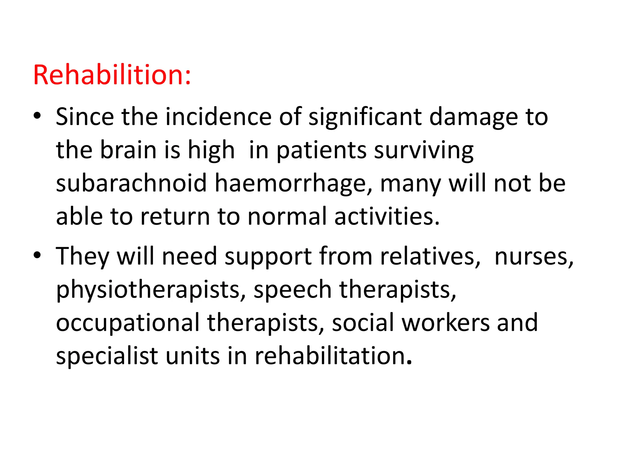
2. Year (2) Physiology 2 016
Day Time Activity Topic
Sat 8: 10 am L1 & L2
Physiology of the NS
General Principles
Sat 8:10 am L3& L4 & L5
Brain Physiology
CSF & Meninges
Sat 8:10 am L6 Cerebral Vascular Accident (Stroke) +
Sat 8: 10 am L7 & L8
Thalamus & Hypothalamus
Basal Nuclei & Reticular Formation &
Brainstem
Sat 8:10 am L9 & L10
Spinal Cord – Sensory and
Motor Spinal Tract & Integrative
Neurophysiology
Sat 8:10 am L11 Cranial Nerves
Sat 8: 10 am L12 & L13 Special Senses (1)
Sat 8:10 am L14 & L15 Special Senses (2)
8- 11 Physiology Practice (Special Senses) 2 Labs
Course Outline
Nervous System
3. Learning Objectives
At the end of this lecture you should be able to
• Define “stroke”
• Discuss incidence & risk factors
• Review Cerebral flow and factors that affect it
• Discuss pathophysiology of CVA
• Describe the Blood Supply (Vascularization) of the brain.
• Differentiate between the ischemic and the hemorrhagic stroke.
• Describe the clinical manifestation of the stroke.
• Describe stroke assessment and patients care.
• Importance of rapid stroke therapy.
4. Stroke: What is it?
• Injury or death of brain
tissue due to oxygen
deprivation; usually due to
an interruption of blood
flow
• Also referred to as “Brain
Attack” or “Cerebrovascular
Accident” (CVA)
• A true emergency!
5. Stroke
• Occurs when the blood supply to the brain is
interrupted
• Thrombus – blood clot
• Embolus – free flowing clot
• Aneurysm – bulging or burst blood vessel
• Transient ischemic attack (TIA) – brief
interruptions that cause temporary
impairment
6. Incidences of Stroke
• Approximately 4 million in USA
– 600,000 experience a new or recurrent stroke each
year
– Females – 61% of all stroke fatalities
– Approximately ½ survive 1st stroke but only 10%
recover completely
– Less than 5% strokes occur in children
• 3rd largest cause of death in USA (behind heart
disease & cancer)
7. Stroke
• General affected areas:
–Motor ability & control
–Sensation & perception
–Communication
–Emotions
–consciousness
16. Transient Ischemic Attack (TIA)
• Transient ischemic
attack (TIA) is a
temporary focal loss of
neurologic function
caused by ischemia
• Most TIAs resolve within
3 hours
• TIAs are a warning sign
of progressive
cerebrovascular disease
17. Types of Stroke
• Occlusive (Ischemic)
– due to the closure of a blood vessel -
• Hemorrhagic
– due to bleeding from a blood vessel
– usually due to either hypertension or an
aneurysm.
19. Ischemic Stroke
• About 80% of all strokes
• Occurs when a cerebral artery is blocked by a
clot or other foreign matter
• Causes ischemia (inadequate blood supply to
tissue)
• Progresses to infarction (death of tissues)
• Classified as:
– Embolic Stroke
– Thrombotic Stroke
20. Ischemic Stroke
• Embolic
– The occlusion is caused by an embolus (solid, liquid, or
gaseous mass) carried to a blood vessel from another area
– Most common emboli are blood clots
– Risk factors for blood clots include Atrial Fibrillation and
diseased or damaged carotid or vertebral arteries
– Rare causes of emboli include air, tumor tissue, and fat
– Occurs suddenly & may rarely be accompanied by
headache
22. Ischemic Strokes
• Thrombotic
– The occlusion is caused by a cerebral thrombus; a
blood clot which develops gradually in a
previously diseased artery and obstructs it
– Caused by atherosclerosis:
• atheromatous plaque deposits form on the inner walls
of arteries, resulting in narrowing and reduction of
blood flow
• platelets adhere to the plaque deposit and a blood clot
is created
23. Hemorrhagic Strokes
• About 20% of all strokes
• Onset usually sudden with severe headache
• Classified as:
– Intracerebral hemorrhage (within the brain)
– Subarachnoid hemorrhage (in the fluid filled
spaces around the blood vessels outside the brain)
24. Hemorrhagic Strokes
• Intracerebral hemorrhage
–Most occur in the hypertensive patient
when a small vessel within the brain tissue
ruptures
–Hemorrhage inside the brain often tears
and separates brain tissue
26. Hemorrhagic Strokes
• Subarachnoid hemorrhage
– Most often result from
congenital blood vessel
abnormalities (e.g.,
aneurysm) or head
trauma
27. dilation, bulging or ballooning out of part of the
wall of a vein or artery in the brain.
Cerebral Aneurysm
28. Hemorrhagic Strokes
• Subarachnoid hemorrhage
– Blood in the subarachnoid space may impair
drainage of cerebrospinal fluid and cause a rise in
intracranial pressure
– Herniation of brain tissue may occur
29. Local infarction:
Cell death ~ 6min
central infarct area
or umbra,
surrounded by a
penumbra of
ischemic tissue
that may recover
30. Left Hemisphere Stroke Causes
• Aphasia
• Right hemiparesis
• Right-sided sensory loss
• Right visual field defect
• Poor right conjugate gaze
• Dysarthria
• Difficulty reading, writing, or calculating
31. Right Hemisphere Stroke Causes
• Defect of left visual field
• Extinction of left-sided stimuli
• Left hemiparesis
• Left-sided sensory loss
• Left visual field defect
• Dysarthria
• Spatial disorientation
32. Stroke Risk Factors
• High blood pressure
• Atrial fibrillation, CHF
• High cholesterol
• Diabetes (twice the risk)
• Smoking (50% higher risk)
• Alcohol or Drug Abuse
• Inactivity or Obesity
• Clotting problems (Sickle Cell)
33. Stroke: Signs & Symptoms
• Paralysis on one side
• Facial Droop
• Limb Weakness
• Paresthesias/Sensory loss
(numbness or tingling)
• Ataxia
– Gait Disturbance
– Uncoordinated fine motor movements
34. Speech Disturbance
• Aphasia
– Inability to speak
• Dysphasia
– Difficulty speaking
• Dysarthria
– Impairment of the tongue muscles essential to
speech
35. Vital Functions
• Airway -
intubate if:coma hypoventilation risk of
aspiration
• Breathing –
pulse oxymetry aim for normoventilation
• Circulation-
i.v. line, Ringer or normal saline, no glucose
- ECG
- BP
36. Investigations:
• CT of the brain without contrast & Magnetic
Resonance Imagine (MRI) location/ext.
• Electrocardiogram - heart
• Chest x-ray - heart
37. Diagnostic Studies
• CT of the brain without contrast & Magnetic
Resonance Imagine (MRI) location/ext.
• Electrocardiogram - heart
• Additional studies
– Complete blood count
– Platelets, prothrombin time, activated partial
thromboplastin time
– Electrolytes, blood glucose
– Renal and hepatic studies
– Lipid profile
38. Treatment : Multidisciplinary Team
• Neurologist, on site
– neurosurgeon,
on duty
– neuropsychiatrist,
on call
• Internist/cardiologist
• Specialist nurses
• Physiotherapists
• Neuropsychologists
• Social workers
• A speech pathologist
and dietitian may
provide advice on diet
both immediately and
in the long term.
40. Rehabilition:
• Since the incidence of significant damage to
the brain is high in patients surviving
subarachnoid haemorrhage, many will not be
able to return to normal activities.
• They will need support from relatives, nurses,
physiotherapists, speech therapists,
occupational therapists, social workers and
specialist units in rehabilitation.
41. Call
emergency
services
ER stroke team
Activated
(15 minutes)
Neuroprotective
drug infused
during transport
Brain scan
Drugs administered
‘stroke-treatment’
cocktail
Full recovery
Stroke onset
Secondary
prevention
Remember