1. THERE’S A FIRST FOR EVERYTHING IN PLASTIC SURGERY
Continual Change and Evolvement in the Aesthetic Industry
Dr. Eric J. Stelnicki, M.D. is the first craniofacial surgeon
to use Virtual Surgical Planning (VSP Orthognathics) in the state of Florida
for treatment of complex jaw deformities related to craniofacial disorders.
By: Elizabeth C. Fassler
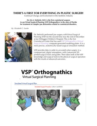
Dr. Stelnicki performed jaw surgery with Virtual Surgical
Planning (VSP) for the second time near the end of December
at Joe DiMaggio Children’s Hospital. This is the first
Craniofacial Center that has used VSP Orthognathics (Virtual
Surgical Planning) computer-generated modeling plates. It is a
more precise, anatomically based surgical simulation method.
VSP provides data in order to accurately plan surgery in a
computerized, digital atmosphere, with customized 3D
printed surgical tools and guides. Then takes both of the VSP
knowledge and surgical tools to the physical surgical operation
with the results of advanced outcomes.
2. Craniofacial Microsomia (CFM) describes a vast array of abnormalities that affect the
skull’s development before birth. Individuals with this possess a facial anatomy of reduced
growth, resulting in various degrees of craniofacial asymmetry that only progresses over
time. Those diagnosed with CFM have differences in size and shape between the right and
left sides of the face. Most cases have abnormalities on both sides of the face where one
side generally differs from the other. Others that are diagnosed with this have
abnormalities only on one side of the face.
The facial features and traits usually include one, underdeveloped side of the jaw
(maxillary or mandibular hypoplasia) that may cause dental problems, eating and speech
complications and in some cases of severe mandibular hypoplasia, difficulties in
breathing. Other abnormalities caused by craniofacial microsomia are the effectiveness of
one or both ears. This can include the possibility in possessing the growth of skin tags,
microtia or anotia or a closed ear canal. These abnormalities can result in the loss of
hearing. The occurrence of eye problems in craniofacial microsomia are less prevalent, but
those affected by this abnormality tend to have microphthalmia or other problems that
result in vision loss. People with craniofacial microsomia can also have abnormalities in
other regions of the body, which include deformed bones of the vertebrae, kidneys with an
unusual shape and heart defects.
The diagnosis is estimated to occur around 1of 3,600-5,600 newborns (Farina, Valladares,
Torrealba, Nuñez, & Uribe, 2015). Although for reasons that are unclear or not yet
3. understood, craniofacial microsomia occurs more in males than females. CFM is very
broad in classifying an array of craniofacial malformation conditions. It is also uncertain
what genes are involved in this disorder, but is caused from complications in the
development of the embryo structures called the branchial and visceral arches, also
known as the first and second pharyngeal arches. Moreover, it is not biologically inherited
(U.S. National Library of Medicine, 2016).
The diagnosis for the second jaw surgery, performed on a 20-year-
old male patient was congenital deformity of the face, and
Hemifacial Microsomia on the right with asymmetry to the
maxilla and mandible. The right appeared hypoplastic relative to
the left. The was more asymmetry included in the lateral wall,
right orbit, right zygomatic arch and right external auditory canal.
Dr. Stelnicki clarifies that this is the "second surgery of its kind in
the state Florida for patients with Craniofacial Microsomia where
we will be correcting the deformity of the jaw…we are using the
computer to reshape the face, taking a crooked face and making
it straight."
Mike Applegate from Kls Martin works exclusively with Dr. Stelnicki in procedures such as
these, using Virtual Surgical Planning, and is also present during the actual surgery.
Applegate describes his part as "basically taking a CT Scan of the patient and through a
virtual surgical webinar we are able to design exactly what movements we want to make
to the maxilla, mandible and bone anatomy. We can do a soft tissue coverage to see what
it’s going to look like post-op, which is very accurate. And that is something we have been
kind of incorporating to Orthognathic surgery as a whole…taking titanium alloy plates
4. that…used to be a stock plate…a surgeon had to bend specific to the anatomy, and we are
now printing them. Now, what was taken on the virtual surgical webinar, is now
completely replicated, every single time in the O.R. with these custom plates."
Dr. Stelnicki’s first patient that received this treatment a few months prior was diagnosed
with left-sided craniofacial microsomia with combined maxillary and mandibular
hypoplasia. In short, the treatment for this surgery is based on skeletal correction. Before
her surgical performance, Dr. Stelnicki participated in extensive pre-operative planning
and modeling. The operation was performed with LeFort-I osteotomy, bilateral sagittal split
osteotomy, nasal endoscopy and internal fixation of both osteotomies. VSP Orthognathics
(the computerized modeled plan and computerized custom modeling) was used for the
staging and creation of the two separate custom oral splints for her procedure.
Furthermore, this same patient created a documentary titled Double Jaw Surgery: Journey
From an Asymmetrical Faced Girl, before undergoing her surgery.
In keeping a positive state-of-mind, she states in her video that "…As teenagers, we tend to
pick at our flaws…and as a 16-year-old, I have never judged my asymmetrical face…In the
6th grade I noticed my smile was crooked on my ID picture. My mom thought I was being
a typical teen by judging my face…Until an ENT found out I had tinnitus. ‘There’s
something wrong with your face,’ he commented…I was diagnosed with a malocclusion
class 3 cross bite. Little did I know then that in two years I would have a new face. I’ll miss
my crooked smile…People ask "...‘Are you nervous?’ No not really. July 23, 2016 will be
the date I’ll never forget."
In conclusion, the Atlantic Center of Aesthetic and Reconstructive Surgery are the only
craniofacial surgeons who’ve performed this type of surgery in the state of Florida today,
using "state-of-the art, computerized planning and intra-operative computer optimization
to treat very complex deformities." Furthermore, Dr. Stelnicki emphasizes that he’s "very
proud" of the Atlantic Center on being "the pioneers of the state offering this type of
surgical direction."
5. REFERENCES
Craniofacial Microsomia. (2016). U.S. National Library of Medicine. Retrieved from
https://ghr.nlm.nih.gov/condition/craniofacial-microsomia
Farina R., Valladares S., Torrealba R., Nuñez M., & Uribe F. (2015). Orthognathic Surgery
in Craniofacial Microsomia: Treatment Algorithm. Plastic Reconstr Surg, 3(1), e294.
VSP Orthognathics. (2016). 3D Systems Healthcare. Retrieved from http://www.med-
icalmodeling.com/solutions-for-surgeons/vsp-technology/vsp-orthognathics