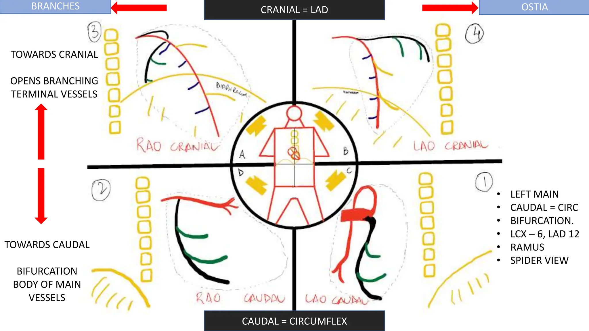
1) The document discusses various angiographic projections used to visualize the coronary arteries during coronary angiography. It provides 10 possible standard views and describes the optimal views for visualizing different coronary artery segments.
2) Tips are provided on how to learn and remember the different views using props like wires and bottles. Terminologies used in angiography and positioning of the image intensifier are also explained.
3) Chamber views used during left ventriculography are demonstrated in the right anterior oblique and left anterior oblique projections. A demonstration of the views is provided using a 3D anatomy atlas.
This document discusses angiographic projections and views used for imaging specific coronary artery segments and congenital heart diseases. It provides the standard angiographic views and degrees of angulation used for visualizing the left main, left anterior descending, circumflex, right coronary arteries, pulmonary arteries, and structures like VSDs and ASDs. The document also lists patient, angiographer, and equipment factors that can cause poor angiograms.
Basics of coronary artery anatomy and angiographic viewsBipul Roy
This document discusses coronary artery anatomy and angiographic views. It describes the normal anatomy of the coronary arteries including the right coronary artery, left main coronary artery, left anterior descending artery, and left circumflex artery. It explains different angiographic views used to visualize each artery including the LAO, RAO, PA, and lateral views. Standard views and supplemental views are outlined for evaluating the left, right, and graft vessels. Diameter measurements of different coronary segments by gender are also provided.
NORMAL CORONARY ANATOMY AND ANGIOGRAPHIC VIEWS SOURCE.pptxJaydeep Malakar
The document discusses normal coronary artery anatomy and angiographic views. It describes the development of the coronary arteries from fish to mammals, coronary blood supply, anatomy including branches and territories, angiographic projections and techniques. Key points include the dual aortic origin of the right and left coronary arteries, their course in the epicardial fat and termination in myocardial capillaries, and the circle and loop theory of coronary artery distribution. Standard angiographic views of the left and right coronary arteries are shown.
VASCULAR RINGS AND SLINGS TYPES HAEMODYNAMICS PRESENTATION AND DIAGNOSIS.pptxJaydeep Malakar
Vascular rings are congenital anomalies where the aorta and its branches completely or incompletely encircle the trachea and esophagus. There are two main types - complete rings which fully encircle the airways and incomplete rings which partially encircle. Common types include double aortic arch, right aortic arch with retroesophageal vessels, and pulmonary artery sling. Clinical features include noisy breathing, cough, wheezing and recurrent respiratory infections in infants and children. Investigations include chest x-ray, CT, MRI and bronchoscopy to identify the specific ring and any tracheal compression. Surgical repair is usually indicated for symptomatic patients to prevent further airway damage.
TRICUSPID VALVE ANATOMY PATHOPHYSIOLOGY INDICATIONS AND INTERVENTIONS.pptxJaydeep Malakar
The document discusses tricuspid valve intervention techniques. It describes Tricuspid Annular Plane Systolic Excursion (TAPSE) which measures tricuspid annular motion and is used to estimate right ventricular function. It discusses approaches to tricuspid valve repair including annuloplasty techniques using rings. It also discusses transcatheter therapies being developed for tricuspid regurgitation including annuloplasty devices and coaptation devices.
This document summarizes the key views and anatomical features seen during coronary angiography. It outlines the 4 main views of the left coronary system - RAO cranial, LAO cranial, RAO caudal, LAO caudal - and describes what branches are seen in each view. It also summarizes the 2 views of the right coronary system - LAO and RAO views. Additional details provided include landmarks for identifying the left main, left circumflex, and left anterior descending coronary arteries. Guidance is given on how the position of the catheter (retracted vs open) determines if the image will be in the RAO or LAO view.
The document discusses inguinal hernia and its management. It defines hernia and inguinal hernia, describing their types as direct or indirect. It details the anatomy of the inguinal region including structures like the inguinal canal, rings, and layers. It also discusses the etiology, risk factors, investigations and classifications of inguinal hernias. The management section summarizes techniques for hernia repair like herniotomy, herniorrhaphy, hernioplasty and laparoscopic repair. It highlights pioneers in the field including Bassini, Shouldice and modifications to their open tension-free techniques.
- Fine-needle aspiration cytology (FNAC) is the most important diagnostic tool for evaluating a solitary thyroid nodule, as it is safe, cost-effective, and reliable for differentiating between benign and malignant diseases of the thyroid. Ultrasound-guided FNAC is more accurate than palpation-guided.
- Thyroid imaging with ultrasound and radioactive iodine uptake scans can identify high-risk features that increase the likelihood of malignancy, such as hypoechogenicity, microcalcifications, irregular shape, and lack of iodine uptake in the nodule.
- Cytology results are categorized using the Bethesda or THY classification systems. Suspicious or malignant results
Adhd Medication Shortage Uk - trinexpharmacy.comreignlana06
The UK is currently facing a Adhd Medication Shortage Uk, which has left many patients and their families grappling with uncertainty and frustration. ADHD, or Attention Deficit Hyperactivity Disorder, is a chronic condition that requires consistent medication to manage effectively. This shortage has highlighted the critical role these medications play in the daily lives of those affected by ADHD. Contact : +1 (747) 209 – 3649 E-mail : sales@trinexpharmacy.com
8 Surprising Reasons To Meditate 40 Minutes A Day That Can Change Your Life.pptxHolistified Wellness
We’re talking about Vedic Meditation, a form of meditation that has been around for at least 5,000 years. Back then, the people who lived in the Indus Valley, now known as India and Pakistan, practised meditation as a fundamental part of daily life. This knowledge that has given us yoga and Ayurveda, was known as Veda, hence the name Vedic. And though there are some written records, the practice has been passed down verbally from generation to generation.
These lecture slides, by Dr Sidra Arshad, offer a quick overview of the physiological basis of a normal electrocardiogram.
Learning objectives:
1. Define an electrocardiogram (ECG) and electrocardiography
2. Describe how dipoles generated by the heart produce the waveforms of the ECG
3. Describe the components of a normal electrocardiogram of a typical bipolar lead (limb II)
4. Differentiate between intervals and segments
5. Enlist some common indications for obtaining an ECG
6. Describe the flow of current around the heart during the cardiac cycle
7. Discuss the placement and polarity of the leads of electrocardiograph
8. Describe the normal electrocardiograms recorded from the limb leads and explain the physiological basis of the different records that are obtained
9. Define mean electrical vector (axis) of the heart and give the normal range
10. Define the mean QRS vector
11. Describe the axes of leads (hexagonal reference system)
12. Comprehend the vectorial analysis of the normal ECG
13. Determine the mean electrical axis of the ventricular QRS and appreciate the mean axis deviation
14. Explain the concepts of current of injury, J point, and their significance
Study Resources:
1. Chapter 11, Guyton and Hall Textbook of Medical Physiology, 14th edition
2. Chapter 9, Human Physiology - From Cells to Systems, Lauralee Sherwood, 9th edition
3. Chapter 29, Ganong’s Review of Medical Physiology, 26th edition
4. Electrocardiogram, StatPearls - https://www.ncbi.nlm.nih.gov/books/NBK549803/
5. ECG in Medical Practice by ABM Abdullah, 4th edition
6. Chapter 3, Cardiology Explained, https://www.ncbi.nlm.nih.gov/books/NBK2214/
7. ECG Basics, http://www.nataliescasebook.com/tag/e-c-g-basics
- Video recording of this lecture in English language: https://youtu.be/Pt1nA32sdHQ
- Video recording of this lecture in Arabic language: https://youtu.be/uFdc9F0rlP0
- Link to download the book free: https://nephrotube.blogspot.com/p/nephrotube-nephrology-books.html
- Link to NephroTube website: www.NephroTube.com
- Link to NephroTube social media accounts: https://nephrotube.blogspot.com/p/join-nephrotube-on-social-media.html
Muktapishti is a traditional Ayurvedic preparation made from Shoditha Mukta (Purified Pearl), is believed to help regulate thyroid function and reduce symptoms of hyperthyroidism due to its cooling and balancing properties. Clinical evidence on its efficacy remains limited, necessitating further research to validate its therapeutic benefits.
One health condition that is becoming more common day by day is diabetes.
According to research conducted by the National Family Health Survey of India, diabetic cases show a projection which might increase to 10.4% by 2030.
13. CAUDAL = CIRCUMFLEX
CRANIAL = LAD
BRANCHES OSTIA
TOWARDS CRANIAL
OPENS BRANCHING
TERMINAL VESSELS
TOWARDS CAUDAL
BIFURCATION
BODY OF MAIN
VESSELS
• CAUDAL = CIRC
• BEST FOR OM
• BODY OF LCx
• PROX LAD –
STRAIGHT AND
ACROSS
30. It takes into account the following
factors before providing a functional
severity of the coronary artery stenosis.
• The length and degree of narrowing of
the coronary stenosis
• Presence of serial stenosis along the
same vessel
• Presence of collateral circulation
• Size of the perfusion territory
• Size of the vessel
Wouldn’t it be nice to see a heart with such perfectly laid out coronaries.
But in reality, this is what we see..
The Aorta, with the RCA going right behind and gives off a Marginal branch at the acute margin of the heart
And the Left main and LAD going behind on the other side and needing to reflect the heart using sponges and hands to visualize it properly.
starting with the first anatomical plane looking from the feet of the patient towards the patient's head.
if the x-ray source is situated directly below the patient with the image intensifier directly above the patient this is referred to as a standard starting position or zero degrees.
You can see that the image intensifier can then rotate around the patient towards the right known as the right anterior oblique or RAO view.
Image intensifier can also rotate to the left known as the left anterior oblique or LAO.
the second plane we can now see that the image intensifier can move towards the head of the patient known as a cranial angulation or towards the feet of the patient known as a caudal angulation
AXIAL VIEW
SAGGITAL VIEW
Lets start with the image labelled 1 here, the LAO Caudal View.
Lao caudal view is BEST for the left main.
Since it is a CAUDAL view, it is best for Circumflex.. Which comes down and is in the centre of the main picture.
It opens up the bifurcation. Left main to lad and circumflex.
It seperates the lad and lcx very well.. Lcx descends down to the 6 0 clock position and lad turns away from you at the 12 o clock position.
If the ramus is present, it can be seen very well.
AKA SPIDER VIEW
ADDITIONAL : OSTIUM OF LAD AND CIRC SEEN WELL, BODY OF MAIN VESSELS, LAD AND LCX SEEN VERY WELL.
Now Coming to the image labelled 2 here, the RAO Caudal View.
Since it is a CAUDAL view, it is best for Circumflex.. Which comes down and is in the centre of the main picture.
OM branches are best seen in this view.
Body of the left circumflex is seen well.
Proximal LAD goes straight, right across the screen.
NOT a good view for the true ostia.
Now Coming to the image labelled 3 here, the RAO CRANIAL View.
Cranial views are good for visualizing the LAD.
LAD goes in the middle of the screen, comes to you at 5 o clock position.
Circumflex artery goes away from you.
Diagonals can be seen but their ostia can hide behind the body of the LAD.
Septals can be seen.
Branching pattern of both LAD and LCX can be seen.
This view is not good for ostium and lcx.
Looking at the last image labelled 4 here, the LAO CRANIAL View.
Since it is a Cranial view, it is good for the LAD.
LAD goes straight down.
View that opens up the diagonals.
Good for terminal portion.
Ostium is clearly seen.
Since it is a Cranial view, it is good for the LAD.
LAD goes straight down.
View that opens up the diagonals.
Good for terminal portion OF BOTH LAD AND LCX.
Ostium is clearly seen.
2 ADDITIONAL VIEWS
AP CAUDAL VIEW
Hybrid between the RAO Caudal and LAO Caudal.
Not a standard view.
Everyones coronary anatomy is not the same and the heart might be rotated.
If we encounter a vessel which is not clear in the RAO or LAO view, we can use the AP caudal view.
Again, since it is a Caudal view, the CIRCumflex is most prominent.
Diaphragm appears small.
AP CRANIAL VIEW.
Can see the LAD clearly.
TIE BREAKER.
Can be used if there is overlap in proximal LAD and standard views are not helping you.
Here Diaphragm appears in the Middle of the Screen.
IN the LAO View which is usually a straight LAO View, RCA Assumes the shape of the Letter C.
Good for the RCA Ostium.
Most importantly, the body of the RCA, if any disease, can be seen easily here,
RV Marginal and Conus branches are well seen.
IN the distal portion, the terminal branches overlap, hence it is not a good view.
R MEIN L L MEIN C
IN the LAO View which is usually a straight LAO View, RCA Assumes the shape of the Letter C.
Good for the RCA Ostium.
Most importantly, the body of the RCA, if any disease, can be seen easily here,
RV Marginal and Conus branches are well seen.
R MEIN L L MEIN C
IN the distal portion, the terminal branches overlap, hence it is not a good view.
RAO View
RCA ASSUMES AN L SHAPE.
Good for the Body of the RCA, which can be seen from a different plane.
Good for bifurcation. PDA and PLV.
PDA provides septals.
R MEIN L L MEIN C
RAO View
RCA ASSUMES AN L SHAPE.
Good for the Body of the RCA, which can be seen from a different plane.
Good for bifurcation. PDA and PLV.
PDA PROVED SEPTALS
R MEIN L L MEIN C
Notice these Judkins LEFT Catheters.
If the Catheter is RETRACTED or FOLDED upon Itself, it is PROBABLY an RAO View.
If the Catheter is OPEN as an L Shape, Probably it is an LAO View.
FFR is a lesion specific physiological index that defines the hemodynamic severity of the coronary stenosis. It accurately identifies blockages responsible for ischemia (lack of oxygen to the heart muscles) that in many cases coronary angiography or intravascular ultrasound (IVUS) would not have detected or correctly assessed.
FFR is determined by a carefully calibrated sensor that measures the blood pressure upstream and downstream (before and after the block) after the administration of medicine like adenosine to induce maximum flow.
FFR measurement correlates to the likelihood of ischemia with a validated cut-off value of:
> 0.75 : Functionally insignificant
If the FFR is >0.75, the interventional cardiologist can safely defer stenting the moderately narrowed but hemodynamically insignificant blocked artery. Medications with blood thinners like Aspirin or Clopidogrel and cholesterol lowering medications like statins will be sufficient.