
Recommended
More Related Content
What's hot
What's hot (20)
Similar to Corneal Abrasion Guide: Causes, Symptoms & Treatments
Similar to Corneal Abrasion Guide: Causes, Symptoms & Treatments (20)
More from LeenaMubiden
Recently uploaded
Recently uploaded (20)
Corneal Abrasion Guide: Causes, Symptoms & Treatments
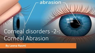
- 1. Corneal disorders -2-: Corneal Abrasion By Leena Rasmi
- 2. Corneal Abrasion • Definition: scrape or scratch injury on the corneal epithelium • Epidemiology: most common eye injury • Etiology: • Injury from an object (e.g., tree branch, fingernail, ball, mascara brushes) hitting, flying into, or poking the eye • Foreign matter stuck under the eyelid • Entropion • Prolonged contact lens wear or improperly fitted lenses • UV light (i.e., UV keratitis) • Thermal burns (e.g., cigarettes) • Dry eyes (e.g., Sjogren syndrome)
- 3. Clinical findings • Foreign body sensation in the eye • Eye pain • Epiphora • Blurred vision • Photophobia • Conjunctival injection
- 4. • Diagnostics: examination of the eye using fluorescein staining • Treatment • Removal of any retained foreign object • Analgesia: oral NSAIDs, topical NSAIDs (e.g., diclofenac, ketorolac), or narcotics, depending on pain severity • Infection prophylaxis • In the general population: antibiotic eye drops or ointment (e.g., erythromycin) • In contact lens wearers: antibiotic eye drops or ointment with antipseudomonal activity (e.g., ciprofloxacin) • Consider eye patching
- 6. Corneal dystrophy • These are rare inherited disorders. They affect different layers of the cornea and often affect corneal transparency • They may be divided into: • Anterior dystrophies involving the epithelium. These may present with recur-rent corneal erosion. • Stromal dystrophies presenting with visual loss. If very anterior they may cause corneal erosion and pain. • Posterior dystrophies which affect the endothelium and cause gradual loss of vision due to corneal oedema. They may also cause pain due to epithelial erosion. Symptoms may include: •Progressive visual impairment •Corneal erosion •Treatment •Treat corneal erosion, as in above. •Keratoplasty in severe cases or when other treatment modalities have not been successful
- 7. Corneal Degeneration •Definition: changes of the cornea that cause corneal deterioration and, potentially, dysfunction •Etiology •Normal aging •Secondary to various pathological processes (e.g., calcium salt deposits in band keratopathy associated with hypercalcemia in the setting of sarcoidosis)
- 8. Band keratopathy • Definition: a type of corneal degeneration that involves the appearance of a band-shaped area of calcification across the central cornea • Etiology • Idiopathic • Risk factors • Hypercalcemia (e.g., hyperparathyroidism, sarcoidosis), hy perphosphatemia (e.g., chronic renal failure) • Chronic, inflammatory eye disease (e.g., uveitis, keratitis) • Exposure to harmful chemicals (e.g., mercury vapors) • Family history of band keratopathy • Pathophysiology: increased serum calcium, serum phosphate, and/or corneal surface pH (caused by chronically inflamed eyes) → change in solubility of calcium and phosphate → calcium phosphate precipitation out of tears, aqueous humor, and corneal tissue → calcium phosphate deposition as salts in the Bowman layer and superficial stroma of the cornea
- 9. • clinical findings • Decreased visual acuity • Foreign body sensation • Photophobia • Diagnostics • Slit lamp examination showing band-like, horizontal, opaque white areas on the cornea • Followup includes investigation of underlying causes and may include: • Serum calcium and phosphate measurement • Parathyroid hormone level measurement • Renal function tests • Sarcoidosis workup (e.g., serum ACE, chest x-ray) •Treatment: •Treatment of the underlying cause (e.g., uveitis, hypercalcemia) •Superficial debridement and lamellar keratectomy •Prognosis: Visual deficits caused by band keratopathy can typically be treated successfully but will recur if the underlying condition is not addressed.
- 11. Keratoconus • Definition: a noninflammatory corneal condition in which the cornea becomes thinner than normal and develops a conic shape, bulging outward at the center • Etiology: unknown; frequently associated with other conditions • Neurodermatitis • Allergic asthma • Marfan syndrome • Ehlers-Danlos syndrome • Down syndrome • A positive family history of keratoconus has been found in a minority of cases (10%) • Clinical findings: symptoms are often initially unilateral; however, they always become bilateral in the further course of disease • Progressive decrease in visual acuity • Myopia • Astigmatism • Photophobia
- 12. • Diagnosis • Slit lamp examination showing protrusion and thinning of the cornea • Ultrasound pachymetry (measurement of corneal thickness via ultrasound) • Computerized corneal topography • Treatment • Correcting astigmatism and myopia: glasses or rigid gas permeable contact lenses • In progressive keratoconus: crosslinking with UV-A light (corneal crosslinking) • If conservative treatment options fail: keratoplasty
- 13. Corneal Graft
- 14. Keratoplasty (corneal transplantation) Indications •Significant visual impairment as a result of pathological changes in the cornea, e.g., opacification, irregular curvature •Keratitis that cannot be controlled by conservative treatment •Procedure: replacement of diseased cornea with cornea harvested from a recently deceased donor •Perforating keratoplasty (replacement of the complete cornea): indicated when both anterior and posterior corneal layers are damaged •Lamellar keratoplasty (replacement of only the anterior or posterior corneal layers): indicated when only anterior or posterior corneal layers are damaged •Follow-up care •Graft rejection prevention: corticosteroid eye drops •Monitoring and treatment for graft rejection
- 15. Corneal deposits
- 16. Arcus senilis (corneal arcus) • Definition: A yellow or gray discoloration of the corneal margin caused by deposits of fat and cholesterol , a condition associated with normal aging, in which annular deposits of lipids appear around the corneal margin • Epidemiology: Incidence increases with age. • 60% in those 50–60 years • Almost 100% in those > 80 years • Clinical findings: asymptomatic • Diagnostics: slit lamp examination • Treatment • In older patients: no treatment necessary • Occurrence before 50 years of age: rule out lipid disorders
- 17. Kayser-Fleischer rings • Copper accumulation in Descemet membrane of the cornea that results in 1–2 mm wide, green-brown rings in the periphery of the iris • Diagnostic sign of Wilson disease
- 18. Sclera
- 21. phenylephrine eye drops test Scleritis complications Scleromalacia ( thinning of the sclera ) , Keratitis , Uveitis , Cataract , Glaucoma
- 22. Blue sclera • Blue discoloration is caused by thinning or transparency of scleral collagen with visualization of the underlying uvea • Important causes include the following: • Osteogenesis imperfecta. • Ehlers–Danlos syndrome type VI • Marshall–Smith syndrome , Russell–Silver syndrome , Hallermann– Streiff–François syndrome.
- 23. Osteogenesis Imperfecta It is an inherited disease of connective tissue, usually caused by defects in the synthesis and structure of Type 1 collagen. There are multiple types, at least two of which have ocular features.
- 24. Thank You