This document discusses congenital heart diseases, including:
- Congenital heart disease occurs in 0.5-0.8% of live births and is more common in stillborns, abortuses, and premature infants. Diagnosis is usually made within the first month of life.
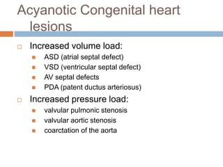
- The most common congenital heart lesions are ventricular septal defects, atrial septal defects, patent ductus arteriosus, coarctation of the aorta, and tetralogy of Fallot.
- While most congenital defects are tolerated in utero due to the fetal circulatory system, after birth when these pathways close the full impact of defects becomes apparent.
- Congenital heart diseases can be