Composizione grafica - punto, linea, superficie, gli elementi della grammatica visiva
•
1 like•360 views

Punto, linea, superficie sono gli elementi minimi della composizione grafica e del pensiero visivo. Come li combini a ha che fare con la tua creatività. Per esercitarti a farlo segui le indicazioni contenute in questo pdf.
Report
Share
Report
Share
Download to read offline

Recommended
Optics of ametropia
This document summarizes key concepts related to ametropia (conditions where the eye fails to focus light properly on the retina). It defines and compares different types of ametropia including myopia, hyperopia, astigmatism, and anisometropia. It also discusses optical correction of ametropia using lenses and the importance of considering lens position and back vertex distance when prescribing high-powered lenses. Key points covered include the differences between axial and refractive ametropia, types of hyperopia and astigmatism, and formulas for calculating effective lens power based on movement relative to the eye.
Posterior segment of eye.pdf
This is a class based presentation presented on Lecture of Shaalakya Tantra guided by Dr. Sadhana Parajuli Mam, H.O.D, Ayurveda Campus & Teaching Hospital, Kirtipur, I.O.M, T.U
Basic anatomy and physiology
The eye is made up of three coats - fibrous, vascular and nervous. The fibrous coat includes the sclera and cornea. The vascular coat provides nutrition via the uveal tract. The nervous coat is the retina which senses light. Within the eyeball are two chambers filled with aqueous humor - the anterior chamber between the cornea and iris and the posterior chamber between the iris and lens. The iris controls the size of the pupil to regulate light. The choroid nourishes the retina and the lens focuses light onto the retina to be transmitted to the brain via the optic nerve.
Astigmatism
astigmatism is a type of refractive error in which parallel ray of light entering the eye can not focus one point but form lines.
Parafoveal telangiectasia-- AJAY DUDANI
This document discusses idiopathic juxtafoveolar telangiectasia (IJFT), including its classification and stages. It describes IJFT types 1 and 2, with type 1 being congenital and aneurysmal and type 2 being acquired and perifoveal. Type 2 is further classified into 5 stages based on angiographic and imaging findings. The document also presents three case studies where anti-VEGF therapy with ranibizumab was used to treat neovascularization associated with types 1 and 2 IJFT, showing improvements in leakage and vision. While anti-VEGF therapy may help reduce leakage, preexisting photoreceptor damage from IJFT may limit improvements in visual acuity.
Astigmatic lens used in ophthalmology and eye
different types and classifications of astigmatic lens used
availability of astigmatic lens
uses of astigmatic lens
advantages and disadvantages of astigmatic lens
Control of ACCOMMODATION
Definition Of The Accommodation
Mechanism Of Accommodation
Triggers Accommodation
Terms Of Accommodation
Accommodative Dysfunction
Spasm Of Accommodation
Accommodative Esotropia
Controlling Accommodation In Vision Test
Tips To Control Accommodation In Lifestyle
References
Clinical Procedures In Optometry By J.D. Bartlett, J.B. Eskridge, J.F. Amos
Theory And Practice Of Squint And Orthoptics By A.K.Khurana
Adler’s Physiology Of The Eye By L.A. Levin, S.F. Nilsson
Borish’s Clinical Refraction By W.J. Benjamin
Duke-elder’s Practice Of Refraction By David Abrams
Optics & Refraction By A.K.Khurana
Textbook Of Ophthalmology By E Ahmed
Clinical Optics By A R. Elkington, Werner L, Trindade F, Pereira F, Werner L
Physiology Of Accommodation And Presbyopia, ARQ. Bras. OFTALMOL, December 2000.
Optometry And Ophthalmology Websites
Anatomy and physiology of cornea
The document provides information on the anatomy and physiology of the cornea. It discusses the gross anatomy including the layers of the cornea - epithelium, Bowman's layer, stroma, Descemet's membrane, and endothelium. The microanatomy and ultrastructure of each layer is described. Key points include the lamellar structure of the stroma providing transparency, regeneration of the epithelium from basal stem cells, and the theories behind corneal transparency relating to the structure of the stromal collagen fibers.
Glaucoma 2 primary angle closure glaucoma,dr.k.n.jha,02.11.16
Angle-closure glaucoma is caused by apposition of the peripheral iris to the trabecular meshwork, reducing drainage of aqueous humor from the eye. Primary angle-closure glaucoma (PACG) has no underlying cause and is due to anatomic factors. It is a leading cause of glaucoma worldwide. PACG presents with acute symptoms like eye pain and blurred vision due to sudden rise in pressure from pupillary block. Treatment involves lowering pressure with medications or iridectomy to prevent future attacks. Long-term management focuses on screening and treatment to prevent angle closure in the fellow eye.
Retina embryology ppt
The retina develops from the evagination and invagination of the optic vesicles from the diencephalon beginning around day 22. The optic vesicles grow toward the ectoderm, inducing lens formation, and invaginate to form the optic cup by day 33. The layers of the optic cup then differentiate into the neural retina and pigmented epithelium, with the anterior portion forming the iris and ciliary body and the posterior portion forming the 10 layers of the retina.
Inscribed Angles
1) An inscribed angle is an angle whose vertex lies on a circle and whose sides contain chords of the circle.
2) The measure of an inscribed angle is equal to one-half the measure of its intercepted arc.
3) If two inscribed angles intercept the same arc or congruent arcs, then the angles are congruent.
ANATOMY AND PHYSIOLOGY OF EXTRAOCULAR MUSCLES.ppt
The presentation begins with an overview of the extraocular muscles, highlighting their crucial role in controlling eye movements and maintaining proper vision. Emphasized the significance of these muscles in daily activities and visual perception.
Vergences of the eye
This document discusses different types of vergence eye movements, including fusional vergence and accommodative convergence. It defines fusional vergence as an optomotor reflex that works to maintain eye alignment and retinal image correspondence. Accommodative convergence is described as a reflex linking convergence and accommodation simultaneously during the near response. The ratio between accommodative convergence and accommodation (AC/A ratio) is also discussed, along with examples of normal and abnormal AC/A ratios.
Astigmatism
Astigmatism is a refractive error where refraction varies in different meridians of the eye. In astigmatism, light rays from one sector focus on one point of the retina while rays from another sector focus on a different point, resulting in blurred vision. Around 60% of refractive error cases involve astigmatism. It is classified as mild, moderate, severe, or extreme depending on the degree of refractive error. Regular astigmatism results from corneal or lenticular shape and irregular astigmatism involves irregular changes in refractive power. Treatment options include optical aids like glasses and contact lenses or refractive surgery procedures.
Extraocular muscles dr.gosai
The document describes the anatomy of the bony orbit and the extraocular muscles within. It discusses the seven bones that make up the bony orbit, including the frontal, zygomatic, maxillary, ethmoid, sphenoid, lacrimal and palatine bones. It describes the roof, floor, medial wall and lateral wall of the orbit. It then discusses the extraocular muscles, including the four rectus muscles, two oblique muscles and levator palpebrae superioris. It details the origin, insertion, nerve supply and actions of each muscle. Finally, it discusses some clinical implications like strabismus and Horner's syndrome.
Mechanisms of angle closure glaucoma
1) Angle-closure glaucoma (ACG) occurs when the drainage angle between the iris and cornea is blocked. It is more common in Asian populations and causes more vision loss than open-angle glaucoma.
2) Risk factors for ACG include older age, female sex, Chinese ethnicity, family history, anatomically shallow anterior chambers, and thick lenses. Precipitating factors are low light, certain drugs, and stress.
3) Pupillary block is the main mechanism of ACG, where the iris blocks the trabecular meshwork due to apposition between the iris and lens at the pupil. Plateau iris is a variant where the peripheral iris is anteriorly displaced onto the angle
Production and flow of aqueous humor
The aqueous humour is a clear fluid produced in the cilliary body that flows through the posterior and anterior chambers of the eye, providing nutrients and removing waste. It is formed primarily via active secretion and diffusion, circulating through the trabecular meshwork and schlemm's canal before draining into episcleral veins. The rate and composition of aqueous humour production and drainage are tightly regulated to maintain intraocular pressure for proper eye function.
21 step-Diagnosis Procedure Refraction
20/12/14-Reference :
-Clinical Procedures for Ocular Examination 3rd edition by Carlson, Kurtz, Heath, Hines
-OPM 303: Diagnostic Procedures - Refraction By: Dr. Pornpatcharin Wongsaisri
Faculty of Optometry.Rangsit University Thailand
Astigmatism complete
Astigmatism is a refractive error where the eye does not focus light evenly on the retina due to an uneven curvature of the cornea or lens. This causes blurred vision. There are different types of astigmatism including with-the-rule, against-the-rule, and oblique astigmatism depending on the axis of the steeper and flatter meridians. Astigmatism is typically corrected using cylindrical lenses in glasses or toric contact lenses to refocus light evenly on the retina. More advanced treatments include refractive surgery such as LASIK.
Orbscan & topo
This document discusses corneal topography and keratometry. It defines topography as determining and describing the features of a surface, specifically the corneal surface. It describes methods of measuring corneal topography including reflection-based methods like keratometry and projection-based methods like slit photography and rasterstereography. It also discusses different topographic maps including axial, tangential, and refractive maps, and indices used to quantify topography such as the simulated keratometry values, surface asymmetry index, and surface regularity index.
Keratoconus
Management of Keratoconus
for more information about icourses
https://www.facebook.com/i.courses.ophthalmology/
https://wa.me/201092909418
https://www.youtube.com/channel/UChSK-t5QtUa7Y6ct889ql7Q?reload=9&
https://t.me/icoursesophthalmology
https://www.instagram.com/i.courses.ophthalmology/
https://www.linkedin.com/in/ahmed-hamdy-626527188/
anatomy of sclera
1. Introduction Gross anatomy Layers Blood supply, drainage and nerve supply
2. INTRODUCTION • Sclera forms posterior 5/6th of external tunic , connective tissue coat of eyeball. • it continues with duramater and cornea • Its whole surface covered by tenon’s capsule • Anteriorly covered by- bulbar conjunctiva • Inner surface lies in contact with choroid • With a potential suprachoroidal space in between
3. Equa THICKNESS OF SCLERA
4. • Thickness varies with individual, with age • Thinner- children, elder, F> M • Thickest posteriorly • Gradually becomes thinner when traced anteriorly • Thin at insertion of extraocular muscle
Anatomy of cornea and sclera
A very helpful concise summary of the anatomy, embryology and histology of cornea for ophthalmology residents!
Extra ocular muscles ppt
The document discusses the extraocular muscles and their nerve supply, actions, and clinical presentations of nerve palsies. It describes the origins, insertions, innervations and primary actions of the 4 recti muscles and 2 oblique muscles. Figure 1 shows ptosis and limitations of eye movements in 3rd nerve palsy. Figures 2 and 3 demonstrate the clinical findings in 4th nerve and 6th nerve palsies, respectively. Ophthalmoplegia is defined as paralysis of the extraocular muscles resulting in double vision.
Ocular motility and gaze
3rd,4th, 6th nerves
Extraocular muscles
How to examine for ocular motility
Ophthalmoplegia
Diplopia and related disorders
Gaze pathway
How to examine for gaze
Gaze palsy
Types of eye movements
How to examine for EM
Nystagmus and non nystagmus ocular oscillation
Anatomy of orbit
The document summarizes the anatomy of the orbit. It is formed by 7 bones and has a quadrangular pyramid shape. The orbit contains 4 walls - medial, inferior, lateral, and roof. Each wall has specific bone formations and relationships to structures like muscles and nerves. The orbit also contains openings like the optic canal and superior orbital fissure that connect to other areas. The periorbita lining and fascia bulbi surrounding the eyeball are described. Finally, the document outlines the 3 surgical spaces within the orbit.
Spherical lenses
This document discusses different types of lenses used in ophthalmology. It describes spherical lenses and how they are either convex or concave, forming converging or diverging images. It also discusses astigmatic lenses, including cylindrical lenses which have one curved and one plane surface, and toric lenses which have two curved surfaces of different curvatures. The key concepts of focal length, power, vergence, and magnification of lenses are defined.
L'infografica
L'infografica è la rappresentazione in forma visiva di informazioni, dati o conoscenze attraverso la forma grafica. E' un modo per comunicare concetti complessi, in modo semplice, chiaro, diretto.
More Related Content
What's hot
Glaucoma 2 primary angle closure glaucoma,dr.k.n.jha,02.11.16
Angle-closure glaucoma is caused by apposition of the peripheral iris to the trabecular meshwork, reducing drainage of aqueous humor from the eye. Primary angle-closure glaucoma (PACG) has no underlying cause and is due to anatomic factors. It is a leading cause of glaucoma worldwide. PACG presents with acute symptoms like eye pain and blurred vision due to sudden rise in pressure from pupillary block. Treatment involves lowering pressure with medications or iridectomy to prevent future attacks. Long-term management focuses on screening and treatment to prevent angle closure in the fellow eye.
Retina embryology ppt
The retina develops from the evagination and invagination of the optic vesicles from the diencephalon beginning around day 22. The optic vesicles grow toward the ectoderm, inducing lens formation, and invaginate to form the optic cup by day 33. The layers of the optic cup then differentiate into the neural retina and pigmented epithelium, with the anterior portion forming the iris and ciliary body and the posterior portion forming the 10 layers of the retina.
Inscribed Angles
1) An inscribed angle is an angle whose vertex lies on a circle and whose sides contain chords of the circle.
2) The measure of an inscribed angle is equal to one-half the measure of its intercepted arc.
3) If two inscribed angles intercept the same arc or congruent arcs, then the angles are congruent.
ANATOMY AND PHYSIOLOGY OF EXTRAOCULAR MUSCLES.ppt
The presentation begins with an overview of the extraocular muscles, highlighting their crucial role in controlling eye movements and maintaining proper vision. Emphasized the significance of these muscles in daily activities and visual perception.
Vergences of the eye
This document discusses different types of vergence eye movements, including fusional vergence and accommodative convergence. It defines fusional vergence as an optomotor reflex that works to maintain eye alignment and retinal image correspondence. Accommodative convergence is described as a reflex linking convergence and accommodation simultaneously during the near response. The ratio between accommodative convergence and accommodation (AC/A ratio) is also discussed, along with examples of normal and abnormal AC/A ratios.
Astigmatism
Astigmatism is a refractive error where refraction varies in different meridians of the eye. In astigmatism, light rays from one sector focus on one point of the retina while rays from another sector focus on a different point, resulting in blurred vision. Around 60% of refractive error cases involve astigmatism. It is classified as mild, moderate, severe, or extreme depending on the degree of refractive error. Regular astigmatism results from corneal or lenticular shape and irregular astigmatism involves irregular changes in refractive power. Treatment options include optical aids like glasses and contact lenses or refractive surgery procedures.
Extraocular muscles dr.gosai
The document describes the anatomy of the bony orbit and the extraocular muscles within. It discusses the seven bones that make up the bony orbit, including the frontal, zygomatic, maxillary, ethmoid, sphenoid, lacrimal and palatine bones. It describes the roof, floor, medial wall and lateral wall of the orbit. It then discusses the extraocular muscles, including the four rectus muscles, two oblique muscles and levator palpebrae superioris. It details the origin, insertion, nerve supply and actions of each muscle. Finally, it discusses some clinical implications like strabismus and Horner's syndrome.
Mechanisms of angle closure glaucoma
1) Angle-closure glaucoma (ACG) occurs when the drainage angle between the iris and cornea is blocked. It is more common in Asian populations and causes more vision loss than open-angle glaucoma.
2) Risk factors for ACG include older age, female sex, Chinese ethnicity, family history, anatomically shallow anterior chambers, and thick lenses. Precipitating factors are low light, certain drugs, and stress.
3) Pupillary block is the main mechanism of ACG, where the iris blocks the trabecular meshwork due to apposition between the iris and lens at the pupil. Plateau iris is a variant where the peripheral iris is anteriorly displaced onto the angle
Production and flow of aqueous humor
The aqueous humour is a clear fluid produced in the cilliary body that flows through the posterior and anterior chambers of the eye, providing nutrients and removing waste. It is formed primarily via active secretion and diffusion, circulating through the trabecular meshwork and schlemm's canal before draining into episcleral veins. The rate and composition of aqueous humour production and drainage are tightly regulated to maintain intraocular pressure for proper eye function.
21 step-Diagnosis Procedure Refraction
20/12/14-Reference :
-Clinical Procedures for Ocular Examination 3rd edition by Carlson, Kurtz, Heath, Hines
-OPM 303: Diagnostic Procedures - Refraction By: Dr. Pornpatcharin Wongsaisri
Faculty of Optometry.Rangsit University Thailand
Astigmatism complete
Astigmatism is a refractive error where the eye does not focus light evenly on the retina due to an uneven curvature of the cornea or lens. This causes blurred vision. There are different types of astigmatism including with-the-rule, against-the-rule, and oblique astigmatism depending on the axis of the steeper and flatter meridians. Astigmatism is typically corrected using cylindrical lenses in glasses or toric contact lenses to refocus light evenly on the retina. More advanced treatments include refractive surgery such as LASIK.
Orbscan & topo
This document discusses corneal topography and keratometry. It defines topography as determining and describing the features of a surface, specifically the corneal surface. It describes methods of measuring corneal topography including reflection-based methods like keratometry and projection-based methods like slit photography and rasterstereography. It also discusses different topographic maps including axial, tangential, and refractive maps, and indices used to quantify topography such as the simulated keratometry values, surface asymmetry index, and surface regularity index.
Keratoconus
Management of Keratoconus
for more information about icourses
https://www.facebook.com/i.courses.ophthalmology/
https://wa.me/201092909418
https://www.youtube.com/channel/UChSK-t5QtUa7Y6ct889ql7Q?reload=9&
https://t.me/icoursesophthalmology
https://www.instagram.com/i.courses.ophthalmology/
https://www.linkedin.com/in/ahmed-hamdy-626527188/
anatomy of sclera
1. Introduction Gross anatomy Layers Blood supply, drainage and nerve supply
2. INTRODUCTION • Sclera forms posterior 5/6th of external tunic , connective tissue coat of eyeball. • it continues with duramater and cornea • Its whole surface covered by tenon’s capsule • Anteriorly covered by- bulbar conjunctiva • Inner surface lies in contact with choroid • With a potential suprachoroidal space in between
3. Equa THICKNESS OF SCLERA
4. • Thickness varies with individual, with age • Thinner- children, elder, F> M • Thickest posteriorly • Gradually becomes thinner when traced anteriorly • Thin at insertion of extraocular muscle
Anatomy of cornea and sclera
A very helpful concise summary of the anatomy, embryology and histology of cornea for ophthalmology residents!
Extra ocular muscles ppt
The document discusses the extraocular muscles and their nerve supply, actions, and clinical presentations of nerve palsies. It describes the origins, insertions, innervations and primary actions of the 4 recti muscles and 2 oblique muscles. Figure 1 shows ptosis and limitations of eye movements in 3rd nerve palsy. Figures 2 and 3 demonstrate the clinical findings in 4th nerve and 6th nerve palsies, respectively. Ophthalmoplegia is defined as paralysis of the extraocular muscles resulting in double vision.
Ocular motility and gaze
3rd,4th, 6th nerves
Extraocular muscles
How to examine for ocular motility
Ophthalmoplegia
Diplopia and related disorders
Gaze pathway
How to examine for gaze
Gaze palsy
Types of eye movements
How to examine for EM
Nystagmus and non nystagmus ocular oscillation
Anatomy of orbit
The document summarizes the anatomy of the orbit. It is formed by 7 bones and has a quadrangular pyramid shape. The orbit contains 4 walls - medial, inferior, lateral, and roof. Each wall has specific bone formations and relationships to structures like muscles and nerves. The orbit also contains openings like the optic canal and superior orbital fissure that connect to other areas. The periorbita lining and fascia bulbi surrounding the eyeball are described. Finally, the document outlines the 3 surgical spaces within the orbit.
Spherical lenses
This document discusses different types of lenses used in ophthalmology. It describes spherical lenses and how they are either convex or concave, forming converging or diverging images. It also discusses astigmatic lenses, including cylindrical lenses which have one curved and one plane surface, and toric lenses which have two curved surfaces of different curvatures. The key concepts of focal length, power, vergence, and magnification of lenses are defined.
What's hot (20)
Glaucoma 2 primary angle closure glaucoma,dr.k.n.jha,02.11.16
Glaucoma 2 primary angle closure glaucoma,dr.k.n.jha,02.11.16
Fizika- Difrakcija svetlosti- Dušan Kostić- Vladimir Milićević
Fizika- Difrakcija svetlosti- Dušan Kostić- Vladimir Milićević
More from Maria Novella Fabiano
L'infografica
L'infografica è la rappresentazione in forma visiva di informazioni, dati o conoscenze attraverso la forma grafica. E' un modo per comunicare concetti complessi, in modo semplice, chiaro, diretto.
Valorizzazione culturale con i media - Canosa di Puglia: una Città-Museo
Tutte le risorse qui contenute sono mostrate a scopo non-commerciale, ma con esclusivo fine educativo e di diffusione culturale, in pieno rispetto delle leggi e dei trattati nazionali e internazionali sulle norme che regolano e tutelano il diritto d'autore.
More from Maria Novella Fabiano (8)
Valorizzazione culturale con i media - Canosa di Puglia: una Città-Museo
Valorizzazione culturale con i media - Canosa di Puglia: una Città-Museo
L'Abc del linguaggio audiovisivo - Le inquadrature - Prima lezione
L'Abc del linguaggio audiovisivo - Le inquadrature - Prima lezione
Composizione grafica - punto, linea, superficie, gli elementi della grammatica visiva
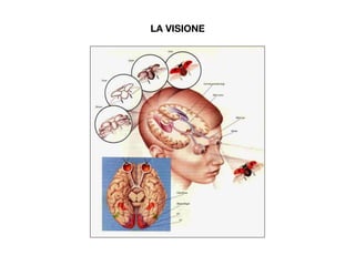
- 1. LA VISIONE
- 2. ELEMENTI MINIMI DELLA GRAMMATICA VISIVA Casa E Punto Linea Superficie BLOCCHI TESTO
- 3. ELEMENTI MINIMI DELLA GRAMMATICA VISIVA Casa E Il punto In grafica si definisce punto un segno limitato che concentra su di sé l’attenzione ed è identificabile tramite coordinate precise all’interno di uno spazio.
- 4. ELEMENTI MINIMI DELLA GRAMMATICA VISIVA La linea In grafica si definisce linea una connessione tra due punti o la trac sia di un punto che si sposta. La linea comunica movimento e direzione. BLOCCHI TESTO
- 5. ELEMENTI MINIMI DELLA GRAMMATICA VISIVA La superficie In grafica si definisce superficie un punto che si allarga in uno spazio, in modo che il suo contorno, ovvero la forma, cominci a diventare significativa e a interagire con lo spazio esterno che lo contiene.
- 6. ELEMENTI MINIMI DELLA GRAMMATICA VISIVA PUNTI, LINEE e SUPERFICI hanno sempre un CAMPO di riferimento.
- 7. ESERCITAZIONE Crea 6 varianti di esempi di punti, linee e superfici per promuovere gli smarties, considerando come varianti per ognuna sia l’aspetto geometrico, sia quello tipografico, sia quello fotografico.
- 8. ISPIRAZIONI
- 9. ISPIRAZIONI