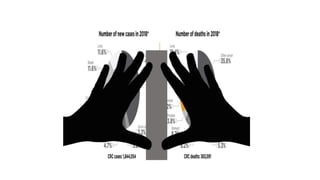
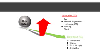
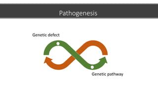
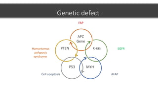
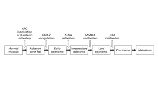
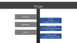
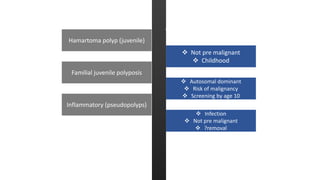
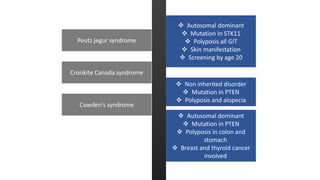
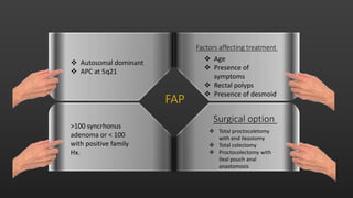
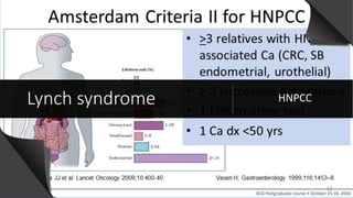
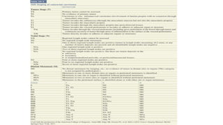
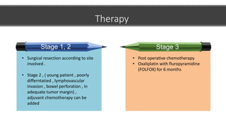
colon cancer
Dr. Ibrahim Albujays
31st Jan 2021
Almoosa specialist hospital
Incidince , risk factors , pathogenesis
Objectives
Polyps
Inherited colon cancer
Staging
Treatment
Risk
factors
Increase risk
 Age
 Personal hx ( colon ca.
, polyposis , IBD)
 Smoking
 Obesity
Decrease risk
 Dietry fibers
 Vitamin
 Good life style
 Antioxident
Pathogenesis
Genetic defect
Genetic pathway
Genetic defect
APC
Gene
K-ras
MYH
P53
PTEN
FAP
EGFR
AFAP
Cell apoptosis
Hamartomus
polyposis
syndrome
 80% of CRC
 Chromosomal
deletion
 APC with FAP
 Most of
remaining
 Error in
mismatch
repair during
DNA
replication
 Lynch 9
 Activate or
inactivate
 Serrated
polyps
Genetic pathway
Loss of heterozygostity Microsatellite instability Methylation
 25%
 >50 yrs
 Malignancy (size /types)
Neoplastic
 Small (<5 mm)
 Hyperplasia without dysplasia
 ? Removal
Hyperplastic
 WHO criteria
 Risk of malignancy 30%
Serrated
Polyps
 Not pre malignant
 Childhood
Hamartoma polyp (juvenile)
 Autosomal dominant
 Risk of malignancy
 Screening by age 10
Familial juvenile polyposis
 Infection
 Not pre malignant
 ?removal
Inflammatory (pseudopolyps)
and Against Argument
 Autosomal dominant
 Mutation in STK11
 Polyposis all GIT
 Skin manifestation
 Screening by age 20
Peutz jegur syndrome
 Non inherited disorder
 Mutation in PTEN
 Polyposis and alopecia
Cronkite Canada syndrome
 Autosomal dominant
 Mutation in PTEN
 Polyposis in colon and
stomach
 Breast and thyroid cancer
involved
Cowden’s syndrome
>100 syncrhonus
adenoma or < 100
with positive family
Hx.
 Autosomal dominant
 APC at 5q21
Surgical option
 Total proctocoletomy
with end ileostomy
 Total colectomy
 Proctocolectomy with
ileal pouch anal
anastomosis
Factors affecting treatment
 Age
 Presence of
symptoms
 Rectal polyps
 Presence of desmoid
FAP
Later presentation , fewer polyps
APC 30%
MYH mutation
AFA
P
Lynch syndrome HNPCC
Therapy
Stage 1, 2
• Surgical resection according to site
involved .
• Stage 2 , ( young patient , poorly
differntatied , lymphovascular
invasion , bowel perforation , in
adequate tumor margin) ,
adjuvant chemotherapy can be
added
Stage 3
• Post operative chemotherapy
• Oxaliplatin with fluropyramidine
(FOLFOX) for 6 months
Stage 4
Pre operative evaluation
• Looking for syncronus disorder
• CT abdomen , pelvis and chest
• CEA level
• In case of obstruction ,
gastrograffin can be used .
Surgical principles
1. Wide resection of involved colon +
lymphatics + mesocolon +enblock
resection of neiboghring
2. Margin around 5 cm is adequate
3. Mininmum of 12 LN for biopsy
4. Postive LN left beyond , consider
inproper resection
5. Inspect abdomen , viserca ,
peritoneum for any Mets
5
4
3
2
1
Surgical Options for Colon
cancer
1-Ca of cecum/ascending colon:
Right Hemicolectomy
2- Ca of hepatic flexure/proximal transverse colon:
Extended right Hemicolectomy
3-Ca of distal transverse colon:
Extended right Hemicolectomy including Splenic flexure OR left
Hemicolectomy
4- Ca of left colon:
Left Hemicolectomy
5-Ca of sigmoid colon:
Sigmoidectomy OR left Hemicolectomy
Specific consideration
 Right side /transverse : right
hemi , extending right hemi
Obstructing colon cancer
 Present with peritonitis
 Hartman is an option
Perforated colon cancer
Follow up
 History, physical examination &
S.CEA: every 3–6 month for 2 y,
then every 6 month for a total
of 5 yr
 Colonoscopy: In 1st yr:
Abnormal repeat in one yr.
Normal: repeat in 3 rd yr then
every 5 yrs If it was not done
before: 3-6 months post
surgery.
 CT scan chest & abdomen:
Annually for pts. with high risk
for recurrence(eg. poorly
differentiated tumors)
Survillance

Colon ca. , presentation , pathophysiology , and treatment

  • 1.
    colon cancer Dr. IbrahimAlbujays 31st Jan 2021 Almoosa specialist hospital
  • 2.
    Incidince , riskfactors , pathogenesis Objectives Polyps Inherited colon cancer Staging Treatment
  • 7.
    Risk factors Increase risk  Age Personal hx ( colon ca. , polyposis , IBD)  Smoking  Obesity Decrease risk  Dietry fibers  Vitamin  Good life style  Antioxident
  • 8.
  • 9.
  • 10.
     80% ofCRC  Chromosomal deletion  APC with FAP  Most of remaining  Error in mismatch repair during DNA replication  Lynch 9  Activate or inactivate  Serrated polyps Genetic pathway Loss of heterozygostity Microsatellite instability Methylation
  • 12.
     25%  >50yrs  Malignancy (size /types) Neoplastic  Small (<5 mm)  Hyperplasia without dysplasia  ? Removal Hyperplastic  WHO criteria  Risk of malignancy 30% Serrated Polyps
  • 13.
     Not premalignant  Childhood Hamartoma polyp (juvenile)  Autosomal dominant  Risk of malignancy  Screening by age 10 Familial juvenile polyposis  Infection  Not pre malignant  ?removal Inflammatory (pseudopolyps) and Against Argument
  • 14.
     Autosomal dominant Mutation in STK11  Polyposis all GIT  Skin manifestation  Screening by age 20 Peutz jegur syndrome  Non inherited disorder  Mutation in PTEN  Polyposis and alopecia Cronkite Canada syndrome  Autosomal dominant  Mutation in PTEN  Polyposis in colon and stomach  Breast and thyroid cancer involved Cowden’s syndrome
  • 15.
    >100 syncrhonus adenoma or< 100 with positive family Hx.  Autosomal dominant  APC at 5q21 Surgical option  Total proctocoletomy with end ileostomy  Total colectomy  Proctocolectomy with ileal pouch anal anastomosis Factors affecting treatment  Age  Presence of symptoms  Rectal polyps  Presence of desmoid FAP
  • 16.
    Later presentation ,fewer polyps APC 30% MYH mutation AFA P
  • 17.
  • 19.
    Therapy Stage 1, 2 •Surgical resection according to site involved . • Stage 2 , ( young patient , poorly differntatied , lymphovascular invasion , bowel perforation , in adequate tumor margin) , adjuvant chemotherapy can be added Stage 3 • Post operative chemotherapy • Oxaliplatin with fluropyramidine (FOLFOX) for 6 months
  • 20.
  • 21.
    Pre operative evaluation •Looking for syncronus disorder • CT abdomen , pelvis and chest • CEA level • In case of obstruction , gastrograffin can be used .
  • 22.
    Surgical principles 1. Wideresection of involved colon + lymphatics + mesocolon +enblock resection of neiboghring 2. Margin around 5 cm is adequate 3. Mininmum of 12 LN for biopsy 4. Postive LN left beyond , consider inproper resection 5. Inspect abdomen , viserca , peritoneum for any Mets 5 4 3 2 1
  • 23.
    Surgical Options forColon cancer 1-Ca of cecum/ascending colon: Right Hemicolectomy 2- Ca of hepatic flexure/proximal transverse colon: Extended right Hemicolectomy 3-Ca of distal transverse colon: Extended right Hemicolectomy including Splenic flexure OR left Hemicolectomy 4- Ca of left colon: Left Hemicolectomy 5-Ca of sigmoid colon: Sigmoidectomy OR left Hemicolectomy
  • 24.
    Specific consideration  Rightside /transverse : right hemi , extending right hemi Obstructing colon cancer  Present with peritonitis  Hartman is an option Perforated colon cancer
  • 25.
    Follow up  History,physical examination & S.CEA: every 3–6 month for 2 y, then every 6 month for a total of 5 yr  Colonoscopy: In 1st yr: Abnormal repeat in one yr. Normal: repeat in 3 rd yr then every 5 yrs If it was not done before: 3-6 months post surgery.  CT scan chest & abdomen: Annually for pts. with high risk for recurrence(eg. poorly differentiated tumors) Survillance