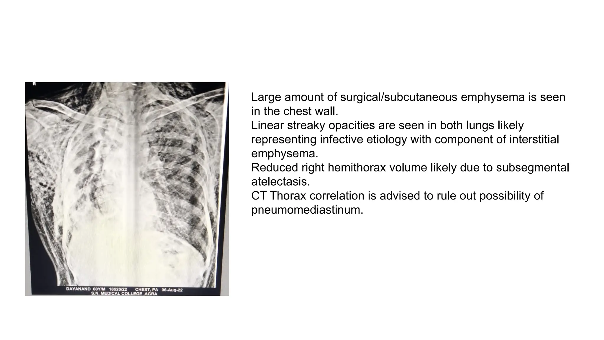
CLINICO-RADIOLOGICAL
MEET
Xrays
Case 1: Virma devi, 60/F
Known hypertensive (on irregular treatment for 5-6 yrs) presented with abdominal
pain, ghabrahat, breathlessness and dry cough
O/E
BP 220/130, PR 74, SPO2 92%RA, RBS 104
Chest: B/L crepts present
Reticulonodular opacities seen in bilateral lung fields
with more predominance towards the lung bases.
Hyperinflated lung fields with flattened domes of
diaphragm.
Findings could represent spectrum of interstitial lung
disease
Case 2: Kiran lal, 64/M
Presented with loss of appetite for 2 months, fever (low grade) for 2 months,
weight loss for 2 months, cough with expectoration for 3 months
H/O 3 episodes of hemoptysis present.
O/E BP 98/50, PR 100, SPO2 92%RA, RBS 108
Chest: decreased breath sounds heard over Rt mammary area
A radio-opaque shadow with relatively defined margins is
seen in the right upper middle zone, with small areas of
breakdown within it.
Possible differentials could include a neoplastic etiology (in
view of patient age), hydatid cyst.
Bilateral lung fields appear hyperinflated
Bilateral apical pleural thickening is seen.
Case 3: Kumar sen, 53/M
℅ breathlessness for 1 yr (increased for 3 days)
Fever for 3 days
Cough with expectoration for 1 yr (on and off)
H/O chronic ganja abuse and bidi smoking present.
Chest: Decreased air entry on right side with B/L crepts
Rotation to left
White out right lung field is seen with air bronchograms
suggestive of consolidation with collapse.
Right CP angle is obscured.
Left lung field is hyperinflated.
Case 4: Kusum singhania, 51/F
K/C/O RHD (Moderate MS, Mild MR, Trivial TR) on anti-coagulant therapy since 2 yrs,
presented with
Fever (mild) for 1yr
Breathlessness for 1yr, increased since last 2 days
(No H/O ATT Intake)
O/E: BP 102/60, PR 88 regular, SPO2 96% O2, RBS 175,
Chest: B/L crepts present
CVS: S1S2 present, Diastolic murmur heard over mitral area
Rotation to left
Cardiomegaly is seen with carinal splaying and
double right heart border suggesting the possibility of
left sided chamber enlargement.
Ill defined infiltrates are seen in both lungs in a
predominant central and peri-bronchovascular
distribution, likely pulmonary edema. Possibility of
superimposed infective etiology cannot be excluded.
Left CP angle is obscured.
Case 5: Arun, 27/M
Presented with sudden onset, abdominal pain which started in Rt Iliac Fossa and progressed to
involve whole abdomen.
O/E: BP 92/56 on Norad, PR 128, SPO2 97% O2, RBS 102
Febrile,
conscious, oriented to T/P/P
Chest: B/L NVBS
P/A: Distended, diffuse tenderness present,
Guarding rigidity in Rt Iliac fossa
Bowel sounds absent.
Large amount of free air is seen under right and left domes of
diaphragms suggestive of pneumo-peritoneum.
Hazy opacities are seen in bilateral lower zones likely postural
atelectatasis.
Case 6: CT Dashrath, 49/M
Presented with ℅:
Abdominal pain for 3 days
Abdominal distension for 3 days,
Inability to pass flatus/feces for 2 days
Nausea for 2 days
O/E: Per abdomen: distended, diffuse tenderness present.
Absent bowel sounds
Multiple air fluid levels are seen in the epigastrium, right and
left hypochondrium with relatively gasless large bowel,
suggestive of acute small bowel obstruction.
Case 7: Subhash chandra, 40/M
Presented with:
Breathlessness for 10 days
Fever for 8 days
Chest pain for 8 days
O/E: BP 114/68, PR 76/min, SPO2 93% RA, RBS 107
Chest: decreased chest expansion on right side,
absent breath sounds in Right mammary and inframammary area, infraaxillary area.
Rotation to right
A large radio-opacity is seen in the right mid lower zone
with sharply defined upper margin, obscuring the right CP
angle, with C/L mediastinal shift suggestive of a large right
sided pleural effusion.
A differential could be a liver abscess with reactionary right
sided pleural effusion.
Case 8: Shikha, 29/F
Presented with:
breathlessness for 5 days, progressive in nature, increased since 1 day
Chest pain on left side
O/E: BP 126/78, PR 132, SPO2 96% RA, RBS 109, RR 30
Reduced lung expansion noted on left side (<2cm)
Hyperresonant note was heard on percussion
Breath sounds absent on left side, crepts heard on Right side.
Large left side pneumothorax is seen causing collapse
of left lung with C/L mediastinal shift and flattened left
hemidiaphragm.
Hazy infiltrates are seen in the right mid and lower
zones ?infective etiology.
Case 9: Sunderlal, 48/M
Presented with ℅:
Fever (mild) for 2months
Cough with expectoration for 1 month
Breathlessness for 1 month, increased for the past 7 days
O/E: BP 128/78, PR 132, SPO2 90%RA, RBS 102, RR 28
Chest: diminished Chest movement on right side,
Reduced Chest expansion on right side (1.5cm)
Shifting dullness noted on percussion on right side
diminished breath sounds over right side, coin test positive on right side.
Normal vesicular sounds on left side
Large right side hydro-pneumothorax is seen with passive
atelectasis of the underlying right lung.
Case 10: Vinod, 57/M
Breathlessness for 3yrs, progressive in nature
Fever (low grade) for 8 days
Cough with expectoration for 7 days
H/O bidi smoking for 12 yrs present (stopped 2 yrs back)
No H/O hemoptysis
O/E BP 100/60, PR 96, SPO2 90% RA, RBS 133, RR 24
R/ S: decreased air entry in left infra axillary and inframammary area,
Bronchial sounds heard over Left infraclavicular area, crepts heard all over
CVS: S1S2 present
Consolidation with breakdown is seen in left lower zone
suggestive of an infective etiology. Left CP angle is obscured.
Fibro-cavitary lesions are seen in right upper zone with tractional
bronchiectasis, pleural thickening and calcific foci with suspicion
for right upper lobe collapse as evidenced by elevated right
hilum and rt sided tracheal deviation.
Bullae are seen in the left upper zone.
Fibrotic lesions are seen in left upper zone with calcific foci.
Pleuroparenchymal bands are seen in right lower zone.
Bilateral hyperinflated lung fields with tubular heart shadow
suggestive of COPD changes.
Case 11: Parwati, 50/F
K/C/O ASD with PAH
℅ B/L lower limb swelling, breathlessness
O/E BP 96/50, PR 124, SPO2 97% O2, RBS 173
PED+/ JVP RAISED
CVS: S1 S2 Present
Systolic murmur heard over tricuspid area.
R/S: B/L crepts prsent
Cardiomegaly is seen with bulging of the pulmonary bay, and
elevated cardiac apex suggestive of predominant right side
chamber enlargement likely due to underlying pulmonary
hypertension.
Ill defined infiltrates are seen in bilateral middle and lower
zones.
Case 12: Wahid khan, 73/M
℅ loss of appetite for 3 months
Productive cough for 1 month
Mild fever for 1 month
Breathlessness for 1 month
O/E: BP 110/66, PR 108, SPO2 91%RA, RBS 165, RR 28
Chest: decreased air entry on left side with bronchial sounds
An ill defined heterogeneous radiodense lesion is seen in the
left para-hilar region with a veil like opacity in the left upper
and mid zone suggestive of a left para-hilar mass lesion
causing left upper lobe collapse with ipsilateral mediastinal
shift.
There is widening of the right paratracheal stripe likely due to
lymphadenopathy.
A thick walled cavitary lesion is seen in the left mid zone.
Case 13:Phoolwati, 58/F
K/C/O RHD presented with
Ghabrahat (on and off) for 1yr
Breathlessness for 1yr, increased since last 4days
O/E: BP 138/76, PR 116, SPO2 95%RA, RBS 123, RR 28
Pedal edema present, JVP raised
Chest: B/L crepts present
CVS: S1S2 heard, systolic murmured hear over tricuspid area, diastolic murmur heard
over apex radiating to axilla
Cardiomegaly is seen with carinal widening and double
cardiac contour suggestive of predominant left atrial
enlargement.
Reticular opacities are seen in both lung fields with
cephalization suggestive of developing pulmonary
edema.
Right CP angle is blunted suggestive of pleural effusion.
Calcific foci are seen in the right upper mid zone and right
axilla.
Case 14:Dharampal, 53/M
K/C/O COPD (for 8yrs, on irregular treatment ), presented with
Breathlessness for 6 days,
Fever for 7 days,
Cough with expectoration for 5 days
H/O bidi smoking for 15yrs present.
O/E: BP 114/72, PR 136, SPO2 95%RA, RBS 174, RR 28
Chest: barrel shaped chest with prominent accessory muscles of respiration,
B/L diminished vesicular sounds heard over B/L lung fields
Bilateral lung fields are hyperinflated with large bullous
lesion seen occupying majority of the lung fields suggestive
of giant bullous emphysema (vanishing lung syndrome).
Retrocardiac opacity is seen in the left lower zone
suggestive of left lower lobe collapse.
Case15: Dayanand,40/M
A K/C/O TB (on ATT for 5 months) presented in gasping state with ℅:
Breathlessness for 10 days (increased since 2 days), Fever (low grade) for 10 days,Chest pain for 8 days, Altered
sensoirum for 1 day,
At the time of presentation-
BP 102/58, PR 132, SPO2 71% RA (92% O2), RBS 99, RR 36,
GCS E3V3M5, Pupil: B/L NSNR, , Plantar: B/NE
Chest: B/L crepts (L>R)
Patient was intubated and on day 2 of hospitalization,
Vitals were as follows: BP 92/50 on Norad support, PR 136, SPO2 91% SIMV, RBS 106,
palpation: crepitus heard on palpating the Chest wall
Auscultation: B/L crepts with decreased air entry
Large amount of surgical/subcutaneous emphysema is seen
in the chest wall.
Linear streaky opacities are seen in both lungs likely
representing infective etiology with component of interstitial
emphysema.
Reduced right hemithorax volume likely due to subsegmental
atelectasis.
CT Thorax correlation is advised to rule out possibility of
pneumomediastinum.

clinico-radiological meet - X rays with clinical

  • 1.
  • 3.
    Case 1: Virmadevi, 60/F Known hypertensive (on irregular treatment for 5-6 yrs) presented with abdominal pain, ghabrahat, breathlessness and dry cough O/E BP 220/130, PR 74, SPO2 92%RA, RBS 104 Chest: B/L crepts present
  • 4.
    Reticulonodular opacities seenin bilateral lung fields with more predominance towards the lung bases. Hyperinflated lung fields with flattened domes of diaphragm. Findings could represent spectrum of interstitial lung disease
  • 5.
    Case 2: Kiranlal, 64/M Presented with loss of appetite for 2 months, fever (low grade) for 2 months, weight loss for 2 months, cough with expectoration for 3 months H/O 3 episodes of hemoptysis present. O/E BP 98/50, PR 100, SPO2 92%RA, RBS 108 Chest: decreased breath sounds heard over Rt mammary area
  • 6.
    A radio-opaque shadowwith relatively defined margins is seen in the right upper middle zone, with small areas of breakdown within it. Possible differentials could include a neoplastic etiology (in view of patient age), hydatid cyst. Bilateral lung fields appear hyperinflated Bilateral apical pleural thickening is seen.
  • 7.
    Case 3: Kumarsen, 53/M ℅ breathlessness for 1 yr (increased for 3 days) Fever for 3 days Cough with expectoration for 1 yr (on and off) H/O chronic ganja abuse and bidi smoking present. Chest: Decreased air entry on right side with B/L crepts
  • 8.
    Rotation to left Whiteout right lung field is seen with air bronchograms suggestive of consolidation with collapse. Right CP angle is obscured. Left lung field is hyperinflated.
  • 9.
    Case 4: Kusumsinghania, 51/F K/C/O RHD (Moderate MS, Mild MR, Trivial TR) on anti-coagulant therapy since 2 yrs, presented with Fever (mild) for 1yr Breathlessness for 1yr, increased since last 2 days (No H/O ATT Intake) O/E: BP 102/60, PR 88 regular, SPO2 96% O2, RBS 175, Chest: B/L crepts present CVS: S1S2 present, Diastolic murmur heard over mitral area
  • 10.
    Rotation to left Cardiomegalyis seen with carinal splaying and double right heart border suggesting the possibility of left sided chamber enlargement. Ill defined infiltrates are seen in both lungs in a predominant central and peri-bronchovascular distribution, likely pulmonary edema. Possibility of superimposed infective etiology cannot be excluded. Left CP angle is obscured.
  • 11.
    Case 5: Arun,27/M Presented with sudden onset, abdominal pain which started in Rt Iliac Fossa and progressed to involve whole abdomen. O/E: BP 92/56 on Norad, PR 128, SPO2 97% O2, RBS 102 Febrile, conscious, oriented to T/P/P Chest: B/L NVBS P/A: Distended, diffuse tenderness present, Guarding rigidity in Rt Iliac fossa Bowel sounds absent.
  • 12.
    Large amount offree air is seen under right and left domes of diaphragms suggestive of pneumo-peritoneum. Hazy opacities are seen in bilateral lower zones likely postural atelectatasis.
  • 13.
    Case 6: CTDashrath, 49/M Presented with ℅: Abdominal pain for 3 days Abdominal distension for 3 days, Inability to pass flatus/feces for 2 days Nausea for 2 days O/E: Per abdomen: distended, diffuse tenderness present. Absent bowel sounds
  • 14.
    Multiple air fluidlevels are seen in the epigastrium, right and left hypochondrium with relatively gasless large bowel, suggestive of acute small bowel obstruction.
  • 15.
    Case 7: Subhashchandra, 40/M Presented with: Breathlessness for 10 days Fever for 8 days Chest pain for 8 days O/E: BP 114/68, PR 76/min, SPO2 93% RA, RBS 107 Chest: decreased chest expansion on right side, absent breath sounds in Right mammary and inframammary area, infraaxillary area.
  • 16.
    Rotation to right Alarge radio-opacity is seen in the right mid lower zone with sharply defined upper margin, obscuring the right CP angle, with C/L mediastinal shift suggestive of a large right sided pleural effusion. A differential could be a liver abscess with reactionary right sided pleural effusion.
  • 17.
    Case 8: Shikha,29/F Presented with: breathlessness for 5 days, progressive in nature, increased since 1 day Chest pain on left side O/E: BP 126/78, PR 132, SPO2 96% RA, RBS 109, RR 30 Reduced lung expansion noted on left side (<2cm) Hyperresonant note was heard on percussion Breath sounds absent on left side, crepts heard on Right side.
  • 18.
    Large left sidepneumothorax is seen causing collapse of left lung with C/L mediastinal shift and flattened left hemidiaphragm. Hazy infiltrates are seen in the right mid and lower zones ?infective etiology.
  • 19.
    Case 9: Sunderlal,48/M Presented with ℅: Fever (mild) for 2months Cough with expectoration for 1 month Breathlessness for 1 month, increased for the past 7 days O/E: BP 128/78, PR 132, SPO2 90%RA, RBS 102, RR 28 Chest: diminished Chest movement on right side, Reduced Chest expansion on right side (1.5cm) Shifting dullness noted on percussion on right side diminished breath sounds over right side, coin test positive on right side. Normal vesicular sounds on left side
  • 20.
    Large right sidehydro-pneumothorax is seen with passive atelectasis of the underlying right lung.
  • 21.
    Case 10: Vinod,57/M Breathlessness for 3yrs, progressive in nature Fever (low grade) for 8 days Cough with expectoration for 7 days H/O bidi smoking for 12 yrs present (stopped 2 yrs back) No H/O hemoptysis O/E BP 100/60, PR 96, SPO2 90% RA, RBS 133, RR 24 R/ S: decreased air entry in left infra axillary and inframammary area, Bronchial sounds heard over Left infraclavicular area, crepts heard all over CVS: S1S2 present
  • 22.
    Consolidation with breakdownis seen in left lower zone suggestive of an infective etiology. Left CP angle is obscured. Fibro-cavitary lesions are seen in right upper zone with tractional bronchiectasis, pleural thickening and calcific foci with suspicion for right upper lobe collapse as evidenced by elevated right hilum and rt sided tracheal deviation. Bullae are seen in the left upper zone. Fibrotic lesions are seen in left upper zone with calcific foci. Pleuroparenchymal bands are seen in right lower zone. Bilateral hyperinflated lung fields with tubular heart shadow suggestive of COPD changes.
  • 23.
    Case 11: Parwati,50/F K/C/O ASD with PAH ℅ B/L lower limb swelling, breathlessness O/E BP 96/50, PR 124, SPO2 97% O2, RBS 173 PED+/ JVP RAISED CVS: S1 S2 Present Systolic murmur heard over tricuspid area. R/S: B/L crepts prsent
  • 24.
    Cardiomegaly is seenwith bulging of the pulmonary bay, and elevated cardiac apex suggestive of predominant right side chamber enlargement likely due to underlying pulmonary hypertension. Ill defined infiltrates are seen in bilateral middle and lower zones.
  • 25.
    Case 12: Wahidkhan, 73/M ℅ loss of appetite for 3 months Productive cough for 1 month Mild fever for 1 month Breathlessness for 1 month O/E: BP 110/66, PR 108, SPO2 91%RA, RBS 165, RR 28 Chest: decreased air entry on left side with bronchial sounds
  • 26.
    An ill definedheterogeneous radiodense lesion is seen in the left para-hilar region with a veil like opacity in the left upper and mid zone suggestive of a left para-hilar mass lesion causing left upper lobe collapse with ipsilateral mediastinal shift. There is widening of the right paratracheal stripe likely due to lymphadenopathy. A thick walled cavitary lesion is seen in the left mid zone.
  • 27.
    Case 13:Phoolwati, 58/F K/C/ORHD presented with Ghabrahat (on and off) for 1yr Breathlessness for 1yr, increased since last 4days O/E: BP 138/76, PR 116, SPO2 95%RA, RBS 123, RR 28 Pedal edema present, JVP raised Chest: B/L crepts present CVS: S1S2 heard, systolic murmured hear over tricuspid area, diastolic murmur heard over apex radiating to axilla
  • 28.
    Cardiomegaly is seenwith carinal widening and double cardiac contour suggestive of predominant left atrial enlargement. Reticular opacities are seen in both lung fields with cephalization suggestive of developing pulmonary edema. Right CP angle is blunted suggestive of pleural effusion. Calcific foci are seen in the right upper mid zone and right axilla.
  • 29.
    Case 14:Dharampal, 53/M K/C/OCOPD (for 8yrs, on irregular treatment ), presented with Breathlessness for 6 days, Fever for 7 days, Cough with expectoration for 5 days H/O bidi smoking for 15yrs present. O/E: BP 114/72, PR 136, SPO2 95%RA, RBS 174, RR 28 Chest: barrel shaped chest with prominent accessory muscles of respiration, B/L diminished vesicular sounds heard over B/L lung fields
  • 30.
    Bilateral lung fieldsare hyperinflated with large bullous lesion seen occupying majority of the lung fields suggestive of giant bullous emphysema (vanishing lung syndrome). Retrocardiac opacity is seen in the left lower zone suggestive of left lower lobe collapse.
  • 31.
    Case15: Dayanand,40/M A K/C/OTB (on ATT for 5 months) presented in gasping state with ℅: Breathlessness for 10 days (increased since 2 days), Fever (low grade) for 10 days,Chest pain for 8 days, Altered sensoirum for 1 day, At the time of presentation- BP 102/58, PR 132, SPO2 71% RA (92% O2), RBS 99, RR 36, GCS E3V3M5, Pupil: B/L NSNR, , Plantar: B/NE Chest: B/L crepts (L>R) Patient was intubated and on day 2 of hospitalization, Vitals were as follows: BP 92/50 on Norad support, PR 136, SPO2 91% SIMV, RBS 106, palpation: crepitus heard on palpating the Chest wall Auscultation: B/L crepts with decreased air entry
  • 32.
    Large amount ofsurgical/subcutaneous emphysema is seen in the chest wall. Linear streaky opacities are seen in both lungs likely representing infective etiology with component of interstitial emphysema. Reduced right hemithorax volume likely due to subsegmental atelectasis. CT Thorax correlation is advised to rule out possibility of pneumomediastinum.