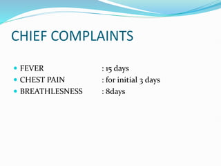
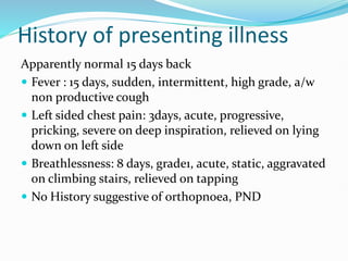
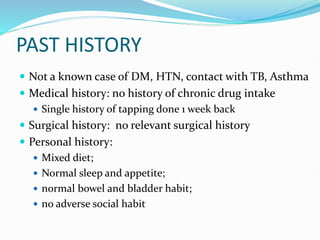
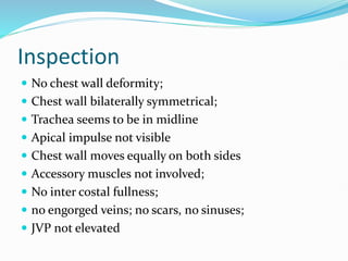
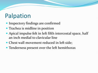
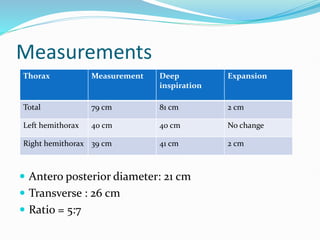
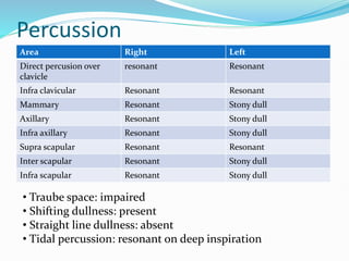
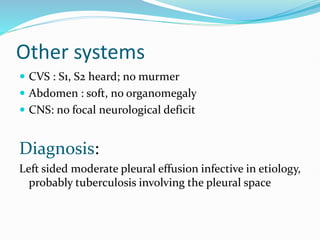
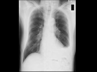
PATIENT DETAILS
 NAME : Mrs Vijaya
 AGE : 43yrs
 GENDER : Female
 ADDRESS : Pallavaram
 OCCUPATION : Homemaker
CHIEF COMPLAINTS
 FEVER : 15 days
 CHEST PAIN : for initial 3 days
 BREATHLESNESS : 8days
History of presenting illness
Apparently normal 15 days back
 Fever : 15 days, sudden, intermittent, high grade, a/w
non productive cough
 Left sided chest pain: 3days, acute, progressive,
pricking, severe on deep inspiration, relieved on lying
down on left side
 Breathlessness: 8 days, grade1, acute, static, aggravated
on climbing stairs, relieved on tapping
 No History suggestive of orthopnoea, PND
PAST HISTORY
 Not a known case of DM, HTN, contact with TB, Asthma
 Medical history: no history of chronic drug intake
 Single history of tapping done 1 week back
 Surgical history: no relevant surgical history
 Personal history:
 Mixed diet;
 Normal sleep and appetite;
 normal bowel and bladder habit;
 no adverse social habit
 Menstrual history:
 Regular cycle – 3-5/28 days;
 No dysmenorrhoea; LMP – 19/10/17
 Family history: Not significant
 Occupational history: Home maker
 Summary:
43 year old female presented with chief complaints of
fever, MMRC grade 1 breathlessness, chest pain for 15
days with one episode of tapping done
General examination
 Patient conscious, oriented to time place and person
 Moderately built, moderately nourished, comfortable;
consent obtained
 Vital signs:
 Pulse: 98 beats/ min; regular; normal volume; no specific
character; no radio radial or radion femoral delay; condition
of vessel wall normal; all peripheral pulses are felt
 BP – 130/90 mmHg measured on right upper arm, sitting
posture
 RR – 26 / min; thoraco – abdominal type
 afebrile
 No pallor
 No icterus
 No cyanosis
 No clubbing
 No generalised lymphadenopathy
 No pedal edema
 No external markers of TB present
Local examination
 consent obtained; Exposed upto umblicus;
 Upper respiratory tract:
 Nose: no DNS, no polyp, no discharge
 Para nasal sinus: no tenderness elicited
 Oral cavity: no tonsilar enlargement; no ulcer; no dental
caries, no loosening of tooth
 Ear - Normal
Inspection
 No chest wall deformity;
 Chest wall bilaterally symmetrical;
 Trachea seems to be in midline
 Apical impulse not visible
 Chest wall moves equally on both sides
 Accessory muscles not involved;
 No inter costal fullness;
 no engorged veins; no scars, no sinuses;
 JVP not elevated
Palpation
 Inspectory findings are confirmed
 Trachea is midline in position
 Apical impulse felt in left fifth intercostal space, half
an inch medial to clavicular line
 Chest wall movement reduced in left side;
 Tenderness present over the left hemithorax
Measurements
 Antero posterior diameter: 21 cm
 Transverse : 26 cm
 Ratio = 5:7
Thorax Measurement Deep
inspiration
Expansion
Total 79 cm 81 cm 2 cm
Left hemithorax 40 cm 40 cm No change
Right hemithorax 39 cm 41 cm 2 cm
Vocal fremitus
Area Right Left
Supra clavicular Normal Normal
Infra clavicular Normal Normal
Mammary Normal Reduced
Axillary Normal Reduced
Infra axillary Normal Reduced
Supra scapular Normal Normal
Inter scapular Normal Reduced
Infra scapular Normal reduced
Percussion
Area Right Left
Direct percusion over
clavicle
resonant Resonant
Infra clavicular Resonant Resonant
Mammary Resonant Stony dull
Axillary Resonant Stony dull
Infra axillary Resonant Stony dull
Supra scapular Resonant Resonant
Inter scapular Resonant Stony dull
Infra scapular Resonant Stony dull
• Traube space: impaired
• Shifting dullness: present
• Straight line dullness: absent
• Tidal percussion: resonant on deep inspiration
Auscultation
 Bilateral air entry present
 Air entry Reduced on left side – mammary, axillary,
infra axillary, inter scapular, infra scapular areas
 Normal vesicular breath sounds heard
 No added sound
Vocal resonance
Area Right Left
Supra clavicular Normal Normal
Infra clavicular Normal Normal
Mammary Normal Reduced
Axillary Normal Reduced
Infra axillary Normal Reduced
Supra scapular Normal Normal
Inter scapular Normal Reduced
Infra scapular Normal Reduced
Other systems
 CVS : S1, S2 heard; no murmer
 Abdomen : soft, no organomegaly
 CNS: no focal neurological deficit
Diagnosis:
Left sided moderate pleural effusion infective in etiology,
probably tuberculosis involving the pleural space
Investigation
 Baseline:
 Complete hemogram: RBC count, TC, DC, Hb, ESR
 Urine routine
 Mantoux test
 Chest X ray
 Diagnostic:
 Diagnostic pleural tapping - gram staining, culture, AFB
smear, cytology, pH, amylase level, glucose, LDH, SFAG
Treatment
 Control infections with antibiotics
 Treat TB – DOTS regimen category I
 Diuretics
 Thoracocentresis and draining of fluid
 Surgical: if needed
Thank you....

CASE PRESENTATION-PLEURAL EFFUSION important topic

  • 2.
    PATIENT DETAILS  NAME: Mrs Vijaya  AGE : 43yrs  GENDER : Female  ADDRESS : Pallavaram  OCCUPATION : Homemaker
  • 3.
    CHIEF COMPLAINTS  FEVER: 15 days  CHEST PAIN : for initial 3 days  BREATHLESNESS : 8days
  • 4.
    History of presentingillness Apparently normal 15 days back  Fever : 15 days, sudden, intermittent, high grade, a/w non productive cough  Left sided chest pain: 3days, acute, progressive, pricking, severe on deep inspiration, relieved on lying down on left side  Breathlessness: 8 days, grade1, acute, static, aggravated on climbing stairs, relieved on tapping  No History suggestive of orthopnoea, PND
  • 5.
    PAST HISTORY  Nota known case of DM, HTN, contact with TB, Asthma  Medical history: no history of chronic drug intake  Single history of tapping done 1 week back  Surgical history: no relevant surgical history  Personal history:  Mixed diet;  Normal sleep and appetite;  normal bowel and bladder habit;  no adverse social habit
  • 6.
     Menstrual history: Regular cycle – 3-5/28 days;  No dysmenorrhoea; LMP – 19/10/17  Family history: Not significant  Occupational history: Home maker  Summary: 43 year old female presented with chief complaints of fever, MMRC grade 1 breathlessness, chest pain for 15 days with one episode of tapping done
  • 7.
    General examination  Patientconscious, oriented to time place and person  Moderately built, moderately nourished, comfortable; consent obtained  Vital signs:  Pulse: 98 beats/ min; regular; normal volume; no specific character; no radio radial or radion femoral delay; condition of vessel wall normal; all peripheral pulses are felt  BP – 130/90 mmHg measured on right upper arm, sitting posture  RR – 26 / min; thoraco – abdominal type  afebrile
  • 8.
     No pallor No icterus  No cyanosis  No clubbing  No generalised lymphadenopathy  No pedal edema  No external markers of TB present
  • 9.
    Local examination  consentobtained; Exposed upto umblicus;  Upper respiratory tract:  Nose: no DNS, no polyp, no discharge  Para nasal sinus: no tenderness elicited  Oral cavity: no tonsilar enlargement; no ulcer; no dental caries, no loosening of tooth  Ear - Normal
  • 10.
    Inspection  No chestwall deformity;  Chest wall bilaterally symmetrical;  Trachea seems to be in midline  Apical impulse not visible  Chest wall moves equally on both sides  Accessory muscles not involved;  No inter costal fullness;  no engorged veins; no scars, no sinuses;  JVP not elevated
  • 11.
    Palpation  Inspectory findingsare confirmed  Trachea is midline in position  Apical impulse felt in left fifth intercostal space, half an inch medial to clavicular line  Chest wall movement reduced in left side;  Tenderness present over the left hemithorax
  • 12.
    Measurements  Antero posteriordiameter: 21 cm  Transverse : 26 cm  Ratio = 5:7 Thorax Measurement Deep inspiration Expansion Total 79 cm 81 cm 2 cm Left hemithorax 40 cm 40 cm No change Right hemithorax 39 cm 41 cm 2 cm
  • 13.
    Vocal fremitus Area RightLeft Supra clavicular Normal Normal Infra clavicular Normal Normal Mammary Normal Reduced Axillary Normal Reduced Infra axillary Normal Reduced Supra scapular Normal Normal Inter scapular Normal Reduced Infra scapular Normal reduced
  • 14.
    Percussion Area Right Left Directpercusion over clavicle resonant Resonant Infra clavicular Resonant Resonant Mammary Resonant Stony dull Axillary Resonant Stony dull Infra axillary Resonant Stony dull Supra scapular Resonant Resonant Inter scapular Resonant Stony dull Infra scapular Resonant Stony dull • Traube space: impaired • Shifting dullness: present • Straight line dullness: absent • Tidal percussion: resonant on deep inspiration
  • 15.
    Auscultation  Bilateral airentry present  Air entry Reduced on left side – mammary, axillary, infra axillary, inter scapular, infra scapular areas  Normal vesicular breath sounds heard  No added sound
  • 16.
    Vocal resonance Area RightLeft Supra clavicular Normal Normal Infra clavicular Normal Normal Mammary Normal Reduced Axillary Normal Reduced Infra axillary Normal Reduced Supra scapular Normal Normal Inter scapular Normal Reduced Infra scapular Normal Reduced
  • 17.
    Other systems  CVS: S1, S2 heard; no murmer  Abdomen : soft, no organomegaly  CNS: no focal neurological deficit Diagnosis: Left sided moderate pleural effusion infective in etiology, probably tuberculosis involving the pleural space
  • 18.
    Investigation  Baseline:  Completehemogram: RBC count, TC, DC, Hb, ESR  Urine routine  Mantoux test  Chest X ray  Diagnostic:  Diagnostic pleural tapping - gram staining, culture, AFB smear, cytology, pH, amylase level, glucose, LDH, SFAG
  • 20.
    Treatment  Control infectionswith antibiotics  Treat TB – DOTS regimen category I  Diuretics  Thoracocentresis and draining of fluid  Surgical: if needed
  • 21.