
Recommended
More Related Content
Similar to Clinical Anatomy 2009 Anatomia de la valvula mitral.pptx
Similar to Clinical Anatomy 2009 Anatomia de la valvula mitral.pptx (20)
More from Juan Diego
More from Juan Diego (20)
Recently uploaded
Recently uploaded (20)
Clinical Anatomy 2009 Anatomia de la valvula mitral.pptx
- 1. HORIA MURESIAN* University Hospital of Bucharest-Cardiovascular Surgery, Bucharest, Romania As a result of the numerous clinicaland surgical data accumulated so far, the classical image of the mitral valve—a bicuspid valve, with two leaflets and two papillary muscles—undergoes significant modifications. The valve, included into the larger concept of the mitral valvular complex unveils numerous important valences and characteristics, among which, some represent newer concepts, of clinicaland surgical significance: the valvular complexis a subtle and finely-tuned system of elements acting in a coordinated manner; the mitral valve is an active valve and not a mere passive flap bordering the atrioventricular junction. Not least, the mitralvalve contributes to the make up and functionof the left ventricu- lar outflow tract. The anatomical and functional interdependence between the mitral valve and the left ventricular myocardium is evident not only followingtheir particularities of vascularization but also it is reflected in morbid states such as ischemic cardiac disease and dilated cardiomyopathy. All the new concepts and ideas, ask for a more profound study of the clinical anatomy of the mitral valve, underscoring the importance of a pertinent dialogue between specialists and by using a more appropriate and unitary terminology. Clin. Anat. 22:85–98, 2009. V C 2008 Wiley-Liss, Inc. Keywords: anatomy mitral valve; echocardiography; valvular plasty coordinated manner. Age and the various disease INTRODUCTION The thorough knowledge of the anatomy of the mitral valve is of utmost importance for diagnostic interrogation and surgery. This review article presents the most recent data and emerging ideas regarding this vast and significant topic and under- lines the clinical and surgical applications. In fact, the mitral valve does not represent solely a passive flap guarding the left atrioventricular orifice, but a finely-tuned and active set of elements acting in a processes change or alter this disposition and ask for a more elaborate medical thinking and for skilled and more complex surgical maneuvers. THE MITRAL VALVULARCOMPLEX Subsequent to the numerous clinical and anatomi- cal data accumulated so far, the mitral valve is best understood and characterizedtogether with the ad- Abbreviations used: Ao, aorta; AoL, aortic leaflet of the mitral valve (anterior leaflet); Ao-M, aorto-mural diameter of the mi- tral annulus; A1– A3, echographical portions of the aortic leaf- let; C-C, intercommissural diameter of the mitral annulus; CS, coronary sinus; Cx, circumflex branch of the left coronary ar- tery; D1, first diagonal branch; FO, fossa ovalis; right fibrous trigone; SC, superior (anterolateral) commissure; S-L, septal- to-lateral diameter of the mitral annulus; SPM, superior (anterolateral) papillary muscle; SVC, superior vena cava; S1, firstanterior septal branch.
- 2. a See text or a further characterizationof ‘‘the annulus.’’ jacent cardiac structures that collectively constitute the mitral valvular complex (Table 1) (Perloff and Roberts, 1972; Ho, 2002). The combined action of the structures comprising the mitral valvular complexmust ensure and facilitate: commissural areas. The anterior mitral leaflet occu- pies roughly one third of the annular circumference and is wider than the posterior leaflet, depicting a trapezoidal or semicircular outline. Because of its the properly-timed passage of blood from the left atrium into the left ventricle, the tight systolic closure of the left atrioventric- ular orifice (preventing thus the backflow of blood from the left ventricle into the atrium), the accommodation of blood, eventually fol- lowed by its rapid, efficient and forceful ejection through the left ventricular outflow tract into the aortic root. The mitral valve consequently has additional physiologic roles different from the tricuspid valve, as part of it contributes to the formation of the left ventricular outflow tract (furthermore, the mitral-aortic curtain is also a component of the aorticroot). • • • Such important functional roles depend on the coordinated action of interrelated anatomical ele- ments: the left atrium, the mitral valve leaflets, ‘‘the annulus,’’ the chordae, the papillary muscles, and the left ventricular wall. Each of the aforementioned components is equally important, as alterations in the structure and function of any of these elements may cause hampered emptying of the left atrium, mitral valvular incompetence, and/or alterations of the left ventricular ejection. Accumulatingdata suggest the fact that mitral valve closure does not represent a passive process; instead, the model of an active valve emerges (Williams and Jew, 2004). The implementation and the use of a correctter- minology are of utmost importance in describing the normal anatomy of the mitral valve or for the diag- nostic interrogation and therapeutic approach in mitral valve disease. (Anderson and Wilcox,1995). The component parts of the mitral valvular complex must be also considered in an attitudinally appropri- ate fashion with the heart oriented as ‘‘in situ’’ (Anderson and Frater, 2006). Fig. 1. Mitral valve leaflets. The mitral valve in closed position as seen from the left atrium. Advanced dissection of an adult heart specimen with only the aor- tic root and mitral valve left in place. Elements in attitu- dinally correct orientation. LFT and RFT ¼ left and right fibrous trigones of the heart. Sinuses of the aorta: NF ¼ nonfacing and 2L ¼ left (or sinus 2) - following the Lei- den nomenclature (Sauer and Gittenberger-de Groot, 1997). LAD, the left anterior descending; Cx, the cir- cumflex branches of the left coronary artery; IPM, infe- rior papillary muscle; M1-M3; the scallops of the mural leaflet; A1-A3, the corresponding echocardiographic portions of the aortic leaflet Aorto-mitral curtain (‘‘mitral-aortic continuity’’)
- 3. material, rendering pliable the posterior segment of the mitral valve allowing thus the sequential dilatation and reduction of the valvular circumference (the sphincteric mechanism). The mitral valvular leaflets close along a solitary zone of apposition; the commissures are no more than the ends of this zone of apposition (as the line of apposition does not extend to reach the annulus) and together with the clefts in the mural leaflet act as pleats providing a tight closure of the valve in systole (Victorand Nayak, 1994). On microscopic examination, the leaflets have a fi- brous skeleton (the fibrosa), covered towards the atrial side by a layer of myxomatous connective tis- sue (the spongiosa). The atrial and ventricular endo- cardium are continued over the leaflet surface; atrial myocardium may stick in between the endocardium and the spongiosa, for varying depths, especially in the middle scallop of the mural leaflet The valve leaflets are nonhomogenous structures and show a nonlinear anisotropic behavior (May- Newman and Yin, 1995). Both leaflets are less extensible in the circumferential than in the radial direction. The mural leaflet shows greater extensibil- ity in both directions, possibly due to the more abun- dant chordal sustain. The aortic leaflet has a reduced circumferential length but is wider than the mural leaflet and usually has no clefts. The conspicuous and less pliable rough zone marks the line of leaflet coaptation. With advancing age, nodules are visible at this level, marking better the area of overlap with the mural leaflet. More peripherally, the clear zone of the aortic leaflet is thinner and more malleable. On the ventric- ular aspect, this corresponds to the gutter formed between the arching chordal attachments, constitut- ing an important portion of the left ventricularout- flow tract (Nayak and Victor,2006). The mural leaflet is a partially divided structure with three or more scallops and the corresponding clefts. In spite of their different shape, both mitral leaflets share similar areas (Roberts and Perloff, 1972; Ho, 2002). Besides a rough and a clear zone analogous to the aortic leaflet, the mural leaflet characteristically has a basal zone often reinforced by atrial myocardium. The commissures and the underlying papillary muscles are traditionally labeled: anterolateral (in fact: superior, posterior, parietal, and left) and post- eromedial (in fact: inferior, anterior, septal, and right) (Kanani and Anderson, 2003; Anderson and Frater, 2006). The more appropriate terminology, which will be A3. Consequent to the fact that papillary and chordal sustain is distributed to corre- sponding parts of the mitral valve leaflets—i.e., those portions that will coapt during valve closure, six anatomic-functional scallops eventually result and these can be systematized in pairs of scallops facing a cleft or a commissure. This nomenclature enables a more precise characterization of mitral valve leaflets, both in normal cases as well as in dis- ease (Fig. 1). The various degrees of severity and patterns of mitral valve prolapse can even be quanti- fied, on a scale ranging from 1 of 6 scallops to 6 of 6 (1/6–6/6), reflecting not only the severity of the dis- ease, but also allowing a more precise localization and orientation during diagnostic interrogation and surgery (Muresian et al., 2006). Other authorities (Kumar et al., 1995) speculate about taking into account the so-called commissural cusps, but this might add further ambiguities and confusion and such details are not absolutely necessary; following Carpentier’s original terminology, a prolapse involv- ing the A1 and M1 implicitly comprises a prolapse of the superior commissure too. However, this simpli- fied scheme will not cover all the variations encoun- tered (Victorand Nayak, 1994). The Annulus The annulus represents a concept rather than a well-defined anatomical structure (McAlpine, 1975; Ho, 2002). It is differently defined from the anatomi- cal, echocardiographical, and respectively, the surgi- cal points of view. Following different landmarks, the annulus appears either more planar or saddle- shaped. Anatomically, at the level of the atrioventric- ular junction, fibro-elastic tissue extends from the left and right fibrous trigones posteriorly describing however an incomplete ring, otherwise completed by myocardium (Angelini et al., 1988). The annulus is deficient towards the aorta, the mitral-aortic curtain extending between the two trigones (McAlpine, 1975). The insertion of the left atrial myocardium at this level may serve as useful landmark for the aortic (‘‘anterior’’) portion of the annulus. Echographically, the
- 4. Fig. 2. Mitral annulus. A superior and basal view of the fibrous skeleton of the heart. The mitral annulus includes an important muscular portion in its posterior aspect. The tricuspid annulus is almost completely mus- cular but it was left in place in order to reveal the origi- nal position of the right atrioventricular junction and the general orientation of the two atrioventricular orifices. The aortic sinuses follow the same Leiden nomenclature (cf. Fig. 1). The nonfacing sinus was scalloped, for a better view. The origin of the two coronary arteries (LCA and RCA) is well seen. The intercommissural diam- eter of the mitral annulus is marked by the C-C double headed arrow. The so- called septal-to-lateral diameter is actually the aorto-mural diameter (Ao-M). Note that the literally-speaking and anatomically-positioned sep- tal-to-lateral diameter (S-L) skirts closer to the inter- commissural one. No matter what definition of the annulus is cho- sen, the orifice at the level of the left atrioventricular junction appears ovoid or D-shaped (Fig. 2), with a longer intercommissural (I- C) and a shorter septal- to-lateral axis (S-L). Literally, the anatomical septal- to-lateral diameter stretches closer to the intercom- missural one. It is hence more correct to define and refer to the aorto-mural diameter, which joins the aortic (anterior) annular midpoint with the mural an- nular midpoint (Ao-M). Body-weight-corrected data pertaining to these dimensions are as follows:0.39– 0.59 mm/kg for the intercommissural and 0.32–0.48 mm/kg for the aorto-mural diameters, respectively (Fyrenius et al., 2001). However, dimensions are underestimated of utmost importance for the prevention of mitral insufficiency (Timek et al., 2002a). The annulus depicts complex modifications during the cardiac cycle. The following clinical-echocardio- graphical terminology is briefly presented. The wid- ening and narrowing define the annular flexion (the sphincteric mechanism). There is a 23–40% varia- tion in the annular circumference between the sys- tolic and diastolic configurations (van Rijk-Zwikker et al., 1994). The excursion or annular descent is the movement in the apical-to-basal direction. annulus excursion toward the apex can be evaluated by using the two-dimensional Mitral better strain imaging technique (Hayashi et al., 2006). Mitral an- nular
- 5. Fig. 3. Mitral valve in situ. a: Lateral view of a heart specimen opened at the level of its left aspect. A part of the mitral valve comprising the M2 scallop was removed. A portion of the left atrial wall was also removed, to reveal the left aortic sinus (2L) with the ori- gin and proximal tract of the left coronary artery (LCA). The accompanying greater cardiac vein (GCV) is also evident. After traveling around the annulus, the GCV becomes the coronary sinus (CS). Note its relationship with the mitral annulus. From the cavity of the left atrium the orifice of the left atrial appendage (LAA) is visible, as well as two of the pulmonary veins (*). Note also the false cords connecting the two papillary muscles (SPM and IPM) between them and with the septum. The actual position of the papillary muscles is: superior, parietal, left, posterior (for the traditionally called anterolateral muscle) and inferior, septal, right, and anterior (for the posteromedial). There is also a visible difference between the whitish leaflet and the darker left atrial myocardium. b: A more advanced dis- section of the same specimen. Same view and coordi- nates as in Figure 3a. The atrial septum was removed except the fossa ovalis (FO); under this, the coronay sinus (CS) which was deliberately unroofed, travels towards its confluence with the right atrium. A valve in the CS is also visible. Note the intimate relationship between the various portions of the mitral valve and the LCA, GCV, and CS. The aortic (anterior) intertrigonal portion of the annulus also depicts a dynamic behavior: studies have revealed changes in response to modifications in hemodynamic loading and ventricular contractility (Parishet al., 2004). The aortic and mitral annular areas change in a reciprocal fashion during late diastole and late sys- tole (32% 6 8% for aortic annular area and 13% 6 13% for mitral annular area) but such dynamic changes seem not to be mediatedthrough the ana- tomic fibrous continuity (Timek et al., 2003a). Proceeding counter clockwise from the left fibrous trigone towards the atrial septum (Figs.3a and 3b), the circumflexbranch and the greater cardiac vein continued by the coronary sinus mark the external position of the annulus—a detail of surgical rele- vance. The reverse and annulus on one hand and the ventricular wall on the other. Different from the tricuspid valve, there is no direct attachment of the mitral valve to the ven- tricular septum, although the papillary muscles are frequently connected to the septum or to the right fi- brous trigone by false cords (Loukas et al., 2007). The length and the reciprocal ratio between papillary muscles and cords show individual variations, but some studies demonstrated similar annular-to-papil- lary muscle tip distances in the 2-, 4-, 8-, and 10- o’clock positions that also correlate with mitral annu- lar diameter (Sakai et al., 1999) and which are rele- vant for the proper choice of cordal length during reparative surgery. Mitral valvular leaflets, scallops, clefts, commis- sures, on one hand, and tendinous cords on the other, are reciprocally definable. The aortic leaflet, and each of the scallops of the mural leaflet, have a convex free margin and receive cordal
- 6. Fig. 4. Definition of the components of the mitral valve. The mitral valvular complex is exposed by cutting the heart specimen at the level of the left aspect, through the M1 scallop of the ML. In this specimen, the ML has four scallops (M1-M4). The inferior papillary muscle (IPM) depicts more fascicles (heads); the supe- rior papillary muscle (SPM) consists of two fascicles one of which lies directly under the superior commissure (SC). Note that the corresponding portions of the leaf- lets that will coapt in systole, have a convexfree margin and receive cordal support from different papillary mus- cle fascicles. The clefts and commissural areas have a concave free margin and receive cordal support from a directly underlying muscular fascicle or head. The ap- proximate position of the annulus is also evident. It is also of clinical significance to note that the papillary muscles form an almost circumferential support for the leaflets, except under the aortic leaflet of the mitral valve, where the left ventricular inflow and outflow tracts communicate. immediately underneath, a cordal support that diverges (dichotomously or in a fan-like manner) while approaching the leaflet. Following their shape and cordal distribution, each scallop of the mural leaflet can be considered as a ‘‘mini- aorticleaflet.’’ The tendinous cords represent complex rope-like structures. Their inner layer is composed of tightly bound collagen; the outer layer consists of loose col- lagen and elastic fibers and contains blood vessels. The tendinous cords are covered by a layer of endo- thelial cells (endocardium). Fibroblasts are evenly distributed through both the inner and outer layers. The presence of blood vessels suggest an additional nutritional role for the leaflets (Ritchie et al., 2005). The cords depict different microstructures according to their type, with higher levels of DNA and collagen at the level of the marginal cords of the size and decreases with age (Lim and Boughner, 1976; Liao and Vesely, 2003). The weakest portion, corresponding to their nar- rowest section and where cords usually tear, is located close to (Sedransk et al., cords varies with their insertion into the leaflet 2002). Tension in the tendinous cordal type (greater in the strut cords) and in line with left ventricular pressure, reaching a maximum in or just prior to early systole (Lomholt et al., 2002). The tendinous cords intermingle within the very substance of the leaflet’s, contributing to their fi- brous framework. The maximal cordal intermingling occurs at the level of the leaflets rough zone. Some of the cordal intraleaflet extensions reach the annu- lus and the two fibrous trigones (Fig. 5b).
- 7. the AoL Main stem of cleft cords of the ML Third orderc Basal cords of ML Basal posterior cord Typeof cord Major role Equivalenttraditional terms Proposed simplifiedclassification Marginal cords Essential for coaptation ‘‘First order’’ Roughzone cords Essential for leaflet geometry ‘‘Second order’’ Strut (sustain) cords Basal cords Essential for ventriculargeometry Annular reinforcement ‘‘Third order’’ AOL, aortic leaflet; ML, mural leaflet. a Inserting on the free edge of the leaflet. b Inserting on the ventricularaspect of the leaflet and contributingto the rough zone. c Originfrom the posterior left ventricular wall. physiologic situations as well as in disease (Scam- pardonis et al., 1973). The picture resulting from the classical works is that of the mitral valve, a bicuspid valve, with two leaflets and two papillary muscles but this is far from what we perceive nowadays. The muscular-cordal arrangement of the subvalvular apparatus must however ensure a circumferentially-constant support and with a resultant even distribution of stresses over the leaflets. In the mean time, the papillary-cor- dal disposition must not interfere with the passage of blood from the atrium towards the ventricle and from the ventricular inflow to the ventricular outflow com- partment. The classical view of two papillary muscles Fig. 5. The tendinous cords of the mitral valve. a: A special anatomical preparation revealing the distribution of the cordal support at the level of the two leaflets: aortic and mural. The wider aortic leaflet allows to iden- tify better the marginal (free adjacent commissural areas, to reveal the intra-leaflet disposition of the cordal support. The tendinous cords proceed in the very substance of the leaflet, contribut- ing to its fibrous framework. Some branches of division reach the level of the annulus. An
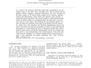
- 8. Fig. 6. The vascularization of the papillary muscles. a: Left view of a heart specimen in which the atrial walls were removed except the junction between the superior vena cava (SVC) with the right atrium. Large parts of the left ventricular walls were removed in order to ex- hibit the papillary muscles. The ventricular septum was also largely dissected, demonstrating the posterior interventricular artery (doubled in this specimen and with origin from the right coronary artery, RCA) and the first anterior septal branch (S1). The close relationship between the Cx and the mitral annulus is evident. The superior papillary muscle (SPM) and adjacent left ven- tricular wall are vascualrized by an obtuse marginal branch (OM), while the inferior papillary muscle (IPM), is vascularized from the distal from the distal branches of the right coronary artery (RCA). A conspicuous sinus node artery with origin from the left coronary artery (LCA) travels posterior to the aortic root, immediately superior to the mitral-aortic curtain (*) - a detail of sur- gical relevance. b: A more advanced dissection reveal- ing the vascularization of the papillary muscles, in a left dominant system. The superior papillary muscle (SPM) is vascularized by a first obtuse marginal branch (OM1) while the IPM is supplied by a second marginal branch (OM2) and further by a retroventricular branch. Note again the almost circumferentially-continuous papillary structure. should consequently be hence adapted: the two muscles represent the ends of a more or less contin- uous column of papillary structures that through a gap between them and under the aortic leaflet, allow the passage of blood towards the left ventricular out- flow tract. The position of the papillary fascicles and their three- dimensional orientation has an important role in the distribution of forces and in assisting the proper closure of the mitral valve (Scampardonis et al., 1973; Jimenez et al., 2005; Chen and May- Newman, 2006). Papillary muscle displacement as occurring in have origin in either or both coronary arteries. The superior papillary muscle is vascularized by branches from diagonal, circumflex, or even obtuse marginal branches of the left coronary artery. The classical assertion regarding the vascularization of the papil- lary muscles must be reconsidered: the superior group that theoretically receives multiple arterial branches can become infarcted even after occlusion of a single diagonal branch (Kim et al., 2005). Fur- thermore, the inferior group is seldom represented by a single fascicle and the vascularization is pro- vided by more than one arterial branch (H. Muresian, 2006, Personal communication). Arterial branches for the
- 9. aforementioned spaces. These two parameters must be congruent though not identical. coaptation is as equally impor- tant as coaptation between the aortic and mural leaf- lets (Lai et al., 2002). Coaptation takes place in a more apical plane ‘‘under’’ the level of the annulus. The resultant coaptation triangle, tenting area, and tenting volume, are all parameters of diagnostic and surgical relevance (Watanabe et al., 2005). By join- ing their rough zones, the valvular leaflets overlap for about 6–8 mm (Crooke et al., 2007). The quanti- The Functional Reserve of the MitralValve The mitral total leaflet area is 1.5–2 times the an- nular area. Annular area values above 2.3 cm/m2 BSA are accepted as normal. The differencebetween the total leaflet and the annular area is to be found in the coaptation surface (i.e., the amount of leaflet overlapping) and this characterizes the functional reserve of the mitral valve, as this apparent leaflet excess makes possible a tight coaptation under vari- ous hemodynamic conditions (Muresian et al., 2006). The ratio between the total leaflet area and annular area is of surgical relevance as any imbal- ance after the mitral repair may cause either poor coaptation or systolic anterior motion of the aortic leaflet The area of the aperture is roughly 2/3 of the annular area. (Tsakiris, 1976). The valvular aperture between the valve leaflets is ovoid in shape but becomes crescent-shaped after ring annuloplasty or rheumatic mitralstenosis. The complex geometric distribution of the tendi- nous cords inserting into the leaflets and the result- ant triangular structures (Fig.8), act both under nor- mal conditions, adapting the valve to the various he- modynamic states and in disease, compensating for the papillary muscle displacement and increased tethering of the leaflets(He et al., 2000). fication of the resultant also diagnostic relevance, performed. coaptation surface has although not yet widely The mechanisms of normal valve closure are com- plex and still incompletely understood. The major processes are: annular reduction (sphincteric mech- anism), followed by the apposition of the mural leaf- let against the elevated aortic leaflet (that functions as a trap door), reduction of all left ventricular dimensions and the subsequent creation of a pres- sure gradient between the ventricle and atrium that eventually brings the leaflets toward the annular plane. Many interesting details are emerging, such as the presence of a rich innervation (Marron et al., 1996) and contractile elements within the leaflets, the contributionof left atrial myocardium to annular dynamics (Glasson et al., 1997; Timeket al., 2002c, 2003b), the architecture of the left ventricular myo- cardium, the spatial disposition of the left ventricular trabeculae and papillary muscles (Loukas et al., 2007), and the presence of receptors in the structure of the leaflets. The greater stress during valve closure is borne by the narrower but better anchored mural leaflet. The aortic leaflet shifts between the closed and respectively, the open position offering a transitory support against which the mural leaflet abuts. The commissural (and cleft) areas allow the apposition and the concomitant reduction of the posterior leaflet and annulus. The submacroscopic structure of the commissural areas and the disposition of their fi- brous framework depict a fan-like pattern which ena- bles the sequential widening and narrowing of these particular areas of the valve. (Fig. 7). Mitral valve plasty with excessive reduction and/or immobiliza- tion of the mural leaflet abolishes this natural pliabil- ity of the commissures causing scissoring stresses over the commissural zones; this mechanism might be responsible for the earlier failure of such techniques. The anatomic aperture of the valve between the two The Normal Asymmetry of the Mitral Valve The mitral valve shows a natural asymmetry. The valve leaflets do not depict a perfect bilateral sym- metry. The scallops of the mural leaflet do not share even leaflet material. The commissural areas are also asymmetric: the inferior commissure has a lon- ger circumferential length and is narrower. Papillary muscle and cordal support are not symmetrically dis- tributed over the two imaginary halves of the valve. (Fig. 9). It appears too simplistic (Kumar et al., 1995) and of less and less clinicalsignificance, to divide the
- 12. Fig. 9. The normal asymmetry of the mitral valve. sought by the surgeon.b: A backlitmitral valve opened
- 13. and surgically corrected if considered into the larger concept of mitral valvular complex. More- over, the mitral valve must be considered not solely as an atrioventricular valve, as it also contributes to the make-up and functionof the left ventricular out- flow tract. Newly-applied and versatile diagnostic principles and the accumulating surgical experience have opened new horizons and have offered new relevance to the anatomy of this important and attractive region; undoubtedly, the reciprocal influence between clinical practice and anatomy will broaden these new frontiers, for the benefit of our patients. Ho SY. 2002. Anatomy of the mitral valve. Heart 88(Suppl 4):iv5– iv10. Jimenez JH, Soerensen DD, He Z, Ritchie J, Yoganathan AP. 2005. Effects of papillary muscle position on chordal force distribution: An in-vitro study. J Heart Valve Dis 14:295–302. Kanani M, Anderson RH. 2003. The anatomy of the mitral valve: A retrospective analysis of yesterday’s future. J Heart Valve Dis 12:543–547. Kim TH, Seung KB, Kim PJ, Baek SH, Chang KY, Shin WS, Choi KB, Moon SW. 2005. Images in cardiovascular medicine. Anterolat- eral papillary muscle rupture complicated by the obstruction of a single diagonal branch. Circulation 112:e269–e270. Kumar N, Kumar M, Duran CM. 1995. A revised terminology for re- cording surgical findings of the mitral valve. J Heart Valve Dis 4:70–75. Lai DT, Tibayan FA, Myrmel T, Timek TA, Dagum P, Daughters GT, Liang D, Ingels NB Jr, Miller DC. 2002. Mechanistic insights into posterior mitral leaflet inter-scallop malcoaptation during acute ischemic mitral regurgitation. Circulation 106(12 Suppl 1):I40– I45. Lam JH, Ranganathan N, Wigle ED, Silver MD. 1970. Morphology of the human mitral valve. I. Chordae tendineae: A new classifica- tion. Circulation 41:449–458. Levine RA, Handschumacher MD, Sanfilippo AJ, Hagege AA, Harri- gan P, Marshall JE, Weyman AE. 1989. Three-dimensional echo- cardiographic reconstruction of the mitral valve, with implica- tions for the diagnosis of mitral valve prolapse. Circulation 80:589–598. Liao J, Vesely I. 2003. A structural basis for the size-related me- chanical properties of mitral valve chordae tendineae. J Biomech 36:1125–1133. Lim KO, Boughner DR. 1976. Morphology and relationship to exten- sibility curves of human mitral valve chordae tendineae. Circ Res 39:580–585. Lim KH, Yeo JH, Duran CM. 2005. Three-dimensional asymmetrical modeling of the mitral valve: A finite element study with dynamic boundaries. J Heart Valve Dis 14:386–392. Lomholt M, Nielsen SL, Hansen SB, Andersen NT, Hasenkam JM. 2002. Differentialtension between secondary and primary mitral chordae in an acute in-vivo porcine model. J Heart Valve Dis 11:337–345. Loukas M, Louis RG Jr, Black B, Pham D, Fudalej M, Sharkees M. 2007. False tendons: An endoscopic and cadaveric approach. Clin Anat 20:163–169. McAlpine WA. 1975. Heart and Coronary Arteries. An Anatomical Atlas for Clinical Diagnosis, Radiological Investigation and Surgi- cal Treatment. New York: Springer Verlag. p 39–56. Marron K, Yacoub MH, Polak JM, Sheppard MN, Fagan D, Whitehead BF, de Leval MR, Anderson RH, Wharton J. 1996. Innervation of human atrioventricular and arterial valves. Circulation 94:368– 375. May-Newman K, Yin FC. 1995. Biaxial mechanical behavior of excised porcine mitral valve leaflets. Am J Physiol 269:H1319– H1327. Millington-Sanders C, Meir A, Lawrence L, Stolinski C. 1998. Struc- ture of chordae tendineae in the left ventricle of the human heart. J Anat REFERENCES Anderson RH, Frater RW. 2006. How can we best describe the com- ponents of the mitral valve? J Heart ValveDis 15:736–739. Anderson RH, Wilcox BR. 1995. Understanding cardiac anatomy: The prerequisite for optimal cardiac surgery. Ann Thorac Surg 59:1366–1375. Angelini A, Ho SY, Anderson RH, Davies MJ, Becker AE. 1988. A his- tological study of the atrioventricular junction in hearts wih nor- mal and prolapsed leaflets of the mitral valve. Br Heart J 59:712–716. Anwar AM, Soliman OI, ten Cate FJ, Nemes A, McGhie JS, Krenning BJ, van Geuns RJ, Galema TW, Geleijnse ML. 2007. True mitral annulus diameter is underestimated by two-dimensional echo- cardiography as evidenced by real- time three-dimensional echo- cardiography and magnetic resonance imaging. Int J Cardiovasc Imaging 23:541–547. Carlhall C, Wigstrom L, Heiberg E, Karlsson M, Bolger AF, Nylander E. 2004. Contribution of mitral annular excursion and shape dy- namics to total left ventricular volume change. Am J Physiol Heart Circ Physiol 287:H1836– H1841. Carpentier A, Branchini B, Cour JC, Asfaou E, Villani M, Deloche A, Relland J, D’Allaines C, Blondeau P, Piwnica A, Parenzan L, Brom G. 1976. Congenital malformations of the mitral valve in chil- dren. Pathology and surgical treatment. J Thorac Cardiovasc Surg 72:854–866. Chen L, May-Newman K. 2006. Effect of strut chordae transection on mitral valve leaflet biomechanics. Ann Biomed Eng 34:917– 926. Crooke GA, Grossi EA, Jorde UP, Colvin SB, Galloway AC. 2007. Functional ischemic mitral regurgitation: A review of the patho- physiology, operative approach and outcomes. Cardiac Surgery Today 3:98–109. Einstein DR, Kunzelman KS, Reinhall PG, Nicosia MA, Cochran RP. 2005. Non-linear fluid-coupled computational model of the mitral valve. J Heart Valve Dis 14:376–385. Fyrenius A, Engvall J, Janerot-Sjoberg B. 2001. Major and minor axes of the normal mitral annulus. J Heart Valve Dis 10:146– 152.
- 14. survey of the conditions causing the mitral valve to func- tion abnormally. Ann Intern Med 77:939–975. Sakai T, Okita Y, Ueda Y, Tahata T, Ogino H, Matsuyama K, Miki S. 1999. Distance between mitral anulus and papillary muscles: Anatomic study in normal human hearts. J Thorac Cardiovasc Surg 118:636–641. Sauer U, Gittenberger-de Groot AC. 1997. Transposition of the great arteries: Anatomic types and coronary artery patterns. In: Eugene Braunwald (Series editor), Robert M. Freedom (Volume editor). Atlas of Heart Diseases, Vol. XII: Congenital Heart Dis- ease. Mosby.p 15.4. Scampardonis G, Yang SS, Maranhao V, Goldberg H, Gooch AS. 1973. Left ventricular abnormalities in prolapsed mitral leaflet syndrome. Review of eighty-seven cases. Circulation 48:287–297. Sedransk KL, Grande-Allen KJ, Vesely I. 2002. Failure mechanics of mitral valve chordae tendineae. J Heart Valve Dis 11:644–650. Timek TA, Miller DC. 2001. Experimental and clinical assessment of mitral annular area and dynamics: What are we actually meas- uring? Ann Thorac Surg 72:966–974. Timek TA, Lai DT, Tibayan F, Liang D, Daughters GT, Dagum P, Ingels NB Jr, Miller DC. 2002a. Septal-lateral annular cinching abolishes acute ischemic mitral regurgitation. J Thorac Cardio- vasc Surg 123:881–888. Timek TA, Lai DT, Dagum P, Tibayan F, Daughters GT, Liang D, Berry GJ, Miller DC, Ingles NB Jr. 2003b. Ablation of mitral annu- lar and leaflet muscle: Effects on annular and leaflet dynamics. Am J Physiol Heart Circ Physiol 285:H1668–H1674. Tsakiris AG. 1976. The physiology of the mitral annulus. In: Kal- manson D, editor. The Mitral Valve. A Pluridisciplinary Approach. London: Edward Arnold. p 21–26. van Rijk-Zwikker GL, Delemarre BJ, Huysmans HA. 1994. Mitral valve anatomy and morphology: Relevance to mitral valve replacement and reconstruction. J Card Surg 9(2 Suppl):255– 261. Victor S, Nayak VM. 1994. Definitions and functions of commis- sures, slits amd scallops of the mitral valve. Analysis of 100 hearts. Asia Pacific J Thorac Cardiovasc Surg 3:10–16. Watanabe N, Ogasawara Y, Yamaura Y, Kawamoto T, Toyota E, Akasaka T, Yoshida K. 2005. Quantitation of mitral valve tenting in ischemic mitral regurgitation by transthoracic real-time three- dimensional echocardiography. J Am Coll Cardiol 45:763–769. Williams TH, Jew JY. 2004. Is the mitral valve passive flap theory overstated? An active valve is hypothesized. Med Hypotheses 62:605–611. Wilcox BR, Cook AC, Anderson RH. 2004. Surgical Anatomy of the Heart. Cambridge: Cambridge UniversityPress. p 59–64.