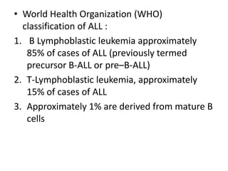
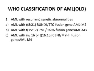
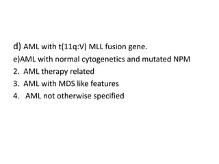
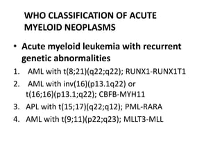
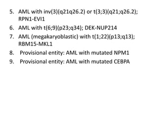
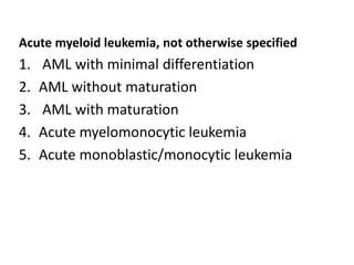
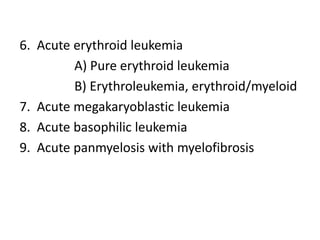
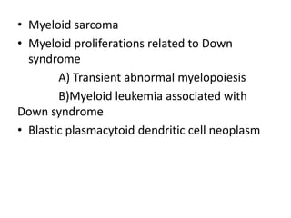
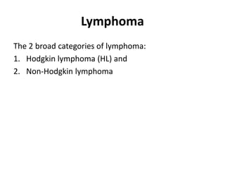
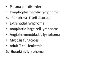
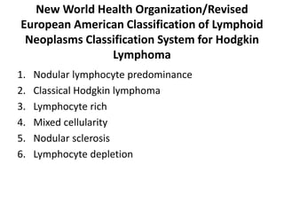
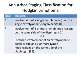
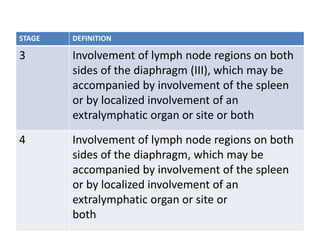
CLASSIFICATION OF LEUKEMIA AND
LYMPHOMA
Dr.Lakshit Bhalala
1st Year Resident
Department Of Paediatrics
Guide By: Dr. Dinesh Patel
Assistant Professor
INTRODUCTION
• The leukemias may be defined as a group of
malignant diseases in which genetic abnormalities in
a hematopoietic cell give rise to an unregulated
clonal proliferation of cells.
• The leukemias are the most common malignant
neoplasms in childhood.
• Accounting for approximately 31% of all malignancies
that occur in children younger than 15 yr of age.
• Acute lymphoblastic leukemia (ALL) accounts
for approximately 77% of cases of childhood
leukemia
• acute myelogenous leukemia (AML) for
approximately 11%
• chronic myelogenous leukemia (CML) for 2-3%
• juvenile myelomonocytic leukemia (JMML) for
1-2%
• The remaining cases consist of a variety of acute
and chronic leukemias that do not fit classic
definitions for ALL, AML, CML, or JMML
Juvenile Myelomonocytic Leukemia
• JMML, formerly termed juvenile CML, is a clonal
proliferation of hematopoietic stem cells
• Typically affects children younger than 2 yr of age.
• Most patients with JMML have been found to have
mutations that lead to activation of the RAS
oncogene pathway including NF1 and PTPN11
• The bone marrow shows a myelodysplastic pattern,
with blasts accounting for <20% of cells.
FAB CLASSIFICATION OF ALL(OLD)
L 1 L 2 L 3
MC type
Best Prognosis
Small Homogenous Blast
Scanty Cytoplasm
Large Heterogenous
Blast,Moderate
Cytoplasm,Prominent
Nucleoli
Least common
Worst prognosis
Large blast
Blue Cytoplasm
WHO CLASSIFICATION OF ALL(OLD)
B-ALL T-ALL
MC (85%)
No Mediastinal invasion
Loss Of Function Mutation In PAX-5,E2A
Or EBF Gene
Better Prognosis
Least common (15%)
Mediastinal invasion Present
Gain Of Function Mutation In Notch 1
Gene
Poor Prognosis
• World Health Organization (WHO)
classification of ALL :
1. B Lymphoblastic leukemia approximately
85% of cases of ALL (previously termed
precursor B-ALL or pre–B-ALL)
2. T-Lymphoblastic leukemia, approximately
15% of cases of ALL
3. Approximately 1% are derived from mature B
cells
FAB CLASSIFICATION OF AML(OLD)
M0 Undifferentiated
M1 Without Maturation
M2 With Maturation
M3 Promyelocytic Leukemia
M4 Myelomonocytic Leukemia
M5 Monocytic Leukemia
M6 Erythro Leukemia
M7 Megakaryoblastic Leukemia
WHO CLASSIFICATION OF AML(OLD)
1. AML with recurrent genetic abnormalities
a) AML with t(8:21) RUN XI/ETO fusion gene:AML-M2
b) AML with t(15:17) PML/RARA fusion gene:AML-M3
c) AML with inv 16 or t(16:16) CBFB/MYHII fusion
gene:AML-M4
d) AML with t(11q:V) MLL fusion gene.
e)AML with normal cytogenetics and mutated NPM
2. AML therapy related
3. AML with MDS like features
4. AML not otherwise specified
• Acute myeloid leukemia with recurrent
genetic abnormalities
1. AML with t(8;21)(q22;q22); RUNX1-RUNX1T1
2. AML with inv(16)(p13.1q22) or
t(16;16)(p13.1;q22); CBFB-MYH11
3. APL with t(15;17)(q22;q12); PML-RARA
4. AML with t(9;11)(p22;q23); MLLT3-MLL
WHO CLASSIFICATION OF ACUTE
MYELOID NEOPLASMS
5. AML with inv(3)(q21q26.2) or t(3;3)(q21;q26.2);
RPN1-EVI1
6. AML with t(6;9)(p23;q34); DEK-NUP214
7. AML (megakaryoblastic) with t(1;22)(p13;q13);
RBM15-MKL1
8. Provisional entity: AML with mutated NPM1
9. Provisional entity: AML with mutated CEBPA
Acute myeloid leukemia, not otherwise specified
1. AML with minimal differentiation
2. AML without maturation
3. AML with maturation
4. Acute myelomonocytic leukemia
5. Acute monoblastic/monocytic leukemia
6. Acute erythroid leukemia
A) Pure erythroid leukemia
B) Erythroleukemia, erythroid/myeloid
7. Acute megakaryoblastic leukemia
8. Acute basophilic leukemia
9. Acute panmyelosis with myelofibrosis
• Myeloid sarcoma
• Myeloid proliferations related to Down
syndrome
A) Transient abnormal myelopoiesis
B)Myeloid leukemia associated with
Down syndrome
• Blastic plasmacytoid dendritic cell neoplasm
Lymphoma
The 2 broad categories of lymphoma:
1. Hodgkin lymphoma (HL) and
2. Non-Hodgkin lymphoma
WHO CLASSIFICATION OF LYMPHOID
DISORDERS(OLD)
1. Precursor b-cell disorders:b-all
2. Precursor t-cell disorders:t-all
3. Peripheral b cell disorders(mature)
• CLL/SLL
• Mantle cell lymphoma
• Follicular lymphoma
• Diffuse large b cell lymphoma
• Marginal zone lymphoma
• Burkitt’s lymphoma
• Hairy cell leukemia
• Plasma cell disorder
• Lymphoplasmacytic lymphoma
4. Peripheral T cell disorder
• Extranodal lymphoma
• Anaplastic large cell lymphoma
• Angioimmunoblastic lymphoma
• Mycosis fungoides
• Adult T cell leukemia
5. Hodgkin’s lymphoma
New World Health Organization/Revised
European American Classification of Lymphoid
Neoplasms Classification System for Hodgkin
Lymphoma
1. Nodular lymphocyte predominance
2. Classical Hodgkin lymphoma
3. Lymphocyte rich
4. Mixed cellularity
5. Nodular sclerosis
6. Lymphocyte depletion
Ann Arbor Staging Classification for
Hodgkin Lymphoma
STAGE DEFINITION
1 Involvement of a single lymph node (I) or of a
single extralymphatic organ or site (IE)
2
Involvement of 2 or more lymph node regions
on the same side of the diaphragm (II)
OR
Localized involvement of an extralymphatic
organ or site and 1 or more lymph
node regions on the same side of the
diaphragm (IIE)
STAGE DEFINITION
3 Involvement of lymph node regions on both
sides of the diaphragm (III), which may be
accompanied by involvement of the spleen
or by localized involvement of an
extralymphatic organ or site or both
4 Involvement of lymph node regions on both
sides of the diaphragm, which may be
accompanied by involvement of the spleen
or by localized involvement of an
extralymphatic organ or site or
both
THANK YOU

CLASSIFICATION OF ALL AND AML.pptx

  • 1.
    CLASSIFICATION OF LEUKEMIAAND LYMPHOMA Dr.Lakshit Bhalala 1st Year Resident Department Of Paediatrics Guide By: Dr. Dinesh Patel Assistant Professor
  • 2.
    INTRODUCTION • The leukemiasmay be defined as a group of malignant diseases in which genetic abnormalities in a hematopoietic cell give rise to an unregulated clonal proliferation of cells. • The leukemias are the most common malignant neoplasms in childhood. • Accounting for approximately 31% of all malignancies that occur in children younger than 15 yr of age.
  • 3.
    • Acute lymphoblasticleukemia (ALL) accounts for approximately 77% of cases of childhood leukemia • acute myelogenous leukemia (AML) for approximately 11% • chronic myelogenous leukemia (CML) for 2-3% • juvenile myelomonocytic leukemia (JMML) for 1-2% • The remaining cases consist of a variety of acute and chronic leukemias that do not fit classic definitions for ALL, AML, CML, or JMML
  • 4.
    Juvenile Myelomonocytic Leukemia •JMML, formerly termed juvenile CML, is a clonal proliferation of hematopoietic stem cells • Typically affects children younger than 2 yr of age. • Most patients with JMML have been found to have mutations that lead to activation of the RAS oncogene pathway including NF1 and PTPN11 • The bone marrow shows a myelodysplastic pattern, with blasts accounting for <20% of cells.
  • 5.
    FAB CLASSIFICATION OFALL(OLD) L 1 L 2 L 3 MC type Best Prognosis Small Homogenous Blast Scanty Cytoplasm Large Heterogenous Blast,Moderate Cytoplasm,Prominent Nucleoli Least common Worst prognosis Large blast Blue Cytoplasm
  • 6.
    WHO CLASSIFICATION OFALL(OLD) B-ALL T-ALL MC (85%) No Mediastinal invasion Loss Of Function Mutation In PAX-5,E2A Or EBF Gene Better Prognosis Least common (15%) Mediastinal invasion Present Gain Of Function Mutation In Notch 1 Gene Poor Prognosis
  • 7.
    • World HealthOrganization (WHO) classification of ALL : 1. B Lymphoblastic leukemia approximately 85% of cases of ALL (previously termed precursor B-ALL or pre–B-ALL) 2. T-Lymphoblastic leukemia, approximately 15% of cases of ALL 3. Approximately 1% are derived from mature B cells
  • 8.
    FAB CLASSIFICATION OFAML(OLD) M0 Undifferentiated M1 Without Maturation M2 With Maturation M3 Promyelocytic Leukemia M4 Myelomonocytic Leukemia M5 Monocytic Leukemia M6 Erythro Leukemia M7 Megakaryoblastic Leukemia
  • 9.
    WHO CLASSIFICATION OFAML(OLD) 1. AML with recurrent genetic abnormalities a) AML with t(8:21) RUN XI/ETO fusion gene:AML-M2 b) AML with t(15:17) PML/RARA fusion gene:AML-M3 c) AML with inv 16 or t(16:16) CBFB/MYHII fusion gene:AML-M4
  • 10.
    d) AML witht(11q:V) MLL fusion gene. e)AML with normal cytogenetics and mutated NPM 2. AML therapy related 3. AML with MDS like features 4. AML not otherwise specified
  • 11.
    • Acute myeloidleukemia with recurrent genetic abnormalities 1. AML with t(8;21)(q22;q22); RUNX1-RUNX1T1 2. AML with inv(16)(p13.1q22) or t(16;16)(p13.1;q22); CBFB-MYH11 3. APL with t(15;17)(q22;q12); PML-RARA 4. AML with t(9;11)(p22;q23); MLLT3-MLL WHO CLASSIFICATION OF ACUTE MYELOID NEOPLASMS
  • 12.
    5. AML withinv(3)(q21q26.2) or t(3;3)(q21;q26.2); RPN1-EVI1 6. AML with t(6;9)(p23;q34); DEK-NUP214 7. AML (megakaryoblastic) with t(1;22)(p13;q13); RBM15-MKL1 8. Provisional entity: AML with mutated NPM1 9. Provisional entity: AML with mutated CEBPA
  • 13.
    Acute myeloid leukemia,not otherwise specified 1. AML with minimal differentiation 2. AML without maturation 3. AML with maturation 4. Acute myelomonocytic leukemia 5. Acute monoblastic/monocytic leukemia
  • 14.
    6. Acute erythroidleukemia A) Pure erythroid leukemia B) Erythroleukemia, erythroid/myeloid 7. Acute megakaryoblastic leukemia 8. Acute basophilic leukemia 9. Acute panmyelosis with myelofibrosis
  • 15.
    • Myeloid sarcoma •Myeloid proliferations related to Down syndrome A) Transient abnormal myelopoiesis B)Myeloid leukemia associated with Down syndrome • Blastic plasmacytoid dendritic cell neoplasm
  • 16.
    Lymphoma The 2 broadcategories of lymphoma: 1. Hodgkin lymphoma (HL) and 2. Non-Hodgkin lymphoma
  • 17.
    WHO CLASSIFICATION OFLYMPHOID DISORDERS(OLD) 1. Precursor b-cell disorders:b-all 2. Precursor t-cell disorders:t-all 3. Peripheral b cell disorders(mature) • CLL/SLL • Mantle cell lymphoma • Follicular lymphoma • Diffuse large b cell lymphoma • Marginal zone lymphoma • Burkitt’s lymphoma • Hairy cell leukemia
  • 18.
    • Plasma celldisorder • Lymphoplasmacytic lymphoma 4. Peripheral T cell disorder • Extranodal lymphoma • Anaplastic large cell lymphoma • Angioimmunoblastic lymphoma • Mycosis fungoides • Adult T cell leukemia 5. Hodgkin’s lymphoma
  • 19.
    New World HealthOrganization/Revised European American Classification of Lymphoid Neoplasms Classification System for Hodgkin Lymphoma 1. Nodular lymphocyte predominance 2. Classical Hodgkin lymphoma 3. Lymphocyte rich 4. Mixed cellularity 5. Nodular sclerosis 6. Lymphocyte depletion
  • 20.
    Ann Arbor StagingClassification for Hodgkin Lymphoma STAGE DEFINITION 1 Involvement of a single lymph node (I) or of a single extralymphatic organ or site (IE) 2 Involvement of 2 or more lymph node regions on the same side of the diaphragm (II) OR Localized involvement of an extralymphatic organ or site and 1 or more lymph node regions on the same side of the diaphragm (IIE)
  • 21.
    STAGE DEFINITION 3 Involvementof lymph node regions on both sides of the diaphragm (III), which may be accompanied by involvement of the spleen or by localized involvement of an extralymphatic organ or site or both 4 Involvement of lymph node regions on both sides of the diaphragm, which may be accompanied by involvement of the spleen or by localized involvement of an extralymphatic organ or site or both
  • 22.