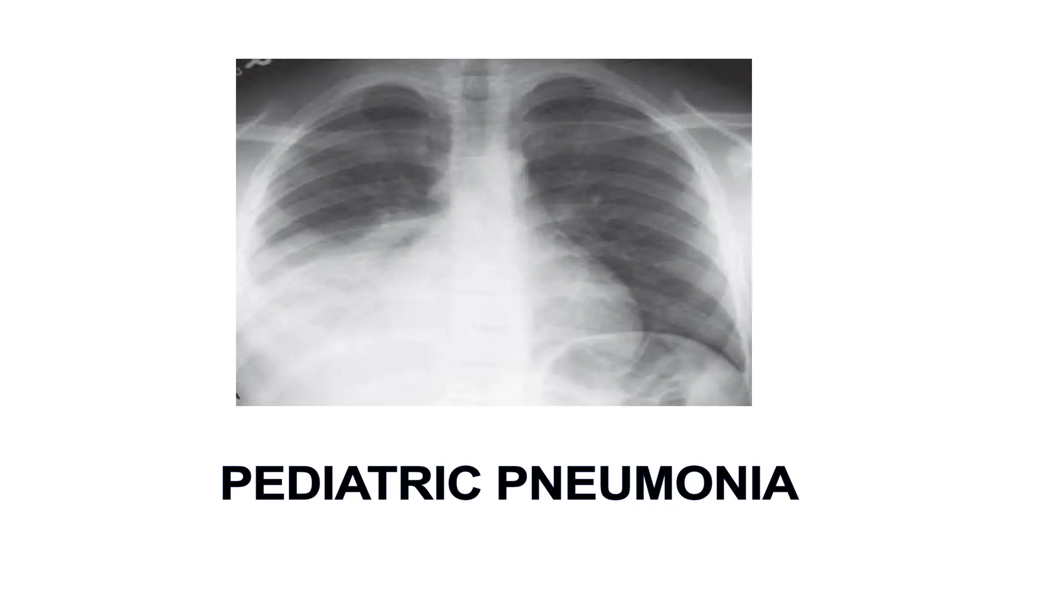
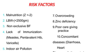
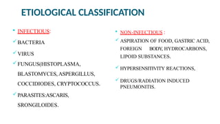
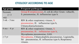
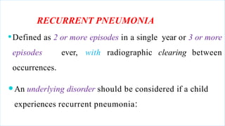
⚫Pneumonia is defined as inflammation
of the lung parenchyma.
(Ref:Nelson Text Book of Pediatrics 20th)
EPIDEMIOLOGY ..
pneumonia
⚫Each year, about 156 million new episodes
of occur world wide.
⚫Among which 151 million episodes in
developing countries.
(Ref: Epidemiology and Etiology of Childhood Pneumonia. Rudan I, Campbell, et al. Bull World
Health Organ 2008, May; 86(5):408-16.)
⚫It is the leading cause of U5 mortality, globally
accounting 16% of all U5 deaths.
Ref: WHO Fact sheet on
Pneumonia.
EPIDEMIOLOGY ..
RISK FACTORS
1. Malnutrition (Z <-2)
2. LBW-(<2500gm)
3. Non exclusive BF
4. Lack of Immunization-
(Measles, Pentavalent Hib,
Varicella)
5. Indoor air Pollution
7.Overcrowding
8.Zinc deficiency
9.Poor care giving
practice
10.Concomitant
diseases (Diarrhoea,
Heart
PNEUMONIA : CLASSIFICATION
Clinical
classification
Etiological
classification
Anatomical
classification
Infectous
Non-
Infectous
1. Community
acquired
2. Nosocomial pneumonia.
3. Pneumonia in
immunocompromised
Typical
Atypical
Pneumonia developed within
48 hours of hospital admission
ETIOLOGICAL CLASSIFICATION
 INFECTIOUS:
BACTERIA
VIRUS
FUNGUS(HISTOPLASMA,
BLASTOMYCES, ASPERGILLUS,
COCCIDIODES, CRYPTOCOCCUS.
PARASITES:ASCARIS,
SRONGILOIDES.
 NON-INFECTIOUS :
 ASPIRATION OF FOOD, GASTRIC ACID,
FOREIGN BODY, HYDROCARBONS,
LIPOID SUBSTANCES.
 HYPERSENSITIVITY REACTIONS,
 DRUGS/RADIATION INDUCED
PNEUMONITIS.
ETIOLOGY ACCORDING TO AGE
Age group Frequent pathogens
Neonate
(< 3
wk)
Group B streptococcus, E. coli & other Gram -vebacilli,
S. pneumoniae, H. influenziae type b.
3 wk – 3 mo RSV & other respiratory viruses, S.
pneumoniae, H. influenziae type b,
Chlamydiatrachomatis.
4 mo – 4 yr RSV & other respiratory viruses, S.
pneumoniae, H. influenziae type b,
Mycoplasma pneumoniae, GAS.
≥ 5 yr Mycoplasma, Chlamydophila pneumoniae, Legionella,
Str pneumoniae, H. influenzae type b, Respiratory
viruses.
RECURRENT PNEUMONIA
⚫Defined as 2 or more episodes in a single year or 3 or more
episodes ever, with radiographic clearing between
occurrences.
⚫An underlying disorder should be considered if a child
experiences recurrent pneumonia:
Recurrent pneumonia causes:
A. HEREDITARY DISORDERS: CYSTIC FIBROSIS, SICKLE CELL
DISEASE.
.B. Disorders of Immunity: HIV/AIDS, Brutons agammaglobinemia,
Selective Ig deficiency, SCID, Chronic Granulomatous disease, Leucocyte
adhesion defect
C. Disorders of cilia: Kartagener syndrome, Immotile cilia syndrome
D. Anatomic Disorders: Pulmonary sequestration, Lobaremphysema,
GER, TEF (H type), Bronchiectasis.
MODE OF TRANSMISSION
1. Droplet Nuclei
2. Nosocomial
3. Endogenous
4. Blood Borne
PATHOGENESIS
•Inhalation of droplet nuclei
•Hematogenous seeding
•Aspiration
Colonization of organism
in respiratory passage
Inflammatory reaction in
respiratory tract including
lung parenchyma
STAGES OF PNEUMONIA
⚫Stage of congestion: Lung parenchyma filled with inflammatory
exudate.
⚫Stage of red hepatization: massive exudation with red cells,
neutrophil & fibrin in alveoli.
⚫Stage of grey hepatization: progressive disintegration of RBC with
greyish brown discoloration.
⚫Stage of resolution: Progressive removal of exudate from alveolar
space.
⚫In VIRAL PNEUMONIA, low grade fever is usually present, along
with other features of respiratory distress:
1. Tachypnea ( mostconsistent C/F)
2. Increasedwork of breathing evident by intercostal,subcostal, and
suprasternal retractions, nasal flaring, and use of accessory
muscles,
3. Cyanosis and lethargy in case of severe infection, with crackles &
wheezing.
4. Hyper resonant chests
Clinical Manifestations
⚫BACTERIAL pneumonia is characterized by:
1. Sudden high grade fever, cough, and chest pain.
2. Drowsiness , occasionally with delirium
3. Along with usual signs of respiratory distress, i.e.
tachypnea, grunting, nasal f laring; retractions of
the supraclavicular, intercostal, and subcostal
areas & often cyanosis
IMCI (2M – 5Y)
IMCI: DAY1 – 2M
 Fast breathing,
 Severe chest indrawing ,
 Grunting,
 hypo/ hyperthermia,
 not feeding well,
 convulsion.
Any of these is classified as very severe disease.
INVESTIGATIONS
⚫X-Ray Chest
⚫CBC
⚫ESR, C-Reactive Proteins.
⚫Blood culture.
⚫Mantoux Test
CHEST X-RAY
⚫Viral pneumonia is usually characterized by:
1. hyperinflation with bilateral interstitial infiltrates and
2. peribronchial cuffing .
⚫Confluent lobar consolidation &/or pleural effusion is
typically seen with pneumococcal pneumonia .
VIRAL VS BACTERIAL PNEUMONIA
CBC
 Inviral pneumonia: WBC-normal or usually not higher
than 20,000/mm3, with a lymphocyte predominance.
 Inbacterial pneumonia: Elevated WBC count,
15,000- 40,000/mm3 with predominance of granulocytes.
⚫Acute phase reactants (ESR, CRP):
Higher in bacterial, normal or slightly raised in viral
pneumonia.
Blood culture: Blood culture results are positive in only
10%.
TREATMENT
⚫Treatment of suspected bacterial pneumonia is based on the presumptive
cause,age and clinical appearance of the child.
⚫For mildly ill children who do not require hospitalization, amoxicillinis
recommended.
⚫With the emergence of penicillin-resistant pneumococci, high doses of
amoxicillin (80-90 mg/kg/24 hr) should be prescribed.
⚫Therapeutic alternatives include cefuroxime axetil and
amoxicillin/clavulanate.
⚫For school-aged children and in children with suggested
infection of M. Pneumoniae or C. pneumoniae , a macrolide
antibiotic such as azithromycin is an appropriate choice
⚫ In adolescents, a respiratory f luoroquinolone (levof loxacin,
moxifloxacin) may be considered as analternative.
⚫The empiric treatment of suspected bacterial pneumoniain a
hospitalized child start on the clinical
manifestations at the time of presentation.
INDICATIONS FOR ADMISSION TO HOSPITAL
⚫Young age - < 6 months of age;
⚫Toxic appearance
⚫Moderate to severe respiratorydistress
⚫Inability of family to providecare at home;
⚫Failure of outpatienttherapy;
⚫Complicated pneumonia
⚫Vomiting or inability to tolerateoral fluid or medications.
⚫Immunocompromised state
TREATMENT AFTER HOSPITAL ADMISSION
Supportive care for children
⚫Oxygen, if needed (SpO2-<92%)
⚫Fluids and ensurehydration
⚫Antipyretics, analgesics
⚫Antibiotics
1. In areas without substantial high-level penicillin resistance among
S.
immunized against H. inf luenzae type b and
S.
fully
pneumoniae,
2. children who
are
pneumoniae and
3. are not severely ill should receiveampicillin or penicillin G.
⚫For children who do not meet these criteria, ceftriaxone or cefotaxime should
be
pneumonia initial antimicrobial
used.
⚫If clinical features suggest
staphylococcal
therapy vancomycin or clindamycin.
If viral pneumonia is suspected,it is reasonable
to withhold antibiotic therapy, especially for those patients
⚫who are mildly ill,
⚫have clinical evidence suggesting viral infection and
⚫are in no respiratorydistress.
⚫The optimal duration of antibiotic treatment for pneumonia has not
been well- established in controlled studies.
⚫Antibiotics should generally be continued until the patient has been
afebrile for 72 hr, and the total duration should not be < 10 days (or 5
days for azithromycin).
⚫Shorter courses (5-7 days) may also be effective, particularly for
children managed on an outpatientbasis.
⚫In developing countries, oral zinc (10 mg/day for <12 mo, 20 mg/day
for ≥12 mo) is advised to reduce mortality among children
COMPLICATIONS
 Pleural effusion
 Empyema
 Lung abscess
 Pneumothorax
 Pneumatocele
 Delayed Resolution
 Respiratory Failure
 Metastatic Septic lesions
 Activation of latentTB
COMPLICATED PNEUMONIA
PROGNOSIS
⚫Typically,
patients
with uncomplicated community-acquired bacterial
cough,
pneumonia show improvement in clinical symptoms (fever,
tachypnea, chest pain), within 48-96 hours of initiation of antibiotics.
⚫Radiographic evidence of improvement lags substantially behind
clinical improvement. It may take 6 to 8 weeks to return
to normal.
⚫When a patient does not improve with appropriate
antibiotic therapy complications, such as
1. empyema
2. bacterial resistance
3. nonbacterial etiologies such as viruses or fungi and aspiration
of foreign bodies or food
4. Preexisting diseases such as immuno deficiencies, ciliary
dyskinesia,cystic fibrosis, pulmonary sequestration or congenital
pulmonary airway malformation and
5. other noninfectious causes including bronchiolitis obliterans,
hypersensitivity pneumonitis, eosinophilic pneumonia,
aspirationand granulomatosis with polyangitis are suspected.
IS DONE TO DETERMINE THE REASON FOR
DELAY IN RESPONSE TO
⚫A repeat chest X-ray after treatment.
⚫BRONCHOALVEOLAR LAVAGE MAY BE INDICATED IN
CHILDREN WITH RESPIRATORY FAILURE.
⚫HIGH-RESOLUTION CT SCANS MAY BETTER TO IDENTIFY
COMPLICATIONS OR AN ANATOMIC REASON.
PREVENTION
1.Exclusive breastfeeding up to 6 mths age
2.Immunization against with-- Hib, PCV,
Measles, Pertussis, Varicella.
3.Adequete Nutrition---Under nutrition causes >1 millions death
under 5 due to Pneumonia.
4.Hand washing, safe water drinking & prevention of Diarrhoea.
5.Avoidance of parental or other sorts of secondary & tertiary
smoking.
6.Free from indoor airpollution.
7.Zinc supplementation.
childhood_Pneumonia-compressed final.pptx

childhood_Pneumonia-compressed final.pptx

  • 2.
    ⚫Pneumonia is definedas inflammation of the lung parenchyma. (Ref:Nelson Text Book of Pediatrics 20th)
  • 3.
    EPIDEMIOLOGY .. pneumonia ⚫Each year,about 156 million new episodes of occur world wide. ⚫Among which 151 million episodes in developing countries. (Ref: Epidemiology and Etiology of Childhood Pneumonia. Rudan I, Campbell, et al. Bull World Health Organ 2008, May; 86(5):408-16.)
  • 4.
    ⚫It is theleading cause of U5 mortality, globally accounting 16% of all U5 deaths. Ref: WHO Fact sheet on Pneumonia. EPIDEMIOLOGY ..
  • 5.
    RISK FACTORS 1. Malnutrition(Z <-2) 2. LBW-(<2500gm) 3. Non exclusive BF 4. Lack of Immunization- (Measles, Pentavalent Hib, Varicella) 5. Indoor air Pollution 7.Overcrowding 8.Zinc deficiency 9.Poor care giving practice 10.Concomitant diseases (Diarrhoea, Heart
  • 6.
    PNEUMONIA : CLASSIFICATION Clinical classification Etiological classification Anatomical classification Infectous Non- Infectous 1.Community acquired 2. Nosocomial pneumonia. 3. Pneumonia in immunocompromised Typical Atypical Pneumonia developed within 48 hours of hospital admission
  • 7.
    ETIOLOGICAL CLASSIFICATION  INFECTIOUS: BACTERIA VIRUS FUNGUS(HISTOPLASMA, BLASTOMYCES,ASPERGILLUS, COCCIDIODES, CRYPTOCOCCUS. PARASITES:ASCARIS, SRONGILOIDES.  NON-INFECTIOUS :  ASPIRATION OF FOOD, GASTRIC ACID, FOREIGN BODY, HYDROCARBONS, LIPOID SUBSTANCES.  HYPERSENSITIVITY REACTIONS,  DRUGS/RADIATION INDUCED PNEUMONITIS.
  • 8.
    ETIOLOGY ACCORDING TOAGE Age group Frequent pathogens Neonate (< 3 wk) Group B streptococcus, E. coli & other Gram -vebacilli, S. pneumoniae, H. influenziae type b. 3 wk – 3 mo RSV & other respiratory viruses, S. pneumoniae, H. influenziae type b, Chlamydiatrachomatis. 4 mo – 4 yr RSV & other respiratory viruses, S. pneumoniae, H. influenziae type b, Mycoplasma pneumoniae, GAS. ≥ 5 yr Mycoplasma, Chlamydophila pneumoniae, Legionella, Str pneumoniae, H. influenzae type b, Respiratory viruses.
  • 9.
    RECURRENT PNEUMONIA ⚫Defined as2 or more episodes in a single year or 3 or more episodes ever, with radiographic clearing between occurrences. ⚫An underlying disorder should be considered if a child experiences recurrent pneumonia:
  • 10.
    Recurrent pneumonia causes: A.HEREDITARY DISORDERS: CYSTIC FIBROSIS, SICKLE CELL DISEASE. .B. Disorders of Immunity: HIV/AIDS, Brutons agammaglobinemia, Selective Ig deficiency, SCID, Chronic Granulomatous disease, Leucocyte adhesion defect C. Disorders of cilia: Kartagener syndrome, Immotile cilia syndrome D. Anatomic Disorders: Pulmonary sequestration, Lobaremphysema, GER, TEF (H type), Bronchiectasis.
  • 11.
    MODE OF TRANSMISSION 1.Droplet Nuclei 2. Nosocomial 3. Endogenous 4. Blood Borne
  • 12.
    PATHOGENESIS •Inhalation of dropletnuclei •Hematogenous seeding •Aspiration Colonization of organism in respiratory passage Inflammatory reaction in respiratory tract including lung parenchyma
  • 13.
    STAGES OF PNEUMONIA ⚫Stageof congestion: Lung parenchyma filled with inflammatory exudate. ⚫Stage of red hepatization: massive exudation with red cells, neutrophil & fibrin in alveoli. ⚫Stage of grey hepatization: progressive disintegration of RBC with greyish brown discoloration. ⚫Stage of resolution: Progressive removal of exudate from alveolar space.
  • 14.
    ⚫In VIRAL PNEUMONIA,low grade fever is usually present, along with other features of respiratory distress: 1. Tachypnea ( mostconsistent C/F) 2. Increasedwork of breathing evident by intercostal,subcostal, and suprasternal retractions, nasal flaring, and use of accessory muscles, 3. Cyanosis and lethargy in case of severe infection, with crackles & wheezing. 4. Hyper resonant chests Clinical Manifestations
  • 15.
    ⚫BACTERIAL pneumonia ischaracterized by: 1. Sudden high grade fever, cough, and chest pain. 2. Drowsiness , occasionally with delirium 3. Along with usual signs of respiratory distress, i.e. tachypnea, grunting, nasal f laring; retractions of the supraclavicular, intercostal, and subcostal areas & often cyanosis
  • 16.
  • 18.
    IMCI: DAY1 –2M  Fast breathing,  Severe chest indrawing ,  Grunting,  hypo/ hyperthermia,  not feeding well,  convulsion. Any of these is classified as very severe disease.
  • 19.
    INVESTIGATIONS ⚫X-Ray Chest ⚫CBC ⚫ESR, C-ReactiveProteins. ⚫Blood culture. ⚫Mantoux Test
  • 20.
    CHEST X-RAY ⚫Viral pneumoniais usually characterized by: 1. hyperinflation with bilateral interstitial infiltrates and 2. peribronchial cuffing . ⚫Confluent lobar consolidation &/or pleural effusion is typically seen with pneumococcal pneumonia .
  • 21.
  • 22.
    CBC  Inviral pneumonia:WBC-normal or usually not higher than 20,000/mm3, with a lymphocyte predominance.  Inbacterial pneumonia: Elevated WBC count, 15,000- 40,000/mm3 with predominance of granulocytes.
  • 23.
    ⚫Acute phase reactants(ESR, CRP): Higher in bacterial, normal or slightly raised in viral pneumonia. Blood culture: Blood culture results are positive in only 10%.
  • 24.
    TREATMENT ⚫Treatment of suspectedbacterial pneumonia is based on the presumptive cause,age and clinical appearance of the child. ⚫For mildly ill children who do not require hospitalization, amoxicillinis recommended. ⚫With the emergence of penicillin-resistant pneumococci, high doses of amoxicillin (80-90 mg/kg/24 hr) should be prescribed. ⚫Therapeutic alternatives include cefuroxime axetil and amoxicillin/clavulanate.
  • 25.
    ⚫For school-aged childrenand in children with suggested infection of M. Pneumoniae or C. pneumoniae , a macrolide antibiotic such as azithromycin is an appropriate choice ⚫ In adolescents, a respiratory f luoroquinolone (levof loxacin, moxifloxacin) may be considered as analternative.
  • 26.
    ⚫The empiric treatmentof suspected bacterial pneumoniain a hospitalized child start on the clinical manifestations at the time of presentation.
  • 27.
    INDICATIONS FOR ADMISSIONTO HOSPITAL ⚫Young age - < 6 months of age; ⚫Toxic appearance ⚫Moderate to severe respiratorydistress ⚫Inability of family to providecare at home; ⚫Failure of outpatienttherapy; ⚫Complicated pneumonia ⚫Vomiting or inability to tolerateoral fluid or medications. ⚫Immunocompromised state
  • 28.
    TREATMENT AFTER HOSPITALADMISSION Supportive care for children ⚫Oxygen, if needed (SpO2-<92%) ⚫Fluids and ensurehydration ⚫Antipyretics, analgesics ⚫Antibiotics
  • 29.
    1. In areaswithout substantial high-level penicillin resistance among S. immunized against H. inf luenzae type b and S. fully pneumoniae, 2. children who are pneumoniae and 3. are not severely ill should receiveampicillin or penicillin G. ⚫For children who do not meet these criteria, ceftriaxone or cefotaxime should be pneumonia initial antimicrobial used. ⚫If clinical features suggest staphylococcal therapy vancomycin or clindamycin.
  • 30.
    If viral pneumoniais suspected,it is reasonable to withhold antibiotic therapy, especially for those patients ⚫who are mildly ill, ⚫have clinical evidence suggesting viral infection and ⚫are in no respiratorydistress.
  • 31.
    ⚫The optimal durationof antibiotic treatment for pneumonia has not been well- established in controlled studies. ⚫Antibiotics should generally be continued until the patient has been afebrile for 72 hr, and the total duration should not be < 10 days (or 5 days for azithromycin). ⚫Shorter courses (5-7 days) may also be effective, particularly for children managed on an outpatientbasis. ⚫In developing countries, oral zinc (10 mg/day for <12 mo, 20 mg/day for ≥12 mo) is advised to reduce mortality among children
  • 32.
    COMPLICATIONS  Pleural effusion Empyema  Lung abscess  Pneumothorax  Pneumatocele  Delayed Resolution  Respiratory Failure  Metastatic Septic lesions  Activation of latentTB
  • 33.
  • 34.
    PROGNOSIS ⚫Typically, patients with uncomplicated community-acquiredbacterial cough, pneumonia show improvement in clinical symptoms (fever, tachypnea, chest pain), within 48-96 hours of initiation of antibiotics. ⚫Radiographic evidence of improvement lags substantially behind clinical improvement. It may take 6 to 8 weeks to return to normal.
  • 35.
    ⚫When a patientdoes not improve with appropriate antibiotic therapy complications, such as 1. empyema 2. bacterial resistance 3. nonbacterial etiologies such as viruses or fungi and aspiration of foreign bodies or food 4. Preexisting diseases such as immuno deficiencies, ciliary dyskinesia,cystic fibrosis, pulmonary sequestration or congenital pulmonary airway malformation and 5. other noninfectious causes including bronchiolitis obliterans, hypersensitivity pneumonitis, eosinophilic pneumonia, aspirationand granulomatosis with polyangitis are suspected.
  • 36.
    IS DONE TODETERMINE THE REASON FOR DELAY IN RESPONSE TO ⚫A repeat chest X-ray after treatment. ⚫BRONCHOALVEOLAR LAVAGE MAY BE INDICATED IN CHILDREN WITH RESPIRATORY FAILURE. ⚫HIGH-RESOLUTION CT SCANS MAY BETTER TO IDENTIFY COMPLICATIONS OR AN ANATOMIC REASON.
  • 37.
    PREVENTION 1.Exclusive breastfeeding upto 6 mths age 2.Immunization against with-- Hib, PCV, Measles, Pertussis, Varicella. 3.Adequete Nutrition---Under nutrition causes >1 millions death under 5 due to Pneumonia. 4.Hand washing, safe water drinking & prevention of Diarrhoea. 5.Avoidance of parental or other sorts of secondary & tertiary smoking. 6.Free from indoor airpollution. 7.Zinc supplementation.