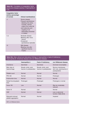
1. Table 26.1 Correlation of coagulation factor
activity and disease severity in haemophilia A
or B.
Coagulation factor
activity (percentage
of normal) Clinical manifestations
<1 Severe disease
Frequent spontaneous
bleeding into joints,
muscles, internal
organs from early life
Joint deformity and
crippling if not
adequately prevented
or treated
1–5 Moderate disease
Bleeding after minor
trauma
Occasional
spontaneous episodes
>5 Mild disease
Bleeding only after
significant trauma,
surgery
Table 26.2 Main clinical and laboratory findings in haemophilia A, factor IX deficiency
(haemophilia B, Christmas disease) and von Willebrand disease.
Haemophilia A Factor IX deficiency von Willebrand disease
Inheritance Sex-linked Sex-linked Dominant (incomplete)
Main sites of
Muscle, joints, post-trauma
Muscle, joints, post-trauma
haemorrhage
or postoperative
or postoperative
Mucous membranes,
skin cuts, post-trauma or
postoperative
Platelet count Normal Normal Normal
PFA-100 Normal Normal Prolonged
Prothrombin time Normal Normal Normal
Partial thromboplastin
time
Prolonged Prolonged Prolonged or normal
Factor VIII Low Normal May be moderately
reduced
Factor IX Normal Low Normal
VWF Normal Normal Low or abnormal
function (Table 26.3)
Ristocetin-induced
platelet aggregation
Normal Normal Impaired
VWF, von Willebrand factor.
2. Table 26.3 Classification of von Willebrand disease.
Type 1 Quantitative partial deficiency
Type 2 Functional abnormality
Type 3 Complete deficiency
Secondary classification of type 2 VWD
Subtype Platelet-associated function Factor VIII binding capacity High MW VWF multimers
2A Decreased Normal Absent
2B Increased affinity for GPIb Normal Usually reduced/absent
2M Decreased Normal Normal
2N Normal Reduced Normal
GPIb, glycoprotein Ib; MW, molecular weight; VWD, von Willebrand disease; VWF, von Willebrand factor.
Table 26.4 The acquired coagulation
disorders.
Deficiency of vitamin K-dependent factors
Haemorrhagic disease of the newborn
Biliary obstruction
Malabsorption of vitamin K (e.g. tropical sprue,
gluten-induced enteropathy)
Vitamin K-antagonist therapy (e.g. coumarins,
indandiones)
Liver disease – complex dysregulation with
synthetic failure of pro- and anticoagulant factors
Disseminated intravascular coagulation –
consumption of all clotting factors and platelets
Inhibition of coagulation
Specific inhibitors (e.g. antibodies against factor
VIII)
Non-specific inhibitors (e.g. antibodies found in
systemic lupus erythematosus, rheumatoid
arthritis which paradoxically cause thrombosis)
Miscellaneous
Diseases with M-protein production that interfere
with haemostasis
L-Asparaginase
Therapy with heparin, defibrinating agents or
thrombolytics
Massive transfusion syndrome
3. Table 26.5 Causes of disseminated
intravascular coagulation.
Infections
Gram-negative and meningococcal septicaemia
Clostridium welchii septicaemia
Severe falciparum malaria
Viral infection – varicella, HIV, hepatitis,
cytomegalovirus
Malignancy
Widespread mucin-secreting adenocarcinoma
Acute promyelocytic leukaemia
Obstetric complications
Amniotic fluid embolism
Premature separation of placenta
Eclampsia; retained placenta
Septic abortion
Hypersensitivity reactions
Anaphylaxis
Incompatible blood transfusion
Widespread tissue damage
Following surgery or trauma
After severe burns
Vascular abnormalities
Kasabach–Merritt syndrome
Leaking prosthetic valves
Cardiac bypass surgery
Vascular aneurysms
Miscellaneous
Liver failure
Pancreatitis
Snake and invertebrate venoms
Hypothermia
Heat stroke
Acute hypoxia
Massive blood loss
4. Table 26.6 Haemostasis tests: typical results in acquired bleeding disorders.
Platelet count Prothrombin time
Activated partial
thromboplastin
time Thrombin time
Liver disease Low Prolonged Prolonged Normal (rarely
prolonged)
DIC Low Prolonged Prolonged Grossly prolonged
Massive
Low Prolonged Prolonged Normal
transfusion
Coumarin
anticoagulants
Normal Grossly prolonged Prolonged Normal
Heparin Normal (rarely low) Mildly prolonged Prolonged Prolonged
Circulating
Normal Normal or
Prolonged Normal
anticoagulant
prolonged
DIC, Disseminated intravascular coagulation.
Table 26.7 Indications for the use of fresh
frozen plasma (National Institutes of Health
Consensus Guidelines).
Coagulation factor deficiency (PCC where
specific or combined factor concentrate is not
available)
Reversal of warfarin effect (PCC if available are
highly effective compared to plasma which has
almost no effect)
Multiple coagulation defects (e.g. in patients
with liver disease, DIC) (PCC are much better,
plasma is virtually useless)
Massive blood transfusion with coagulopathy
and clinical bleeding
Thrombotic thrombocytopenic purpura
Deficiencies of antithrombin*, protein C* or
protein S
Some patients with immunodeficiency
syndromes
DIC, disseminated intravascular coagulation; PCC,
prothrombin complex concentrates.
* Antithrombin and protein C concentrates now available.