1. Receptor binding site
MATERIAL
12 Pathology at a Glance. By C.J. Finlayson and B.A.T. Newell. Published 2009 by Blackwell Publishing. ISBN: 978-1-4051-3650-1
General pathology
1 The normal human cell
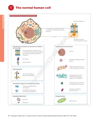
Controlling the intracellular environment: cell membrane
and ion pumps
Phospholipid bilayer: hydrophilic ends
on outer and inner aspects. Hydrophobic
ends inside membrane, stabilised by
cholesterol
Receiving signals
Cell surface receptors
and second messengers
Intracellular transport, cell movement and mitosis
Centriole: spindle formation
(cell division)
Cytoskeletal components:
microtubules and filaments
Degradation/destruction
Lysosome
Peroxisome
Protein production and secretion
Nucleus
Ribosome and newly produced
peptide
Rough (with ribosomes) and
smooth endoplasmic reticulum
Golgi apparatus: protein
modification (folding and
addition of carbohydrate)
Secretory vesicle containing
protein/glycoprotein product
Proteosome: degrades
defective protein
Energy production
Mitochondrion
Na/Cl pump
Calcium channel
Cell membrane: a phospholipid bilayer,
containing ion channels and receptor
molecules
Cytosol
Signalling molecules
transmit signal to nucleus
Overview of the structure of normal human cells
COPYRIGHTED
2. The normal human cell Introduction 13
General pathology
The important functions of the cell are: manufacture of proteins for
local or distant use, energy generation, functions appropriate to tissue
type and replication.
The main elements are the nucleus, the cytoplasm (cytosol), the
cytoskeleton and the subcellular organelles, all bound by membranes.
Nucleus
The nuclear membrane contains pores to permit metabolites, RNA and
ribosomal subunits in or out. It contains:
• DNA, the nuclear chromatin, which only forms about 20% of the
nuclear mass.
• Nucleoli – ribosomal RNA synthesis and ribosome subunit assembly.
• Nucleoprotein, e.g. synthetic enzymes for DNA, RNA and regulatory
proteins, all made in the cytoplasm and imported into the nucleus.
• Messenger, transfer and ribosomal RNA en route for the cytoplasm.
Cytosol
The nutritious fluid medium that bathes and supports the organelles,
through which the cytoskeleton ramifies. Many reactions take place here.
Cytoskeleton
• Microtubules: organelles such as secretory vesicles or internalised
receptors can be transported through the cell via the cytoskeleton.
• Microfilaments (actin, myosin): these stabilise cell shape and act as
contractile proteins in muscle.
• Intermediate filaments, e.g. cytokeratin, desmin, neurofilament pro-teins
and glial fibrillary acidic protein (the types differ between tissues
and all are structural).
Organelles
Mitochondria
These are the main ATP/energy generating organelles and house the
Krebs cycle and oxidative phosphorylation. They have their own
ssDNA (maternally derived) which codes a minority of their proteins. A
porous outer membrane and folded inner membrane are present.
Ribosomes
Nucleolus-produced ribosomal subunits aggregate in the cytosol and
attach to the endoplasmic reticulum or lie loose in the cytosol, depend-ing
on the destination of the protein to be made (free ribosomes make
proteins for inside the cell itself). Ribosomes translate RNA strands
into a correctly assembled amino acid sequence (peptide molecule).
Endoplasmic reticulum (ER)
The ER is an irregular maze of membrane-bound tubules, saccules and
cisterns which ramifies through the cell.
• Rough ER is studded with ribosomes. Proteins made by the rough
ER pass into the rough ER cisternae and undergo secondary folding
and early glycosylation before being incorporated into membranes for
export from the cell, receptor molecules on the cell, or components such
as lysosomes within the cell.
• Smooth ER: there is a further addition of carbohydrate moieties to
protein, folding to achieve tertiary structure.
Golgi apparatus – see diagram.
Secretory vesicles
These membrane bound packets are moved via the cytoskeleton to fuse
with the cell membrane to expel their contents outside.
Lysosomes
These are intracellular membrane-bound vesicles, containing destructive
chemicals and enzymes, which fuse with phagosomes to release their
contents into the phagolysosome and destroy pathogens. Lysosomes
also degrade worn-out cell organelles (autophagy).
Peroxisomes
These small membrane-bound granules contain oxidative enzymes
which make hydrogen peroxide plus its regulator catalase.
Proteasomes
These identify defective proteins and degrade them into their component
peptides and amino acids for re-use by the cell. Portions of broken-down
protein are bound by MHC class I molecules and displayed on the cell
surface to Tc cells.
Centrosome
This contains the two linked centrioles, from which microtubules
radiate into the cell. The centrioles duplicate and migrate to oppo-site
ends of the cell during cell division, separating the duplicated
chromosomes.
Membranes
Membranes are phospholipid barriers surrounding the cell itself and
certain organelles. They isolate portions of the cell and permit several,
often incompatible, metabolic processes to take place simultaneously.
The cell membrane
This phospholipid bilayer interacts with the extracellular world by
assorted surface molecules. The centre is lipophilic and the surfaces
hydrophilic, with cholesterol as a stabilising ‘spacer’ between them.
The ‘raft theory’ suggests that intramembrane structures can float and
be cross-linked around the perimeter of the cell.
Membrane proteins: proteins that project through the membrane
outside the cell usually have attached carbohydrates. Glycolipids are
carbohydrates attached to the lipid membrane and are important in
cell recognition, cell–cell bonds and adsorbing molecules. Some tissues
have a protective glycocalyx.
Transport through the cell membrane: The main mechanisms are:
• Passive diffusion (needs only a concentration gradient), e.g. lipids
and lipid-soluble agents like ethanol.
• Facilitated diffusion: the binding of a molecule triggers a conforma-tional
change which moves the molecule across the membrane.
• Active transport: against a concentration gradient to maintain ion
concentrations within the cell, e.g. the Na+/K+/ATPase complex.
• Bulk transport: endocytosis, transcytosis and exocytosis. Endocytosis
includes receptor-mediated endocytosis (ligands or viral particles)
and phagocytosis (engulfing of particles). Pinocytosis, dendritic cell
sampling of small quantities of tissue fluid is not receptor mediated.
Transmission of messages across the cell membrane:
• Lipid-soluble agents (e.g. steroids) diffuse directly across cell
memtranes.
• Receptor binding and activation of secondary messengers: applies
to protein messenger molecules, which bind to a specific cell surface
receptor (ligand), resulting in active transport of the molecule through
the membrane or the triggering of intracellular cascade reactions.
Neurotransmitters: these are chemical messengers for neurones or
myocytes that cause an electrical response in the target by receptor-mediated
opening of an ion channel.