SlideShare a Scribd company logo
1 of 53
Copyright © John Wiley & Sons, Inc. All rights reserved.
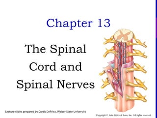
Chapter 13
The Spinal
Cord and
Spinal Nerves
Lecture slides prepared by Curtis DeFriez, Weber State University
Copyright © John Wiley & Sons, Inc. All rights reserved.
Introduction to the Spinal Cord
 About 100 million neuronsand even moreneuroglia
comprisethespinal cord, thepart of thecentral nervous
system that extendsfrom thebrain.
 Thespinal cord and itsassociated spinal nervescontain
reflex circuits that control someof your most rapid
reactionsto environmental changes.
 Thegray matter of thecord isasitefor integrationof
postsynaptic potentials(IPSPsand EPSPs).
 Thewhitematter of thecord containsmajor sensory and
motortracts to and from thebrain.
Copyright © John Wiley & Sons, Inc. All rights reserved.
 Thespinal cord beginsasacontinuation of themedulla
oblongata(themost inferior portion of the
brain stem) extending
from theforamen
magnum of the
occipital boneto its
termination asthe
conus medullaris
between L1 - L2.
External Cord Anatomy
Copyright © John Wiley & Sons, Inc. All rights reserved.
External Cord Anatomy
 Thespinal cord isoval in shapeand slightly flattened
anteriorly and posteriorly.
 Two typesof connectivetissue
coveringsprotect thecord
and providephysical stability:
Thebony vertebral column
providesthebackbone.
Thespinal meninges surround
thecord asacontinuation of
thecranial meningesthat encirclethebrain.
Copyright © John Wiley & Sons, Inc. All rights reserved.
External Cord Anatomy
 Thesethreemembranes(each called ameninx) are
labeled from superficial to deep asfollows:
Theoutermost dura mater(tough mother) formsasac that
enclosestheentirecord.
Themiddlemeninx isadelicateavascular covering
called thearachnoid mater. It isattached to theinside
of theduraand formstheroof of thesubarachnoid space
(SAS) in which cerebral spinal fluid (CSF) circulates.
Thetransparent pia materispressed-up against thecord and
isfilled with blood vesselsthat supply nutrientsto it.
Copyright © John Wiley & Sons, Inc. All rights reserved.
 Theepidural space runsbetween theduramater and the
moresuperficial ligamentum flavum (which linesthe
undersideof thebony vertebral lamina).
 Thesubdural space liesbetween theduraand the
arachnoid. In thespinal column, theduraand arachnoid
membranes
areheld firmly together
so that thesubdural
spaceisoften no more
than apotential space.
External Cord Anatomy
Copyright © John Wiley & Sons, Inc. All rights reserved.
 Thepiamater has21 pairsof denticulate ligaments which
attach it to thearachnoid and duramaters. Named for their
tooth-likeappearance, thedenticulateligamentsare
traditionally believed to providestability for thespinal cord
against sudden
shock and
displacement within
thevertebral column.
External Cord Anatomy
Copyright © John Wiley & Sons, Inc. All rights reserved.
 Arising from theconusmedullarisisthefilum terminale,
an extension of thepiamater that extendsinferiorly and
blendswith the
arachnoid and durato
anchor thespinal cord to
thecoccyx.
 Thecaudaequinaor “horses
tail” aretherootsof the
lower spinal nervesthat angle
down alongsidethefilum terminale.
External Cord Anatomy
Copyright © John Wiley & Sons, Inc. All rights reserved.
External Cord Anatomy
 Thespinal cord hastwo enlargements, onein thecervical
areafrom C4–T1, and another in thelumbar areabetween
T9–T12.
 Thecervical enlargement
correlateswith thesensory
input and motor output to the
upper extremities.
 Thelumbar enlargement handles
motor output and sensory input
to and from thelegs.
Copyright © John Wiley & Sons, Inc. All rights reserved.
External Cord Anatomy
 From superior to inferior, thespinal
cord becomesprogressively smaller.
 Thereislessand lesswhitematter
aswedescend becausethereare
fewer sensory tractsgoing up
(they haven’t “jumped on” yet),
and therearefewer motor tracts
going down (they’ve“jumped
off” already.)
Copyright © John Wiley & Sons, Inc. All rights reserved.
 Two bundlesof axons, called roots, connect each spinal
nerveto asegment of thecord by even smaller bundlesof
axonscalled rootlets.
 The posterior(dorsal) root and rootletscontain only
sensory axons, which conduct
nerveimpulsesfrom sensory
receptorsin theskin, muscles,
and internal organsinto the
central nervoussystem.
External Cord Anatomy
Copyright © John Wiley & Sons, Inc. All rights reserved.
External Cord Anatomy
 Each posterior root hasaswelling, theposterior(dorsal)
root ganglion, which containsthecell bodiesof sensory
neurons. Theanterior(ventral) root and rootlets contain
axonsof motorneurons, which conduct nerveimpulsesfrom
theCNSto
effectors(muscles
and glands).
Copyright © John Wiley & Sons, Inc. All rights reserved.
Epidural Anesthesia
 Epidural anesthesiaiscommonly administered to women
about to go into labor. In thisprocedure, aneedleisplaced
between thebonesof theposterior spineuntil it just
penetratestheligamentum flavum yet remainssuperficial to
theduramater.
 Local anesthetic isused to providepain relief –
even completeanesthesiaif acesarean section isrequired.
Copyright © John Wiley & Sons, Inc. All rights reserved.
Lumbar Puncture
 A needleinserted into thesubarachnoid spacefor thepurpose
of withdrawing CSF (for diagnosisor to reducepressure) or
to introduceadrug or contrast agent iscalled alumbar
puncture.
 CSF isoften collected to diagnosemeningitisor some
other diseaseof theCNS.
 Agentsinjected into theSASincludedrugssuch as
antibiotics, chemotherapeutic agents, or analgesics, or
contrast mediafor radiographic procedures.
• Thepressureof CSF in theSAScan also bemeasured
during alumbar puncture.
Copyright © John Wiley & Sons, Inc. All rights reserved.
Lumbar Puncture
 Thesiteused for most lumbar puncturesisbetween the3rd
and 4th
(or 4th
and 5th
) lumbar vertebrae- below thetermination
of theactual cord in theregion of thecaudaequina. With the
needlein theSAS, CSF can besampled.
 Anestheticscan also
begiven in thisway,
but using 1/10 the
doserequired for
epidural anesthesia.
Copyright © John Wiley & Sons, Inc. All rights reserved.
Internal Cord Anatomy
 In thespinal cord, thewhitematter ison theoutside, and the
gray matter ison theinside. In thebrain thewhitematter is
on theinside, and thegray matter ison theoutside.
Copyright © John Wiley & Sons, Inc. All rights reserved.
 Thewhite matterof thecord consistsof millionsof nerve
fibers which transmit electrical information between the
limbs, trunk and organsof thebody, and thebrain.
 Internal to thisperipheral region, and surrounding the
central canal, isthe
butterfly-shaped
central region made
up of nerve cell
bodies (gray matter–
herestained brown).
Internal Cord Anatomy
Copyright © John Wiley & Sons, Inc. All rights reserved.
Internal Cord Anatomy
 Anterior(ventral) gray horns consist of somatic motor
neurons.
 Posterior(dorsal) gray horns consist of somatic and
autonomic sensory nuclei.
 Theposteriorgray horn isthesiteof synapsebetween
first-order sensory neuronscoming in from theperiphery,
and second-order neuronswhich either ascend in thecord
or exit back out aspartsof reflex arcs.
Copyright © John Wiley & Sons, Inc. All rights reserved.
Internal Cord Anatomy
 Thelateral gray horns arefound only in thethoracic,
upper lumbar, and sacral segmentsof thecord.
 They contain cell bodiesof autonomic motor neurons
• Someof thesefibersascend outsidethedurabut closeto
thecord to supply sympathetic
innervation to thehead.
• Otherstravel in sympathetic
trunksto theorgansand
glandsof thethorax, abdomen,
and pelvis.
Copyright © John Wiley & Sons, Inc. All rights reserved.
Internal Cord Anatomy
 Other notablefeaturesvisualized on atransected cord arethe
anterior median fissure, theposterior median sulcus, thegray
and whitecommissures, and thecentral canal.
 Thecentral canal extendstheentirelength of thespinal
cord and
isfilled
with CSF.
Copyright © John Wiley & Sons, Inc. All rights reserved.
Internal Cord Anatomy
 A tract isabundleof
neuronal axonsthat areall
located in aspecific areaof
thecord and all traveling to
thesameplace(higher or
lower in thebrain or cord).
Copyright © John Wiley & Sons, Inc. All rights reserved.
Internal Cord Anatomy
 Thewhitematter of thecord isdivided into anterior,
posterior, and lateral columnsin which ascending sensory
tractsaretraveling to someplacein thebrain and descending
motor tracts(red) aretraveling to alocation in thecord.
The afferent tracts are noted here in blue, while the efferent tracts are shown in red.
Copyright © John Wiley & Sons, Inc. All rights reserved.
Internal Cord Anatomy
 Names of tracts areformed by using compound wordsthat
denotetheoriginof thetract, and theplacewhereit ends.
 Thespinothalamic tract goesfrom thespinal cord to the
brain – it isan afferent tract.
 Thecorticospinal tract goesfrom thecortex of thebrain to
thespinal cord – it isan efferent tract.
 Thevestibulospinal tract originatesfrom an areain the
brain which you probably don’t recognize; however, you
can recognizethedestination in thespine, and therefore
deducethat it isamotor tract.
Copyright © John Wiley & Sons, Inc. All rights reserved.
Internal Cord Anatomy
 Theposteriorcolumns areafferent tractsthat convey
nerveimpulsesfor discriminativetouch, light pressure,
vibration, and consciousproprioception (awarenessof
tendon and joint position in spaceand their
relativemovements).
 Thespinothalamic tract isan
afferent tract that transmitssensations
of pain, warmth, coolness, itching,
tickling, deep pressure, and
crudetouch.
Copyright © John Wiley & Sons, Inc. All rights reserved.
Internal Cord Anatomy
 Thelateral and anteriorcorticospinal tracts aremajor
pathwaysfor carrying signalsfrom thecerebral cortex
that result in voluntary movement of skeletal muscles.
 Other motortracts convey nerveimpulsesfrom thebrain
stem that coordinate
visual stimuli with body
movements, maintain
postureand regulate
skeletal muscletone.
Copyright © John Wiley & Sons, Inc. All rights reserved.
 A dermatomeisan areaof skin that is
innervated by asinglespinal nerve,
indicated by thelettersand number of a
particular segmental nerve.
 Important dermatomesinclude:
 C6/C7 - thumb and index
finger (“six-shooter”)
 T4 - nippleline
 T10 - umbilicus
 L1-L5 - lower extremities
“L for legs”)
Dermatomes
Copyright © John Wiley & Sons, Inc. All rights reserved.
Damage to the Cord
 “Transection” of the spinal cord meansthat ascending
and descending tractsarepartially or completely severed.
 If transection occurs, say in amotor vehicleor diving
accident, paralysiswill occur depending on thelevel of the
injury. Transection:
 at thebaseof skull resultsin death by asphyxiation
 in theupper cervical arearesultsin quadriplegia
 between thecord enlargementsresultsin someform of
paraplegia
Copyright © John Wiley & Sons, Inc. All rights reserved.
Peripheral Nerves
Copyright © John Wiley & Sons, Inc. All rights reserved.
Peripheral Nerves
 Spinal nerves arethepathsof communication between the
spinal cord and specific regionsof thebody.
 Nervesarearranged in fasciclessurrounded by a
perineurium, with theentirenervesheathed by aCT
epineurium.
Copyright © John Wiley & Sons, Inc. All rights reserved.
Peripheral Nerves
 31 left-right pairs of spinal nerves emergefrom the
cord at regular intervals(called segments). Except for the
first cervical pair thespinal nervesleavethevertebral
column from theintervertebral foramen between adjoining
vertebrae– thefirst pair leavesbetween the
skull and thefirst cervical vertebrae.
Cervical – 8 pairs, C1-C8
Thoracic – 12 pairs, T1-T12
Lumbar – 5 pairs, L1-L5
Sacral - 5 pairs, S1-S5

Copyright © John Wiley & Sons, Inc. All rights reserved.
 Piercing the dura, thesegmental (spinal) nervesexit the
central nervoussystem into theperipheral nervoussystem
and almost immediately split into 3 major branches: An
anterior ramus, posterior ramus, and rami communicantes
(connectionsto
sympathetic
ganglia).
Peripheral Nerves
Copyright © John Wiley & Sons, Inc. All rights reserved.
Peripheral Nerves
 Theanterior rami of thesegmental nervesmay travel alone
(such astheintercostal nerveswhich run underneath each of
the12 ribs), or they can join together to form large“braided
ropes” – a plexus of nerves.
 Thereareanumber of major nerveplexuses, all formed
from anterior rami of spinal nerves, and all located
anterior to thespine: Thecervical plexus, brachial
plexus, celiac (solar) plexus, lumbarplexus, sacral
plexus, and coccygeal plexus
Copyright © John Wiley & Sons, Inc. All rights reserved.
Nerve Plexuses
 Thecervical plexus, formed by theanterior rami of C1-C5,
servesthehead, neck, and diaphragm.
 Thephrenic nerves arisefrom thecervical plexusto
supply themajor muscleof
respiration (“C3,4,5 keep
thediaphragm alive”).
Copyright © John Wiley & Sons, Inc. All rights reserved.
Nerve Plexuses
 The brachial plexus isformed
by theanterior rami of C5-C8 and
T1. It isdivided into roots→
trunks→ divisions→ cords
→ nerves.
 Thenervesfrom thebrachial
plexussupply theshoulders
and upper limbs.
Copyright © John Wiley & Sons, Inc. All rights reserved.
 Someof themajor nervesthat arisefrom thebrachial
plexusesarethe:
 musculocutaneous
nerve
 axillary nerve
 radial nerve
 median nerve
 ulnarnerve
 long thoracic nerve
Nerve Plexuses
Copyright © John Wiley & Sons, Inc. All rights reserved.
Nerve Plexuses
 Injuriesto thebrachial plexusarenot uncommon:
 Erb’s palsy isaparalysisof thearm that most often
occursasan infant'shead and neck arepulled toward
thesideat thesametimeastheshoulderspass
through thebirth canal.
• A similar injury may beobserved at any age,
including adults, following atraumatic fall
or other traumawhereby
thenervesof theplexus
areviolently stretched.
Copyright © John Wiley & Sons, Inc. All rights reserved.
Nerve Plexuses
 Injuriesto thebrachial plexusor peripheral nerves:
 Median nerve injury, either at theplexusor occurring
moredistally, resultsin numbness, tingling and pain in the
palm and fingers.
• Carpal tunnel syndromeisacommon typeof median
nerveinjury that isseen in peoplewho perform
repetitivemotionsof thehand and
wrist liketyping on acomputer
keyboard.
Copyright © John Wiley & Sons, Inc. All rights reserved.
 Theulnarnerve isthelargest unprotected (by muscleor
bone) nervein thehuman body. It emergesfrom themedial
and lateral cordsof thebrachial plexusto supply themedial
half of thehand. Striking themedial epicondyleof the
humeruswherethenerveisexposed
isreferred to asbumping one’s“funny bone”.
 Damageto thenerveleadsto
abnormal sensationsin the4–5th
fingersand an inability to abduct
or adduct thelittleand ring fingers.
Nerve Plexuses
Copyright © John Wiley & Sons, Inc. All rights reserved.
Nerve Plexuses
 Thelong thoracic nerve emergesfrom thecordsof the
brachial plexusto supply theserratusanterior muscle.
 Becauseof itslong, relatively superficial course, it is
susceptibleto injury either through direct traumaor stretch
of theplexus. Injury (resulting in
a“winged scapula” in which the
arm cannot beabducted beyond the
horizontal position) hasbeen reported
in almost all sports.
Copyright © John Wiley & Sons, Inc. All rights reserved.
Nerve Plexuses
 Thelumbarplexus isformed by theanterior rami of L1-L4
to supply theanterolateral abdominal wall, external genitalia,
and part of thelower limbs.
 Thefemoral and obturator
nerves comefrom the
lumbar plexus.
Copyright © John Wiley & Sons, Inc. All rights reserved.
Nerve Plexuses
 Thesacral plexus isformed by theanterior rami of L4-L5
and S1-S4. It suppliesthebuttocks, perineum, and part of the
lower limbs.
 It givesriseto thelargest
nervein thebody, the
sciatic nerve.
Copyright © John Wiley & Sons, Inc. All rights reserved.
Nerve Plexuses
 Thecoccygeal plexus isformed by theanterior rami of S4-
S5 and thecoccygeal nerves. It isasmall plexusfrom which
theanococcygeal nerveexitsto supply asmall areaof skin in
the
coccygeal region.
Copyright © John Wiley & Sons, Inc. All rights reserved.
Nerve Terminology Summary
 Rootlets roots(ant. and post.)  segmental nerves
 anterior ramus form largenerveplexuses
 posterior ramus
 rami communicantes
Copyright © John Wiley & Sons, Inc. All rights reserved.
Reflexes
 A reflex isafast, involuntary responseto astimulus. In a
spinal reflex theintegration takesplacein thespinal cord,
not thebrain.
 Spinal reflexescan bemonosynaptic (sensory neuron
with motor neuron) or polysynaptic (involving
interneurons), and they can go in and out on thesame, or
on theopposite
sideof the
cord.
Copyright © John Wiley & Sons, Inc. All rights reserved.
Reflexes
 A reflex arc isapathway that anerveimpulsefollowsto
produceareflex. Componentsof areflex arc includea
sensory receptorand asensory neuron, an integrating
centerinsidethecord, an exiting motorneuron, and an
effector(which isusually some
sort of muscleor agland
which makessome-
thing moveor secrete
“involuntarily”).
Copyright © John Wiley & Sons, Inc. All rights reserved.
Reflexes
 Sensory receptors involved in reflex arcsarespecifically
adapted to perceivethestimulusand initiatean impulse.
 Themotor neuron
becomesstimulated
without any processing
in thebrain (that comes
later)… first you kick the
person examining you
(patellarreflex), then you feel
thetap of thehammer!
Copyright © John Wiley & Sons, Inc. All rights reserved.
Reflexes
Interactions Animation
 Componentsof aReflex Arc Animations
You must be connected to the internet to run this animation
Copyright © John Wiley & Sons, Inc. All rights reserved.
 Reflex arcscan beipsilateral (all neuronsand effectorson
thesamesideof thebody) or contralateral (thereceptors
and afferent neuronsareon theopposite
sideof thebody astheefferent
neuronsand effectors.)
Theflexor (withdrawal)
reflex isagood example
of acontralateral reflex
(stepping on atack).
Reflexes
Copyright © John Wiley & Sons, Inc. All rights reserved.
Reflexes
 In addition to initiating theflexor
reflex that causesyou to
withdraw thelimb, thepain
impulsesfrom stepping
on thetack also
initiateacrossed-
extensorreflex to
help you maintain
your balance.
Copyright © John Wiley & Sons, Inc. All rights reserved.
Reflexes
 Someimportant spinal reflexes include:
 Thepatellarreflex in which theleg extendsin response
to stretch of thepatellar tendon. Thisreflex can be
blocked by damagein thecorticospinal tractsfrom
diabetes, neurosyphilis, or damageto thelumbar region
of thespinal cord.
 TheAchilles reflex causescontraction of thecalf when
aforceisapplied to theAchillestendon. It isabsent after
damageto thelower cord or lumbosacral plexus.
Copyright © John Wiley & Sons, Inc. All rights reserved.
Reflexes
 Someimportant spinal reflexes include:
 TheBabinski, orplantarflexion reflex isconsidered
normal in adultsif they flex (curl) thebig toewhen the
soleof thefoot isstimulated. If thesoleof thefoot is
stimulated and thepatient extendsthebig toe, it would
indicatedamagein thecorticospinal tract.
 Infantsnormally extend their toeswhen stimulated in this
way; so an “abnormal Babinski” doesnot indicateany
diseaseor damagein thisagegroup.
Copyright © John Wiley & Sons, Inc. All rights reserved.
 Summary of ReflexesAnimation
Reflexes
Interactions Animation
You must be connected to the internet to run this animation
Copyright © John Wiley & Sons, Inc. All rights reserved.
End of Chapter 13
Copyright 2012 John Wiley & Sons, Inc. All rightsreserved.
Reproduction or translation of thiswork beyond that permitted in
section 117 of the1976 United StatesCopyright Act without
expresspermission of thecopyright owner isunlawful. Request for
further information should beaddressed to thePermission
Department, John Wiley & Sons, Inc. Thepurchaser may make
back-up copiesfor his/her own useonly and not for distribution or
resale. ThePublisher assumesno responsibility for errors,
omissions, or damagescaused by theuseof theseprogramsor from
theuseof theinformation herein.

More Related Content

What's hot

14 [chapter 14 the brain and cranial nerves]
14 [chapter 14 the brain and cranial nerves]14 [chapter 14 the brain and cranial nerves]
14 [chapter 14 the brain and cranial nerves]Sompoch Thanachaikan
 
Development of Nervous System (Special Embryology)
Development of Nervous System (Special Embryology)Development of Nervous System (Special Embryology)
Development of Nervous System (Special Embryology)Dr. Sherif Fahmy
 
VESTIBULOCOCHLEAR NERVE.pptx
VESTIBULOCOCHLEAR NERVE.pptxVESTIBULOCOCHLEAR NERVE.pptx
VESTIBULOCOCHLEAR NERVE.pptxDr. sana yaseen
 
15 [chapter 15 the autonomic nervous system]
15 [chapter 15 the autonomic nervous system]15 [chapter 15 the autonomic nervous system]
15 [chapter 15 the autonomic nervous system]Sompoch Thanachaikan
 
Brain computer interface
Brain computer interfaceBrain computer interface
Brain computer interfaceRahul Baghla
 
(18)hypoglossal nerve
(18)hypoglossal nerve(18)hypoglossal nerve
(18)hypoglossal nervehamoody9999
 
Introduction to cns
Introduction to cnsIntroduction to cns
Introduction to cnsUrooj Arif
 
Meninges of brain & spinal cord
Meninges of brain & spinal cordMeninges of brain & spinal cord
Meninges of brain & spinal cordABDUL QADEER MEMON
 
REFLEX ( Physiology)
REFLEX ( Physiology)REFLEX ( Physiology)
REFLEX ( Physiology)DrANITA4
 
Functional organization of the cerebral cortex i sensory & motor cortex
Functional organization of the cerebral cortex i sensory & motor cortexFunctional organization of the cerebral cortex i sensory & motor cortex
Functional organization of the cerebral cortex i sensory & motor cortexFatima Mangrio
 
Brachial plexus in the Upper Extremity. By Dr. G Kamau
Brachial plexus in the Upper Extremity. By Dr. G KamauBrachial plexus in the Upper Extremity. By Dr. G Kamau
Brachial plexus in the Upper Extremity. By Dr. G KamauMathewJude
 

What's hot (20)

Auditory senses AND PATHWAY OF HEARING
Auditory senses AND PATHWAY OF HEARINGAuditory senses AND PATHWAY OF HEARING
Auditory senses AND PATHWAY OF HEARING
 
Cerebellum 1
Cerebellum 1Cerebellum 1
Cerebellum 1
 
Chapter 12
Chapter 12Chapter 12
Chapter 12
 
Exam cranialnerves
Exam cranialnervesExam cranialnerves
Exam cranialnerves
 
12 [chapter 12 nervous tissue]
12 [chapter 12 nervous tissue]12 [chapter 12 nervous tissue]
12 [chapter 12 nervous tissue]
 
14 [chapter 14 the brain and cranial nerves]
14 [chapter 14 the brain and cranial nerves]14 [chapter 14 the brain and cranial nerves]
14 [chapter 14 the brain and cranial nerves]
 
Development of Nervous System (Special Embryology)
Development of Nervous System (Special Embryology)Development of Nervous System (Special Embryology)
Development of Nervous System (Special Embryology)
 
VESTIBULOCOCHLEAR NERVE.pptx
VESTIBULOCOCHLEAR NERVE.pptxVESTIBULOCOCHLEAR NERVE.pptx
VESTIBULOCOCHLEAR NERVE.pptx
 
15 [chapter 15 the autonomic nervous system]
15 [chapter 15 the autonomic nervous system]15 [chapter 15 the autonomic nervous system]
15 [chapter 15 the autonomic nervous system]
 
10 [chapter 10 muscular tissue]
10 [chapter 10 muscular tissue]10 [chapter 10 muscular tissue]
10 [chapter 10 muscular tissue]
 
Brain computer interface
Brain computer interfaceBrain computer interface
Brain computer interface
 
NEURAL LACES
NEURAL LACESNEURAL LACES
NEURAL LACES
 
(18)hypoglossal nerve
(18)hypoglossal nerve(18)hypoglossal nerve
(18)hypoglossal nerve
 
Introduction to cns
Introduction to cnsIntroduction to cns
Introduction to cns
 
Meninges of brain & spinal cord
Meninges of brain & spinal cordMeninges of brain & spinal cord
Meninges of brain & spinal cord
 
09 [chapter 9 joints]
09 [chapter 9 joints]09 [chapter 9 joints]
09 [chapter 9 joints]
 
REFLEX ( Physiology)
REFLEX ( Physiology)REFLEX ( Physiology)
REFLEX ( Physiology)
 
Functional organization of the cerebral cortex i sensory & motor cortex
Functional organization of the cerebral cortex i sensory & motor cortexFunctional organization of the cerebral cortex i sensory & motor cortex
Functional organization of the cerebral cortex i sensory & motor cortex
 
Brachial plexus in the Upper Extremity. By Dr. G Kamau
Brachial plexus in the Upper Extremity. By Dr. G KamauBrachial plexus in the Upper Extremity. By Dr. G Kamau
Brachial plexus in the Upper Extremity. By Dr. G Kamau
 
Cerebellum
CerebellumCerebellum
Cerebellum
 

Similar to Chapter 13

8 nervous system
8  nervous system8  nervous system
8 nervous systemHome
 
презентация Accesorius
презентация Accesoriusпрезентация Accesorius
презентация AccesoriusAjaindu Shrivastava
 
SPINAL CORD PRESENTATION (1) (1).pptx
SPINAL CORD PRESENTATION (1) (1).pptxSPINAL CORD PRESENTATION (1) (1).pptx
SPINAL CORD PRESENTATION (1) (1).pptxganta rajasekhar
 
21 spinal cord tracts iby prc
21 spinal cord tracts iby prc21 spinal cord tracts iby prc
21 spinal cord tracts iby prcprema5252
 
4 q periodic exam reviewer (answers to d. t. crisologo's guide questions)
4 q   periodic exam reviewer (answers to d. t. crisologo's guide questions)4 q   periodic exam reviewer (answers to d. t. crisologo's guide questions)
4 q periodic exam reviewer (answers to d. t. crisologo's guide questions)Ferdinand, Jr. Sta. Ana
 
Lab 14 Sheepbraindiss 2
Lab 14 Sheepbraindiss 2Lab 14 Sheepbraindiss 2
Lab 14 Sheepbraindiss 2bholmes
 
15 Cranial Nerves
15 Cranial Nerves15 Cranial Nerves
15 Cranial Nervesguest334add
 
Lab 14 Sheepbraindiss 2
Lab 14 Sheepbraindiss 2Lab 14 Sheepbraindiss 2
Lab 14 Sheepbraindiss 2bholmes
 
Craniovertebral junction
Craniovertebral junction Craniovertebral junction
Craniovertebral junction abinash66
 
Chapter 10 central nervous system, spinal nerves, and cranial nerves
Chapter 10  central nervous system, spinal nerves, and cranial nervesChapter 10  central nervous system, spinal nerves, and cranial nerves
Chapter 10 central nervous system, spinal nerves, and cranial nervesNada G.Youssef
 
Normal & abnormal radiology of brain part i
Normal & abnormal radiology of brain part iNormal & abnormal radiology of brain part i
Normal & abnormal radiology of brain part iMohammed Fathy
 
Anatomy & embryology of vestibular system
Anatomy & embryology of vestibular systemAnatomy & embryology of vestibular system
Anatomy & embryology of vestibular systemsarita pandey
 
white fibres of cns modified
white fibres of cns modifiedwhite fibres of cns modified
white fibres of cns modifiedMed Study
 
Peripheral and autonomic nervous system of vertebrates
Peripheral and autonomic nervous system of vertebratesPeripheral and autonomic nervous system of vertebrates
Peripheral and autonomic nervous system of vertebratesGovt.college,Nagda, ujjain.M.P
 

Similar to Chapter 13 (20)

Orbital anatomy
Orbital anatomyOrbital anatomy
Orbital anatomy
 
Chapter13 15(one section)
Chapter13 15(one section)Chapter13 15(one section)
Chapter13 15(one section)
 
8 nervous system
8  nervous system8  nervous system
8 nervous system
 
презентация Accesorius
презентация Accesoriusпрезентация Accesorius
презентация Accesorius
 
SPINAL CORD PRESENTATION (1) (1).pptx
SPINAL CORD PRESENTATION (1) (1).pptxSPINAL CORD PRESENTATION (1) (1).pptx
SPINAL CORD PRESENTATION (1) (1).pptx
 
Spinal cord
Spinal cordSpinal cord
Spinal cord
 
Central nervous system
Central nervous systemCentral nervous system
Central nervous system
 
Lecture piramidal system
Lecture piramidal systemLecture piramidal system
Lecture piramidal system
 
21 spinal cord tracts iby prc
21 spinal cord tracts iby prc21 spinal cord tracts iby prc
21 spinal cord tracts iby prc
 
Chapter 12
Chapter 12Chapter 12
Chapter 12
 
4 q periodic exam reviewer (answers to d. t. crisologo's guide questions)
4 q   periodic exam reviewer (answers to d. t. crisologo's guide questions)4 q   periodic exam reviewer (answers to d. t. crisologo's guide questions)
4 q periodic exam reviewer (answers to d. t. crisologo's guide questions)
 
Lab 14 Sheepbraindiss 2
Lab 14 Sheepbraindiss 2Lab 14 Sheepbraindiss 2
Lab 14 Sheepbraindiss 2
 
15 Cranial Nerves
15 Cranial Nerves15 Cranial Nerves
15 Cranial Nerves
 
Lab 14 Sheepbraindiss 2
Lab 14 Sheepbraindiss 2Lab 14 Sheepbraindiss 2
Lab 14 Sheepbraindiss 2
 
Craniovertebral junction
Craniovertebral junction Craniovertebral junction
Craniovertebral junction
 
Chapter 10 central nervous system, spinal nerves, and cranial nerves
Chapter 10  central nervous system, spinal nerves, and cranial nervesChapter 10  central nervous system, spinal nerves, and cranial nerves
Chapter 10 central nervous system, spinal nerves, and cranial nerves
 
Normal & abnormal radiology of brain part i
Normal & abnormal radiology of brain part iNormal & abnormal radiology of brain part i
Normal & abnormal radiology of brain part i
 
Anatomy & embryology of vestibular system
Anatomy & embryology of vestibular systemAnatomy & embryology of vestibular system
Anatomy & embryology of vestibular system
 
white fibres of cns modified
white fibres of cns modifiedwhite fibres of cns modified
white fibres of cns modified
 
Peripheral and autonomic nervous system of vertebrates
Peripheral and autonomic nervous system of vertebratesPeripheral and autonomic nervous system of vertebrates
Peripheral and autonomic nervous system of vertebrates
 

Chapter 13

  • 1. Copyright © John Wiley & Sons, Inc. All rights reserved. Chapter 13 The Spinal Cord and Spinal Nerves Lecture slides prepared by Curtis DeFriez, Weber State University
  • 2. Copyright © John Wiley & Sons, Inc. All rights reserved. Introduction to the Spinal Cord  About 100 million neuronsand even moreneuroglia comprisethespinal cord, thepart of thecentral nervous system that extendsfrom thebrain.  Thespinal cord and itsassociated spinal nervescontain reflex circuits that control someof your most rapid reactionsto environmental changes.  Thegray matter of thecord isasitefor integrationof postsynaptic potentials(IPSPsand EPSPs).  Thewhitematter of thecord containsmajor sensory and motortracts to and from thebrain.
  • 3. Copyright © John Wiley & Sons, Inc. All rights reserved.  Thespinal cord beginsasacontinuation of themedulla oblongata(themost inferior portion of the brain stem) extending from theforamen magnum of the occipital boneto its termination asthe conus medullaris between L1 - L2. External Cord Anatomy
  • 4. Copyright © John Wiley & Sons, Inc. All rights reserved. External Cord Anatomy  Thespinal cord isoval in shapeand slightly flattened anteriorly and posteriorly.  Two typesof connectivetissue coveringsprotect thecord and providephysical stability: Thebony vertebral column providesthebackbone. Thespinal meninges surround thecord asacontinuation of thecranial meningesthat encirclethebrain.
  • 5. Copyright © John Wiley & Sons, Inc. All rights reserved. External Cord Anatomy  Thesethreemembranes(each called ameninx) are labeled from superficial to deep asfollows: Theoutermost dura mater(tough mother) formsasac that enclosestheentirecord. Themiddlemeninx isadelicateavascular covering called thearachnoid mater. It isattached to theinside of theduraand formstheroof of thesubarachnoid space (SAS) in which cerebral spinal fluid (CSF) circulates. Thetransparent pia materispressed-up against thecord and isfilled with blood vesselsthat supply nutrientsto it.
  • 6. Copyright © John Wiley & Sons, Inc. All rights reserved.  Theepidural space runsbetween theduramater and the moresuperficial ligamentum flavum (which linesthe undersideof thebony vertebral lamina).  Thesubdural space liesbetween theduraand the arachnoid. In thespinal column, theduraand arachnoid membranes areheld firmly together so that thesubdural spaceisoften no more than apotential space. External Cord Anatomy
  • 7. Copyright © John Wiley & Sons, Inc. All rights reserved.  Thepiamater has21 pairsof denticulate ligaments which attach it to thearachnoid and duramaters. Named for their tooth-likeappearance, thedenticulateligamentsare traditionally believed to providestability for thespinal cord against sudden shock and displacement within thevertebral column. External Cord Anatomy
  • 8. Copyright © John Wiley & Sons, Inc. All rights reserved.  Arising from theconusmedullarisisthefilum terminale, an extension of thepiamater that extendsinferiorly and blendswith the arachnoid and durato anchor thespinal cord to thecoccyx.  Thecaudaequinaor “horses tail” aretherootsof the lower spinal nervesthat angle down alongsidethefilum terminale. External Cord Anatomy
  • 9. Copyright © John Wiley & Sons, Inc. All rights reserved. External Cord Anatomy  Thespinal cord hastwo enlargements, onein thecervical areafrom C4–T1, and another in thelumbar areabetween T9–T12.  Thecervical enlargement correlateswith thesensory input and motor output to the upper extremities.  Thelumbar enlargement handles motor output and sensory input to and from thelegs.
  • 10. Copyright © John Wiley & Sons, Inc. All rights reserved. External Cord Anatomy  From superior to inferior, thespinal cord becomesprogressively smaller.  Thereislessand lesswhitematter aswedescend becausethereare fewer sensory tractsgoing up (they haven’t “jumped on” yet), and therearefewer motor tracts going down (they’ve“jumped off” already.)
  • 11. Copyright © John Wiley & Sons, Inc. All rights reserved.  Two bundlesof axons, called roots, connect each spinal nerveto asegment of thecord by even smaller bundlesof axonscalled rootlets.  The posterior(dorsal) root and rootletscontain only sensory axons, which conduct nerveimpulsesfrom sensory receptorsin theskin, muscles, and internal organsinto the central nervoussystem. External Cord Anatomy
  • 12. Copyright © John Wiley & Sons, Inc. All rights reserved. External Cord Anatomy  Each posterior root hasaswelling, theposterior(dorsal) root ganglion, which containsthecell bodiesof sensory neurons. Theanterior(ventral) root and rootlets contain axonsof motorneurons, which conduct nerveimpulsesfrom theCNSto effectors(muscles and glands).
  • 13. Copyright © John Wiley & Sons, Inc. All rights reserved. Epidural Anesthesia  Epidural anesthesiaiscommonly administered to women about to go into labor. In thisprocedure, aneedleisplaced between thebonesof theposterior spineuntil it just penetratestheligamentum flavum yet remainssuperficial to theduramater.  Local anesthetic isused to providepain relief – even completeanesthesiaif acesarean section isrequired.
  • 14. Copyright © John Wiley & Sons, Inc. All rights reserved. Lumbar Puncture  A needleinserted into thesubarachnoid spacefor thepurpose of withdrawing CSF (for diagnosisor to reducepressure) or to introduceadrug or contrast agent iscalled alumbar puncture.  CSF isoften collected to diagnosemeningitisor some other diseaseof theCNS.  Agentsinjected into theSASincludedrugssuch as antibiotics, chemotherapeutic agents, or analgesics, or contrast mediafor radiographic procedures. • Thepressureof CSF in theSAScan also bemeasured during alumbar puncture.
  • 15. Copyright © John Wiley & Sons, Inc. All rights reserved. Lumbar Puncture  Thesiteused for most lumbar puncturesisbetween the3rd and 4th (or 4th and 5th ) lumbar vertebrae- below thetermination of theactual cord in theregion of thecaudaequina. With the needlein theSAS, CSF can besampled.  Anestheticscan also begiven in thisway, but using 1/10 the doserequired for epidural anesthesia.
  • 16. Copyright © John Wiley & Sons, Inc. All rights reserved. Internal Cord Anatomy  In thespinal cord, thewhitematter ison theoutside, and the gray matter ison theinside. In thebrain thewhitematter is on theinside, and thegray matter ison theoutside.
  • 17. Copyright © John Wiley & Sons, Inc. All rights reserved.  Thewhite matterof thecord consistsof millionsof nerve fibers which transmit electrical information between the limbs, trunk and organsof thebody, and thebrain.  Internal to thisperipheral region, and surrounding the central canal, isthe butterfly-shaped central region made up of nerve cell bodies (gray matter– herestained brown). Internal Cord Anatomy
  • 18. Copyright © John Wiley & Sons, Inc. All rights reserved. Internal Cord Anatomy  Anterior(ventral) gray horns consist of somatic motor neurons.  Posterior(dorsal) gray horns consist of somatic and autonomic sensory nuclei.  Theposteriorgray horn isthesiteof synapsebetween first-order sensory neuronscoming in from theperiphery, and second-order neuronswhich either ascend in thecord or exit back out aspartsof reflex arcs.
  • 19. Copyright © John Wiley & Sons, Inc. All rights reserved. Internal Cord Anatomy  Thelateral gray horns arefound only in thethoracic, upper lumbar, and sacral segmentsof thecord.  They contain cell bodiesof autonomic motor neurons • Someof thesefibersascend outsidethedurabut closeto thecord to supply sympathetic innervation to thehead. • Otherstravel in sympathetic trunksto theorgansand glandsof thethorax, abdomen, and pelvis.
  • 20. Copyright © John Wiley & Sons, Inc. All rights reserved. Internal Cord Anatomy  Other notablefeaturesvisualized on atransected cord arethe anterior median fissure, theposterior median sulcus, thegray and whitecommissures, and thecentral canal.  Thecentral canal extendstheentirelength of thespinal cord and isfilled with CSF.
  • 21. Copyright © John Wiley & Sons, Inc. All rights reserved. Internal Cord Anatomy  A tract isabundleof neuronal axonsthat areall located in aspecific areaof thecord and all traveling to thesameplace(higher or lower in thebrain or cord).
  • 22. Copyright © John Wiley & Sons, Inc. All rights reserved. Internal Cord Anatomy  Thewhitematter of thecord isdivided into anterior, posterior, and lateral columnsin which ascending sensory tractsaretraveling to someplacein thebrain and descending motor tracts(red) aretraveling to alocation in thecord. The afferent tracts are noted here in blue, while the efferent tracts are shown in red.
  • 23. Copyright © John Wiley & Sons, Inc. All rights reserved. Internal Cord Anatomy  Names of tracts areformed by using compound wordsthat denotetheoriginof thetract, and theplacewhereit ends.  Thespinothalamic tract goesfrom thespinal cord to the brain – it isan afferent tract.  Thecorticospinal tract goesfrom thecortex of thebrain to thespinal cord – it isan efferent tract.  Thevestibulospinal tract originatesfrom an areain the brain which you probably don’t recognize; however, you can recognizethedestination in thespine, and therefore deducethat it isamotor tract.
  • 24. Copyright © John Wiley & Sons, Inc. All rights reserved. Internal Cord Anatomy  Theposteriorcolumns areafferent tractsthat convey nerveimpulsesfor discriminativetouch, light pressure, vibration, and consciousproprioception (awarenessof tendon and joint position in spaceand their relativemovements).  Thespinothalamic tract isan afferent tract that transmitssensations of pain, warmth, coolness, itching, tickling, deep pressure, and crudetouch.
  • 25. Copyright © John Wiley & Sons, Inc. All rights reserved. Internal Cord Anatomy  Thelateral and anteriorcorticospinal tracts aremajor pathwaysfor carrying signalsfrom thecerebral cortex that result in voluntary movement of skeletal muscles.  Other motortracts convey nerveimpulsesfrom thebrain stem that coordinate visual stimuli with body movements, maintain postureand regulate skeletal muscletone.
  • 26. Copyright © John Wiley & Sons, Inc. All rights reserved.  A dermatomeisan areaof skin that is innervated by asinglespinal nerve, indicated by thelettersand number of a particular segmental nerve.  Important dermatomesinclude:  C6/C7 - thumb and index finger (“six-shooter”)  T4 - nippleline  T10 - umbilicus  L1-L5 - lower extremities “L for legs”) Dermatomes
  • 27. Copyright © John Wiley & Sons, Inc. All rights reserved. Damage to the Cord  “Transection” of the spinal cord meansthat ascending and descending tractsarepartially or completely severed.  If transection occurs, say in amotor vehicleor diving accident, paralysiswill occur depending on thelevel of the injury. Transection:  at thebaseof skull resultsin death by asphyxiation  in theupper cervical arearesultsin quadriplegia  between thecord enlargementsresultsin someform of paraplegia
  • 28. Copyright © John Wiley & Sons, Inc. All rights reserved. Peripheral Nerves
  • 29. Copyright © John Wiley & Sons, Inc. All rights reserved. Peripheral Nerves  Spinal nerves arethepathsof communication between the spinal cord and specific regionsof thebody.  Nervesarearranged in fasciclessurrounded by a perineurium, with theentirenervesheathed by aCT epineurium.
  • 30. Copyright © John Wiley & Sons, Inc. All rights reserved. Peripheral Nerves  31 left-right pairs of spinal nerves emergefrom the cord at regular intervals(called segments). Except for the first cervical pair thespinal nervesleavethevertebral column from theintervertebral foramen between adjoining vertebrae– thefirst pair leavesbetween the skull and thefirst cervical vertebrae. Cervical – 8 pairs, C1-C8 Thoracic – 12 pairs, T1-T12 Lumbar – 5 pairs, L1-L5 Sacral - 5 pairs, S1-S5 
  • 31. Copyright © John Wiley & Sons, Inc. All rights reserved.  Piercing the dura, thesegmental (spinal) nervesexit the central nervoussystem into theperipheral nervoussystem and almost immediately split into 3 major branches: An anterior ramus, posterior ramus, and rami communicantes (connectionsto sympathetic ganglia). Peripheral Nerves
  • 32. Copyright © John Wiley & Sons, Inc. All rights reserved. Peripheral Nerves  Theanterior rami of thesegmental nervesmay travel alone (such astheintercostal nerveswhich run underneath each of the12 ribs), or they can join together to form large“braided ropes” – a plexus of nerves.  Thereareanumber of major nerveplexuses, all formed from anterior rami of spinal nerves, and all located anterior to thespine: Thecervical plexus, brachial plexus, celiac (solar) plexus, lumbarplexus, sacral plexus, and coccygeal plexus
  • 33. Copyright © John Wiley & Sons, Inc. All rights reserved. Nerve Plexuses  Thecervical plexus, formed by theanterior rami of C1-C5, servesthehead, neck, and diaphragm.  Thephrenic nerves arisefrom thecervical plexusto supply themajor muscleof respiration (“C3,4,5 keep thediaphragm alive”).
  • 34. Copyright © John Wiley & Sons, Inc. All rights reserved. Nerve Plexuses  The brachial plexus isformed by theanterior rami of C5-C8 and T1. It isdivided into roots→ trunks→ divisions→ cords → nerves.  Thenervesfrom thebrachial plexussupply theshoulders and upper limbs.
  • 35. Copyright © John Wiley & Sons, Inc. All rights reserved.  Someof themajor nervesthat arisefrom thebrachial plexusesarethe:  musculocutaneous nerve  axillary nerve  radial nerve  median nerve  ulnarnerve  long thoracic nerve Nerve Plexuses
  • 36. Copyright © John Wiley & Sons, Inc. All rights reserved. Nerve Plexuses  Injuriesto thebrachial plexusarenot uncommon:  Erb’s palsy isaparalysisof thearm that most often occursasan infant'shead and neck arepulled toward thesideat thesametimeastheshoulderspass through thebirth canal. • A similar injury may beobserved at any age, including adults, following atraumatic fall or other traumawhereby thenervesof theplexus areviolently stretched.
  • 37. Copyright © John Wiley & Sons, Inc. All rights reserved. Nerve Plexuses  Injuriesto thebrachial plexusor peripheral nerves:  Median nerve injury, either at theplexusor occurring moredistally, resultsin numbness, tingling and pain in the palm and fingers. • Carpal tunnel syndromeisacommon typeof median nerveinjury that isseen in peoplewho perform repetitivemotionsof thehand and wrist liketyping on acomputer keyboard.
  • 38. Copyright © John Wiley & Sons, Inc. All rights reserved.  Theulnarnerve isthelargest unprotected (by muscleor bone) nervein thehuman body. It emergesfrom themedial and lateral cordsof thebrachial plexusto supply themedial half of thehand. Striking themedial epicondyleof the humeruswherethenerveisexposed isreferred to asbumping one’s“funny bone”.  Damageto thenerveleadsto abnormal sensationsin the4–5th fingersand an inability to abduct or adduct thelittleand ring fingers. Nerve Plexuses
  • 39. Copyright © John Wiley & Sons, Inc. All rights reserved. Nerve Plexuses  Thelong thoracic nerve emergesfrom thecordsof the brachial plexusto supply theserratusanterior muscle.  Becauseof itslong, relatively superficial course, it is susceptibleto injury either through direct traumaor stretch of theplexus. Injury (resulting in a“winged scapula” in which the arm cannot beabducted beyond the horizontal position) hasbeen reported in almost all sports.
  • 40. Copyright © John Wiley & Sons, Inc. All rights reserved. Nerve Plexuses  Thelumbarplexus isformed by theanterior rami of L1-L4 to supply theanterolateral abdominal wall, external genitalia, and part of thelower limbs.  Thefemoral and obturator nerves comefrom the lumbar plexus.
  • 41. Copyright © John Wiley & Sons, Inc. All rights reserved. Nerve Plexuses  Thesacral plexus isformed by theanterior rami of L4-L5 and S1-S4. It suppliesthebuttocks, perineum, and part of the lower limbs.  It givesriseto thelargest nervein thebody, the sciatic nerve.
  • 42. Copyright © John Wiley & Sons, Inc. All rights reserved. Nerve Plexuses  Thecoccygeal plexus isformed by theanterior rami of S4- S5 and thecoccygeal nerves. It isasmall plexusfrom which theanococcygeal nerveexitsto supply asmall areaof skin in the coccygeal region.
  • 43. Copyright © John Wiley & Sons, Inc. All rights reserved. Nerve Terminology Summary  Rootlets roots(ant. and post.)  segmental nerves  anterior ramus form largenerveplexuses  posterior ramus  rami communicantes
  • 44. Copyright © John Wiley & Sons, Inc. All rights reserved. Reflexes  A reflex isafast, involuntary responseto astimulus. In a spinal reflex theintegration takesplacein thespinal cord, not thebrain.  Spinal reflexescan bemonosynaptic (sensory neuron with motor neuron) or polysynaptic (involving interneurons), and they can go in and out on thesame, or on theopposite sideof the cord.
  • 45. Copyright © John Wiley & Sons, Inc. All rights reserved. Reflexes  A reflex arc isapathway that anerveimpulsefollowsto produceareflex. Componentsof areflex arc includea sensory receptorand asensory neuron, an integrating centerinsidethecord, an exiting motorneuron, and an effector(which isusually some sort of muscleor agland which makessome- thing moveor secrete “involuntarily”).
  • 46. Copyright © John Wiley & Sons, Inc. All rights reserved. Reflexes  Sensory receptors involved in reflex arcsarespecifically adapted to perceivethestimulusand initiatean impulse.  Themotor neuron becomesstimulated without any processing in thebrain (that comes later)… first you kick the person examining you (patellarreflex), then you feel thetap of thehammer!
  • 47. Copyright © John Wiley & Sons, Inc. All rights reserved. Reflexes Interactions Animation  Componentsof aReflex Arc Animations You must be connected to the internet to run this animation
  • 48. Copyright © John Wiley & Sons, Inc. All rights reserved.  Reflex arcscan beipsilateral (all neuronsand effectorson thesamesideof thebody) or contralateral (thereceptors and afferent neuronsareon theopposite sideof thebody astheefferent neuronsand effectors.) Theflexor (withdrawal) reflex isagood example of acontralateral reflex (stepping on atack). Reflexes
  • 49. Copyright © John Wiley & Sons, Inc. All rights reserved. Reflexes  In addition to initiating theflexor reflex that causesyou to withdraw thelimb, thepain impulsesfrom stepping on thetack also initiateacrossed- extensorreflex to help you maintain your balance.
  • 50. Copyright © John Wiley & Sons, Inc. All rights reserved. Reflexes  Someimportant spinal reflexes include:  Thepatellarreflex in which theleg extendsin response to stretch of thepatellar tendon. Thisreflex can be blocked by damagein thecorticospinal tractsfrom diabetes, neurosyphilis, or damageto thelumbar region of thespinal cord.  TheAchilles reflex causescontraction of thecalf when aforceisapplied to theAchillestendon. It isabsent after damageto thelower cord or lumbosacral plexus.
  • 51. Copyright © John Wiley & Sons, Inc. All rights reserved. Reflexes  Someimportant spinal reflexes include:  TheBabinski, orplantarflexion reflex isconsidered normal in adultsif they flex (curl) thebig toewhen the soleof thefoot isstimulated. If thesoleof thefoot is stimulated and thepatient extendsthebig toe, it would indicatedamagein thecorticospinal tract.  Infantsnormally extend their toeswhen stimulated in this way; so an “abnormal Babinski” doesnot indicateany diseaseor damagein thisagegroup.
  • 52. Copyright © John Wiley & Sons, Inc. All rights reserved.  Summary of ReflexesAnimation Reflexes Interactions Animation You must be connected to the internet to run this animation
  • 53. Copyright © John Wiley & Sons, Inc. All rights reserved. End of Chapter 13 Copyright 2012 John Wiley & Sons, Inc. All rightsreserved. Reproduction or translation of thiswork beyond that permitted in section 117 of the1976 United StatesCopyright Act without expresspermission of thecopyright owner isunlawful. Request for further information should beaddressed to thePermission Department, John Wiley & Sons, Inc. Thepurchaser may make back-up copiesfor his/her own useonly and not for distribution or resale. ThePublisher assumesno responsibility for errors, omissions, or damagescaused by theuseof theseprogramsor from theuseof theinformation herein.

Editor's Notes

  1. Deep to the dura mater is the arachnoid. This layer has an appearance of cobwebs (hence the name) and its interstices are filled with fluid to act as a shock-absorbing cushion between the brain and the hard cranial case. There are blood vessels running through.The pia mater is not easily visualized because it is so thin and delicate. It is the innermost of the meninges, pressed up against every curve and convolution of the cord and brain.
  2. Deep to the dura mater is the arachnoid. This layer has an appearance of cobwebs (hence the name) and its interstices are filled with fluid to act as a shock-absorbing cushion between the brain and the hard cranial case. There are blood vessels running through.The pia mater is not easily visualized because it is so thin and delicate. It is the innermost of the meninges, pressed up against every curve and convolution of the cord and brain.
  3. 12_09
  4. Dermatomal charts display a lot of overlap and individual differences.
  5. Erb’s palsy results in a wrist drop and inability to extend the wrist and fingers.
  6. Other common causes of CTS include: •Sewing •Driving •Assembly line work •Painting •Writing •Use of tools (especially hand tools or tools that vibrate) •Sports such as racquetball or handball •Playing some musical instruments
  7. The normal and abnormal Babinski reflex are hyperlinked to demonstrations on YouTube.