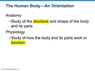
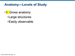
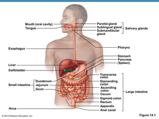
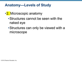
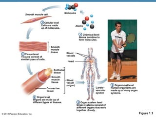
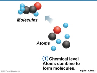
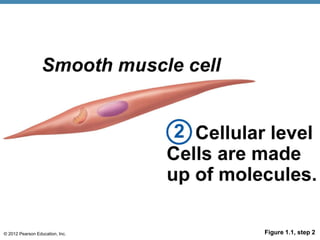
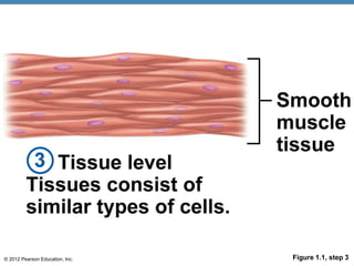
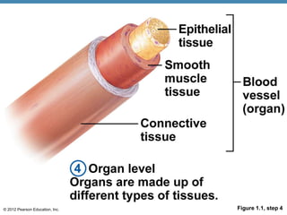
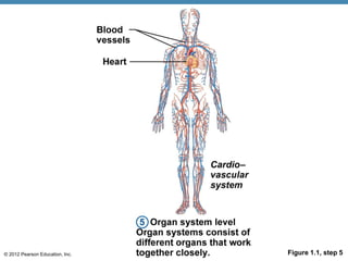
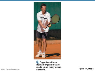
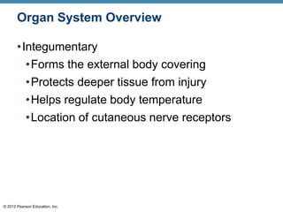
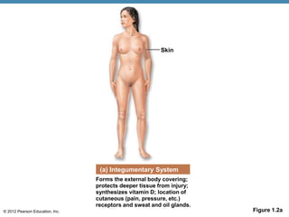
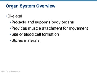
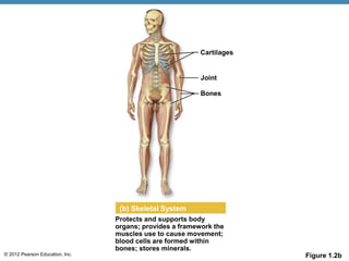
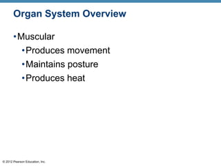
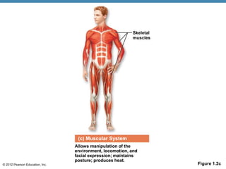
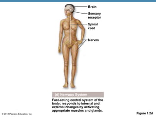
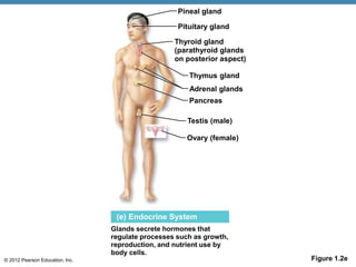
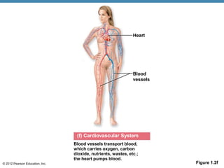
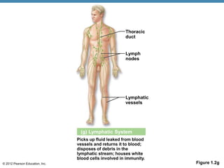
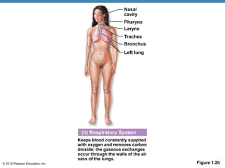
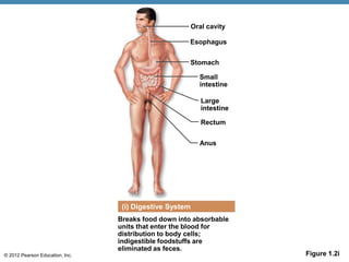
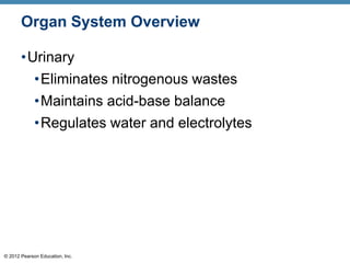
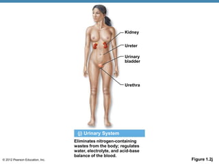
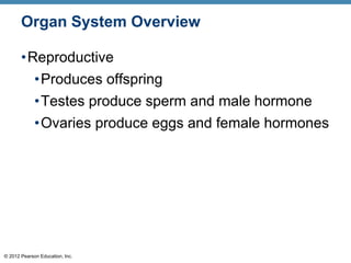
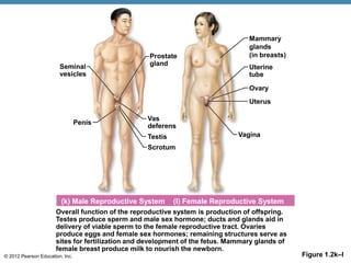
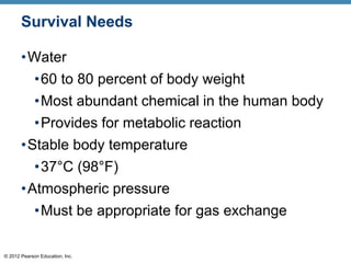
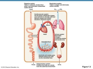
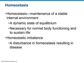
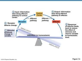
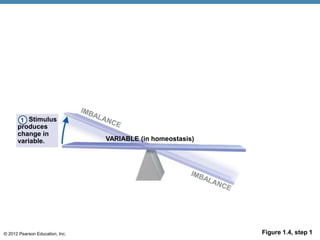
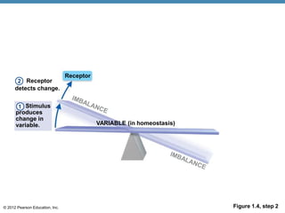
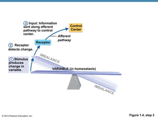
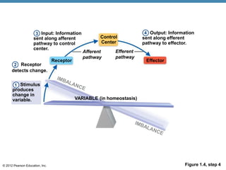
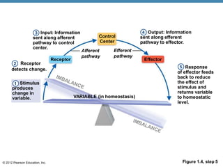
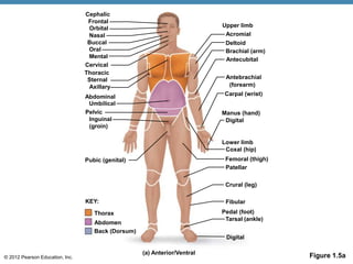
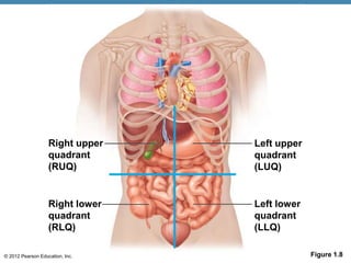
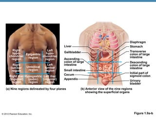
© 2012 Pearson Education, Inc.
PowerPoint® Lecture Slides
Prepared by Patty Bostwick-Taylor,
Florence-Darlington Technical College
C H A P T E R 1
The Human Body:
An Orientation
© 2012 Pearson Education, Inc.
The Human Body—An Orientation
Anatomy
•Study of the structure and shape of the body
and its parts
Physiology
•Study of how the body and its parts work or
function
© 2012 Pearson Education, Inc.
Anatomy—Levels of Study
•1. Gross anatomy
•Large structures
•Easily observable
© 2012 Pearson Education, Inc. Figure 14.1
Mouth (oral cavity)
Tongue
Esophagus
Liver
Gallbladder
Small intestine
Duodenum
Jejunum
lleum
Anus
Parotid gland
Salivary glands
Sublingual gland
Submandibular
gland
Pharynx
Stomach
Pancreas
(Spleen)
Large intestine
Descending
colon
Cecum
Sigmoid colon
Rectum
Appendix
Anal canal
Transverse
colon
Ascending
colon
© 2012 Pearson Education, Inc.
Anatomy—Levels of Study
•2. Microscopic anatomy
•Structures cannot be seen with the
naked eye
•Structures can only be viewed with a
microscope
© 2012 Pearson Education, Inc. Figure 14.4c
Pyloric
sphincter
Gastric pits
Surface
epithelium
Mucous
neck cells
Parietal cells
Gastric
glands
Chief cells
Gastric
pit
Gastric
gland
(c)
© 2012 Pearson Education, Inc. Figure 14.4d
Pepsinogen Pepsin
HCl
Parietal cells
Chief cells
Enteroendocrine
cell
(d)
© 2012 Pearson Education, Inc. Figure 1.1
Organismal level
Human organisms are
made up of many organ
systems.
Organ system level
Organ systems consist of
different organs that work
together closely.
Organ level
Organs are made up of
different types of tissues.
Chemical level
Atoms combine to
form molecules.
Smooth muscle cell
Cellular level
Cells are made
up of molecules.
Molecules
2
Atoms
1
Tissue level
Tissues consist of
similar types of cells.
3
Smooth
muscle
tissue
Epithelial
tissue
Smooth
muscle
tissue
Connective
tissue
Blood
vessel
(organ)
4
5
Cardio–
vascular
system
6
Blood
vessels
Heart
© 2012 Pearson Education, Inc. Figure 1.1, step 1
© 2012 Pearson Education, Inc. Figure 1.1, step 2
© 2012 Pearson Education, Inc. Figure 1.1, step 3
© 2012 Pearson Education, Inc. Figure 1.1, step 4
© 2012 Pearson Education, Inc. Figure 1.1, step 5
© 2012 Pearson Education, Inc. Figure 1.1, step 6
© 2012 Pearson Education, Inc.
Organ System Overview
•Integumentary
•Forms the external body covering
•Protects deeper tissue from injury
•Helps regulate body temperature
•Location of cutaneous nerve receptors
© 2012 Pearson Education, Inc. Figure 1.2a
Skin
(a) Integumentary System
Forms the external body covering;
protects deeper tissue from injury;
synthesizes vitamin D; location of
cutaneous (pain, pressure, etc.)
receptors and sweat and oil glands.
© 2012 Pearson Education, Inc.
Organ System Overview
•Skeletal
•Protects and supports body organs
•Provides muscle attachment for movement
•Site of blood cell formation
•Stores minerals
© 2012 Pearson Education, Inc. Figure 1.2b
(b) Skeletal System
Protects and supports body
organs; provides a framework the
muscles use to cause movement;
blood cells are formed within
bones; stores minerals.
Cartilages
Joint
Bones
© 2012 Pearson Education, Inc.
Organ System Overview
•Muscular
•Produces movement
•Maintains posture
•Produces heat
© 2012 Pearson Education, Inc. Figure 1.2c
(c) Muscular System
Allows manipulation of the
environment, locomotion, and
facial expression; maintains
posture; produces heat.
Skeletal
muscles
© 2012 Pearson Education, Inc.
Organ System Overview
•Nervous
•Fast-acting control system
•Responds to internal and external change
•Activates muscles and glands
© 2012 Pearson Education, Inc. Figure 1.2d
(d) Nervous System
Fast-acting control system of the
body; responds to internal and
external changes by activating
appropriate muscles and glands.
Brain
Sensory
receptor
Spinal
cord
Nerves
© 2012 Pearson Education, Inc.
Organ System Overview
•Endocrine
•Secretes regulatory hormones
•Growth
•Reproduction
•Metabolism
© 2012 Pearson Education, Inc. Figure 1.2e
(e) Endocrine System
Glands secrete hormones that
regulate processes such as growth,
reproduction, and nutrient use by
body cells.
Ovary (female)
Testis (male)
Pancreas
Adrenal glands
Thymus gland
Thyroid gland
(parathyroid glands
on posterior aspect)
Pituitary gland
Pineal gland
© 2012 Pearson Education, Inc.
Organ System Overview
•Cardiovascular
•Transports materials in body via blood
pumped by heart
•Oxygen
•Carbon dioxide
•Nutrients
•Wastes
© 2012 Pearson Education, Inc. Figure 1.2f
(f) Cardiovascular System
Heart
Blood vessels transport blood,
which carries oxygen, carbon
dioxide, nutrients, wastes, etc.;
the heart pumps blood.
Blood
vessels
© 2012 Pearson Education, Inc.
Organ System Overview
•Lymphatic
•Returns fluids to blood vessels
•Cleanses the blood
•Involved in immunity
© 2012 Pearson Education, Inc. Figure 1.2g
Picks up fluid leaked from blood
vessels and returns it to blood;
disposes of debris in the
lymphatic stream; houses white
blood cells involved in immunity.
Lymph
nodes
(g) Lymphatic System
Lymphatic
vessels
Thoracic
duct
© 2012 Pearson Education, Inc.
Organ System Overview
•Respiratory
•Keeps blood supplied with oxygen
•Removes carbon dioxide
© 2012 Pearson Education, Inc. Figure 1.2h
Keeps blood constantly supplied
with oxygen and removes carbon
dioxide; the gaseous exchanges
occur through the walls of the air
sacs of the lungs.
Pharynx
(h) Respiratory System
Nasal
cavity
Larynx
Trachea
Bronchus
Left lung
© 2012 Pearson Education, Inc.
Organ System Overview
•Digestive
•Breaks down food
•Allows for nutrient absorption into blood
•Eliminates indigestible material as feces
© 2012 Pearson Education, Inc. Figure 1.2i
Breaks food down into absorbable
units that enter the blood for
distribution to body cells;
indigestible foodstuffs are
eliminated as feces.
Oral cavity
(i) Digestive System
Esophagus
Stomach
Small
intestine
Anus
Rectum
Large
intestine
© 2012 Pearson Education, Inc.
Organ System Overview
•Urinary
•Eliminates nitrogenous wastes
•Maintains acid-base balance
•Regulates water and electrolytes
© 2012 Pearson Education, Inc. Figure 1.2j
Eliminates nitrogen-containing
wastes from the body; regulates
water, electrolyte, and acid-base
balance of the blood.
(j) Urinary System
Kidney
Ureter
Urinary
bladder
Urethra
© 2012 Pearson Education, Inc.
•Reproductive
•Produces offspring
•Testes produce sperm and male hormone
•Ovaries produce eggs and female hormones
Organ System Overview
© 2012 Pearson Education, Inc.
Overall function of the reproductive system is production of offspring.
Testes produce sperm and male sex hormone; ducts and glands aid in
delivery of viable sperm to the female reproductive tract. Ovaries
produce eggs and female sex hormones; remaining structures serve as
sites for fertilization and development of the fetus. Mammary glands of
female breast produce milk to nourish the newborn.
(k) Male Reproductive System (l) Female Reproductive System
Prostate
gland
Penis
Seminal
vesicles
Vas
deferens
Testis
Scrotum
Vagina
Ovary
Uterine
tube
Mammary
glands
(in breasts)
Uterus
Figure 1.2k–l
© 2012 Pearson Education, Inc.
Necessary Life Functions
•Maintain boundaries
•Movement
•Locomotion
•Movement of substances
•Responsiveness
•Ability to sense changes and react
•Digestion
•Breakdown and absorption of nutrients
© 2012 Pearson Education, Inc.
Necessary Life Functions
•Metabolism—chemical reactions within the
body
•Break down complex molecules into smaller
ones
•Build larger molecules from smaller ones
•Produces energy
•Regulated by hormones
•Excretion
•Eliminates waste from metabolic reactions
•Wastes may be removed in urine or feces
© 2012 Pearson Education, Inc.
Necessary Life Functions
•Reproduction
•Occurs on cellular level or organismal level
•Produces future generation
•Growth
•Increases cell size and number of cells
© 2012 Pearson Education, Inc.
Survival Needs
•Nutrients
•Chemicals for energy and cell building
•Includes carbohydrates, proteins, lipids,
vitamins, and minerals
•Oxygen
•Required for chemical reactions
© 2012 Pearson Education, Inc.
Survival Needs
•Water
•60 to 80 percent of body weight
•Most abundant chemical in the human body
•Provides for metabolic reaction
•Stable body temperature
•37°C (98°F)
•Atmospheric pressure
•Must be appropriate for gas exchange
© 2012 Pearson Education, Inc.
Nutrients
Heart
Interstitial fluid
Blood
Food
Digestive system
Takes in nutrients, breaks them
down, and eliminates unabsorbed
matter (feces)
Respiratory system
Takes in oxygen and eliminates
carbon dioxide
Cardiovascular system
Via the blood, distributes oxygen
and nutrients to all body cells and
delivers wastes and carbon
dioxide to disposal organs
Urinary system
Eliminates
nitrogen-
containing wastes
and excess ions
Feces Urine
Nutrients and wastes pass
between blood and cells
via the interstitial fluid
O2
CO2
Integumentary system
Protects the body as a whole
from the external environment
CO2
O2
Figure 1.3
© 2012 Pearson Education, Inc.
Homeostasis
•Homeostasis—maintenance of a stable
internal environment
•A dynamic state of equilibrium
•Necessary for normal body functioning and
to sustain life
•Homeostatic imbalance
•A disturbance in homeostasis resulting in
disease
© 2012 Pearson Education, Inc. Figure 1.4
Input: Information
sent along afferent
pathway to control
center.
Receptor
Control
Center
Effector
Receptor
detects change.
Afferent
pathway
Efferent
pathway
VARIABLE (in homeostasis)
Stimulus
produces
change in
variable.
Output: Information
sent along efferent
pathway to effector.
Response
of effector feeds
back to reduce
the effect of
stimulus and
returns variable
to homeostatic
level.
1
2
3 4
5
© 2012 Pearson Education, Inc. Figure 1.4, step 1
VARIABLE (in homeostasis)
Stimulus
produces
change in
variable.
1
© 2012 Pearson Education, Inc. Figure 1.4, step 2
Receptor
Receptor
detects change.
Stimulus
produces
change in
variable.
1
2
VARIABLE (in homeostasis)
© 2012 Pearson Education, Inc. Figure 1.4, step 3
Input: Information
sent along afferent
pathway to control
center.
Receptor
Control
Center
Receptor
detects change.
Afferent
pathway
VARIABLE (in homeostasis)
Stimulus
produces
change in
variable.
1
3
2
© 2012 Pearson Education, Inc. Figure 1.4, step 4
Input: Information
sent along afferent
pathway to control
center.
Receptor
Control
Center
Effector
Receptor
detects change.
Afferent
pathway
Efferent
pathway
VARIABLE (in homeostasis)
Stimulus
produces
change in
variable.
Output: Information
sent along efferent
pathway to effector.
1
3 4
2
© 2012 Pearson Education, Inc. Figure 1.4, step 5
Input: Information
sent along afferent
pathway to control
center.
Receptor
Control
Center
Effector
Receptor
detects change.
Afferent
pathway
Efferent
pathway
VARIABLE (in homeostasis)
Stimulus
produces
change in
variable.
Output: Information
sent along efferent
pathway to effector.
Response
of effector feeds
back to reduce
the effect of
stimulus and
returns variable
to homeostatic
level.
1
3 4
5
2
© 2012 Pearson Education, Inc.
Maintaining Homeostasis
•The body communicates through neural and
hormonal control systems
•Receptor
•Responds to changes in the environment
(stimuli)
•Sends information to control center
© 2012 Pearson Education, Inc.
Maintaining Homeostasis
•Control center
•Determines set point
•Analyzes information
•Determines appropriate response
•Effector
•Provides a means for response to the
stimulus
© 2012 Pearson Education, Inc.
Feedback Mechanisms
•Negative feedback
•Includes most homeostatic control
mechanisms
•Shuts off the original stimulus, or reduces its
intensity
•Works like a household thermostat
© 2012 Pearson Education, Inc.
Feedback Mechanisms
•Positive feedback
•Increases the original stimulus to push the
variable farther
•In the body this only occurs in blood clotting
and during the birth of a baby
© 2012 Pearson Education, Inc.
The Language of Anatomy
•Special terminology is used to prevent
misunderstanding
•Exact terms are used for
•Position
•Direction
•Regions
•Structures
© 2012 Pearson Education, Inc.
Regional Terms
•Anterior body landmarks
© 2012 Pearson Education, Inc.
Cervical
Umbilical
Pubic (genital)
Pelvic
Inguinal
(groin)
(a) Anterior/Ventral
KEY:
Deltoid
Digital
Lower limb
Coxal (hip)
Patellar
Fibular
Pedal (foot)
Digital
Upper limb
Acromial
Brachial (arm)
Antecubital
Antebrachial
Carpal (wrist)
Manus (hand)
Crural (leg)
Tarsal (ankle)
Femoral (thigh)
Cephalic
Frontal
Orbital
Nasal
Buccal
Oral
Mental
Thorax
Abdomen
Back (Dorsum)
Thoracic
Sternal
Axillary
Abdominal
(forearm)
Figure 1.5a
© 2012 Pearson Education, Inc.
Regional Terms
•Posterior body landmarks
© 2012 Pearson Education, Inc. Figure 1.5b
(b) Posterior/Dorsal
Gluteal
Sacral
Lumbar
Vertebral
Back (dorsal)
Scapular
Cervical
Cephalic
Occipital (back
of head)
Acromial
Brachial (arm)
Olecranal
Antebrachial
Manus (hand)
Digital
Femoral (thigh)
Popliteal
Sural (calf)
Fibular
Pedal (foot)
Calcaneal
Plantar
Upper limb
KEY:
Thorax
Abdomen
Back (Dorsum)
(forearm)
© 2012 Pearson Education, Inc.
Directional Terms
•Superior (cranial or cephalad): toward the
head end or upper part of a structure or the
body; above
•Inferior (caudal): away from the head end or
toward the lower part of a structure or the
body; below
© 2012 Pearson Education, Inc. Table 1.1
© 2012 Pearson Education, Inc.
Directional Terms
•Ventral (anterior): toward or at the front of
the body; in front of
•Dorsal (posterior): toward or at the backside
of the body; behind
© 2012 Pearson Education, Inc. Table 1.1
© 2012 Pearson Education, Inc.
Directional Terms
•Medial: toward or at the midline of the body;
on the inner side of
•Lateral: away from the midline of the body;
on the outer side of
•Intermediate: between a more medial and a
more lateral structure
© 2012 Pearson Education, Inc. Table 1.1
© 2012 Pearson Education, Inc.
Directional Terms
•Proximal: close to the origin of the body
part or point of attachment to a limb to the
body trunk
•Distal: farther from the origin of a body part
or the point of attachment of a limb to the
body trunk
© 2012 Pearson Education, Inc. Table 1.1
© 2012 Pearson Education, Inc.
Directional Terms
•Superficial: toward or at the body surface
•Deep: away from the body surface; more
internal
© 2012 Pearson Education, Inc. Table 1.1
© 2012 Pearson Education, Inc.
Body Planes and Sections
•A sagittal section divides the body (or organ)
into left and right parts.
•A median, or midsagittal, section divides the
body (or organ) into equal left and right parts.
•A frontal, or coronal, section divides the body
(or organ) into anterior and posterior parts.
•A transverse, or cross, section divides the
body (or organ) into superior and inferior parts.
© 2012 Pearson Education, Inc.
(a) Median (midsagittal)
Vertebral
column
(b) Frontal (coronal) plane
Right
lung Heart
Left
lung
(c) Transverse plane
Liver Aorta Spleen
Spinal
cord
Rectum Intestines Liver Stomach Spleen Subcutaneous
fat layer
Stomach
Figure 1.6
© 2012 Pearson Education, Inc.
Body Cavities
•Dorsal body cavity
•Cranial cavity houses the brain
•Spinal cavity houses the spinal cord
•Ventral body cavity
•Thoracic cavity houses heart, lungs, and
others
•Abdominopelvic cavity houses digestive
system and most urinary system organs
© 2012 Pearson Education, Inc.
Cranial
cavity
Spinal
cavity
Thoracic
cavity
Diaphragm
Abdominal
cavity
Pelvic
cavity
Abdominopelvic
cavity
KEY:
Dorsal body cavity Ventral body cavity
Figure 1.7
© 2012 Pearson Education, Inc.
Right upper
quadrant
(RUQ)
Right lower
quadrant
(RLQ)
Left upper
quadrant
(LUQ)
Left lower
quadrant
(LLQ)
Figure 1.8
© 2012 Pearson Education, Inc. Figure 1.9a-b
Right
hypo-
chondriac
region
Epigastric
region
Right
lumbar
region
Umbilical
region
Right iliac
(inguinal)
region
Hypogastric
(pubic)
region
Left iliac
(inguinal)
region
Left
lumbar
region
Left
hypo-
chondriac
region
(a) Nine regions delineated by four planes (b) Anterior view of the nine regions
showing the superficial organs
Liver
Gallbladder
Ascending
colon of large
intestine
Small intestine
Cecum
Appendix
Diaphragm
Stomach
Transverse
colon of large
intestine
Descending
colon of large
intestine
Initial part of
sigmoid colon
Urinary
bladder

ch_01-the-human-body-organization (1).ppt

  • 1.
    © 2012 PearsonEducation, Inc. PowerPoint® Lecture Slides Prepared by Patty Bostwick-Taylor, Florence-Darlington Technical College C H A P T E R 1 The Human Body: An Orientation
  • 2.
    © 2012 PearsonEducation, Inc. The Human Body—An Orientation Anatomy •Study of the structure and shape of the body and its parts Physiology •Study of how the body and its parts work or function
  • 3.
    © 2012 PearsonEducation, Inc. Anatomy—Levels of Study •1. Gross anatomy •Large structures •Easily observable
  • 4.
    © 2012 PearsonEducation, Inc. Figure 14.1 Mouth (oral cavity) Tongue Esophagus Liver Gallbladder Small intestine Duodenum Jejunum lleum Anus Parotid gland Salivary glands Sublingual gland Submandibular gland Pharynx Stomach Pancreas (Spleen) Large intestine Descending colon Cecum Sigmoid colon Rectum Appendix Anal canal Transverse colon Ascending colon
  • 5.
    © 2012 PearsonEducation, Inc. Anatomy—Levels of Study •2. Microscopic anatomy •Structures cannot be seen with the naked eye •Structures can only be viewed with a microscope
  • 6.
    © 2012 PearsonEducation, Inc. Figure 14.4c Pyloric sphincter Gastric pits Surface epithelium Mucous neck cells Parietal cells Gastric glands Chief cells Gastric pit Gastric gland (c)
  • 7.
    © 2012 PearsonEducation, Inc. Figure 14.4d Pepsinogen Pepsin HCl Parietal cells Chief cells Enteroendocrine cell (d)
  • 8.
    © 2012 PearsonEducation, Inc. Figure 1.1 Organismal level Human organisms are made up of many organ systems. Organ system level Organ systems consist of different organs that work together closely. Organ level Organs are made up of different types of tissues. Chemical level Atoms combine to form molecules. Smooth muscle cell Cellular level Cells are made up of molecules. Molecules 2 Atoms 1 Tissue level Tissues consist of similar types of cells. 3 Smooth muscle tissue Epithelial tissue Smooth muscle tissue Connective tissue Blood vessel (organ) 4 5 Cardio– vascular system 6 Blood vessels Heart
  • 9.
    © 2012 PearsonEducation, Inc. Figure 1.1, step 1
  • 10.
    © 2012 PearsonEducation, Inc. Figure 1.1, step 2
  • 11.
    © 2012 PearsonEducation, Inc. Figure 1.1, step 3
  • 12.
    © 2012 PearsonEducation, Inc. Figure 1.1, step 4
  • 13.
    © 2012 PearsonEducation, Inc. Figure 1.1, step 5
  • 14.
    © 2012 PearsonEducation, Inc. Figure 1.1, step 6
  • 15.
    © 2012 PearsonEducation, Inc. Organ System Overview •Integumentary •Forms the external body covering •Protects deeper tissue from injury •Helps regulate body temperature •Location of cutaneous nerve receptors
  • 16.
    © 2012 PearsonEducation, Inc. Figure 1.2a Skin (a) Integumentary System Forms the external body covering; protects deeper tissue from injury; synthesizes vitamin D; location of cutaneous (pain, pressure, etc.) receptors and sweat and oil glands.
  • 17.
    © 2012 PearsonEducation, Inc. Organ System Overview •Skeletal •Protects and supports body organs •Provides muscle attachment for movement •Site of blood cell formation •Stores minerals
  • 18.
    © 2012 PearsonEducation, Inc. Figure 1.2b (b) Skeletal System Protects and supports body organs; provides a framework the muscles use to cause movement; blood cells are formed within bones; stores minerals. Cartilages Joint Bones
  • 19.
    © 2012 PearsonEducation, Inc. Organ System Overview •Muscular •Produces movement •Maintains posture •Produces heat
  • 20.
    © 2012 PearsonEducation, Inc. Figure 1.2c (c) Muscular System Allows manipulation of the environment, locomotion, and facial expression; maintains posture; produces heat. Skeletal muscles
  • 21.
    © 2012 PearsonEducation, Inc. Organ System Overview •Nervous •Fast-acting control system •Responds to internal and external change •Activates muscles and glands
  • 22.
    © 2012 PearsonEducation, Inc. Figure 1.2d (d) Nervous System Fast-acting control system of the body; responds to internal and external changes by activating appropriate muscles and glands. Brain Sensory receptor Spinal cord Nerves
  • 23.
    © 2012 PearsonEducation, Inc. Organ System Overview •Endocrine •Secretes regulatory hormones •Growth •Reproduction •Metabolism
  • 24.
    © 2012 PearsonEducation, Inc. Figure 1.2e (e) Endocrine System Glands secrete hormones that regulate processes such as growth, reproduction, and nutrient use by body cells. Ovary (female) Testis (male) Pancreas Adrenal glands Thymus gland Thyroid gland (parathyroid glands on posterior aspect) Pituitary gland Pineal gland
  • 25.
    © 2012 PearsonEducation, Inc. Organ System Overview •Cardiovascular •Transports materials in body via blood pumped by heart •Oxygen •Carbon dioxide •Nutrients •Wastes
  • 26.
    © 2012 PearsonEducation, Inc. Figure 1.2f (f) Cardiovascular System Heart Blood vessels transport blood, which carries oxygen, carbon dioxide, nutrients, wastes, etc.; the heart pumps blood. Blood vessels
  • 27.
    © 2012 PearsonEducation, Inc. Organ System Overview •Lymphatic •Returns fluids to blood vessels •Cleanses the blood •Involved in immunity
  • 28.
    © 2012 PearsonEducation, Inc. Figure 1.2g Picks up fluid leaked from blood vessels and returns it to blood; disposes of debris in the lymphatic stream; houses white blood cells involved in immunity. Lymph nodes (g) Lymphatic System Lymphatic vessels Thoracic duct
  • 29.
    © 2012 PearsonEducation, Inc. Organ System Overview •Respiratory •Keeps blood supplied with oxygen •Removes carbon dioxide
  • 30.
    © 2012 PearsonEducation, Inc. Figure 1.2h Keeps blood constantly supplied with oxygen and removes carbon dioxide; the gaseous exchanges occur through the walls of the air sacs of the lungs. Pharynx (h) Respiratory System Nasal cavity Larynx Trachea Bronchus Left lung
  • 31.
    © 2012 PearsonEducation, Inc. Organ System Overview •Digestive •Breaks down food •Allows for nutrient absorption into blood •Eliminates indigestible material as feces
  • 32.
    © 2012 PearsonEducation, Inc. Figure 1.2i Breaks food down into absorbable units that enter the blood for distribution to body cells; indigestible foodstuffs are eliminated as feces. Oral cavity (i) Digestive System Esophagus Stomach Small intestine Anus Rectum Large intestine
  • 33.
    © 2012 PearsonEducation, Inc. Organ System Overview •Urinary •Eliminates nitrogenous wastes •Maintains acid-base balance •Regulates water and electrolytes
  • 34.
    © 2012 PearsonEducation, Inc. Figure 1.2j Eliminates nitrogen-containing wastes from the body; regulates water, electrolyte, and acid-base balance of the blood. (j) Urinary System Kidney Ureter Urinary bladder Urethra
  • 35.
    © 2012 PearsonEducation, Inc. •Reproductive •Produces offspring •Testes produce sperm and male hormone •Ovaries produce eggs and female hormones Organ System Overview
  • 36.
    © 2012 PearsonEducation, Inc. Overall function of the reproductive system is production of offspring. Testes produce sperm and male sex hormone; ducts and glands aid in delivery of viable sperm to the female reproductive tract. Ovaries produce eggs and female sex hormones; remaining structures serve as sites for fertilization and development of the fetus. Mammary glands of female breast produce milk to nourish the newborn. (k) Male Reproductive System (l) Female Reproductive System Prostate gland Penis Seminal vesicles Vas deferens Testis Scrotum Vagina Ovary Uterine tube Mammary glands (in breasts) Uterus Figure 1.2k–l
  • 37.
    © 2012 PearsonEducation, Inc. Necessary Life Functions •Maintain boundaries •Movement •Locomotion •Movement of substances •Responsiveness •Ability to sense changes and react •Digestion •Breakdown and absorption of nutrients
  • 38.
    © 2012 PearsonEducation, Inc. Necessary Life Functions •Metabolism—chemical reactions within the body •Break down complex molecules into smaller ones •Build larger molecules from smaller ones •Produces energy •Regulated by hormones •Excretion •Eliminates waste from metabolic reactions •Wastes may be removed in urine or feces
  • 39.
    © 2012 PearsonEducation, Inc. Necessary Life Functions •Reproduction •Occurs on cellular level or organismal level •Produces future generation •Growth •Increases cell size and number of cells
  • 40.
    © 2012 PearsonEducation, Inc. Survival Needs •Nutrients •Chemicals for energy and cell building •Includes carbohydrates, proteins, lipids, vitamins, and minerals •Oxygen •Required for chemical reactions
  • 41.
    © 2012 PearsonEducation, Inc. Survival Needs •Water •60 to 80 percent of body weight •Most abundant chemical in the human body •Provides for metabolic reaction •Stable body temperature •37°C (98°F) •Atmospheric pressure •Must be appropriate for gas exchange
  • 42.
    © 2012 PearsonEducation, Inc. Nutrients Heart Interstitial fluid Blood Food Digestive system Takes in nutrients, breaks them down, and eliminates unabsorbed matter (feces) Respiratory system Takes in oxygen and eliminates carbon dioxide Cardiovascular system Via the blood, distributes oxygen and nutrients to all body cells and delivers wastes and carbon dioxide to disposal organs Urinary system Eliminates nitrogen- containing wastes and excess ions Feces Urine Nutrients and wastes pass between blood and cells via the interstitial fluid O2 CO2 Integumentary system Protects the body as a whole from the external environment CO2 O2 Figure 1.3
  • 43.
    © 2012 PearsonEducation, Inc. Homeostasis •Homeostasis—maintenance of a stable internal environment •A dynamic state of equilibrium •Necessary for normal body functioning and to sustain life •Homeostatic imbalance •A disturbance in homeostasis resulting in disease
  • 44.
    © 2012 PearsonEducation, Inc. Figure 1.4 Input: Information sent along afferent pathway to control center. Receptor Control Center Effector Receptor detects change. Afferent pathway Efferent pathway VARIABLE (in homeostasis) Stimulus produces change in variable. Output: Information sent along efferent pathway to effector. Response of effector feeds back to reduce the effect of stimulus and returns variable to homeostatic level. 1 2 3 4 5
  • 45.
    © 2012 PearsonEducation, Inc. Figure 1.4, step 1 VARIABLE (in homeostasis) Stimulus produces change in variable. 1
  • 46.
    © 2012 PearsonEducation, Inc. Figure 1.4, step 2 Receptor Receptor detects change. Stimulus produces change in variable. 1 2 VARIABLE (in homeostasis)
  • 47.
    © 2012 PearsonEducation, Inc. Figure 1.4, step 3 Input: Information sent along afferent pathway to control center. Receptor Control Center Receptor detects change. Afferent pathway VARIABLE (in homeostasis) Stimulus produces change in variable. 1 3 2
  • 48.
    © 2012 PearsonEducation, Inc. Figure 1.4, step 4 Input: Information sent along afferent pathway to control center. Receptor Control Center Effector Receptor detects change. Afferent pathway Efferent pathway VARIABLE (in homeostasis) Stimulus produces change in variable. Output: Information sent along efferent pathway to effector. 1 3 4 2
  • 49.
    © 2012 PearsonEducation, Inc. Figure 1.4, step 5 Input: Information sent along afferent pathway to control center. Receptor Control Center Effector Receptor detects change. Afferent pathway Efferent pathway VARIABLE (in homeostasis) Stimulus produces change in variable. Output: Information sent along efferent pathway to effector. Response of effector feeds back to reduce the effect of stimulus and returns variable to homeostatic level. 1 3 4 5 2
  • 50.
    © 2012 PearsonEducation, Inc. Maintaining Homeostasis •The body communicates through neural and hormonal control systems •Receptor •Responds to changes in the environment (stimuli) •Sends information to control center
  • 51.
    © 2012 PearsonEducation, Inc. Maintaining Homeostasis •Control center •Determines set point •Analyzes information •Determines appropriate response •Effector •Provides a means for response to the stimulus
  • 52.
    © 2012 PearsonEducation, Inc. Feedback Mechanisms •Negative feedback •Includes most homeostatic control mechanisms •Shuts off the original stimulus, or reduces its intensity •Works like a household thermostat
  • 53.
    © 2012 PearsonEducation, Inc. Feedback Mechanisms •Positive feedback •Increases the original stimulus to push the variable farther •In the body this only occurs in blood clotting and during the birth of a baby
  • 54.
    © 2012 PearsonEducation, Inc. The Language of Anatomy •Special terminology is used to prevent misunderstanding •Exact terms are used for •Position •Direction •Regions •Structures
  • 55.
    © 2012 PearsonEducation, Inc. Regional Terms •Anterior body landmarks
  • 56.
    © 2012 PearsonEducation, Inc. Cervical Umbilical Pubic (genital) Pelvic Inguinal (groin) (a) Anterior/Ventral KEY: Deltoid Digital Lower limb Coxal (hip) Patellar Fibular Pedal (foot) Digital Upper limb Acromial Brachial (arm) Antecubital Antebrachial Carpal (wrist) Manus (hand) Crural (leg) Tarsal (ankle) Femoral (thigh) Cephalic Frontal Orbital Nasal Buccal Oral Mental Thorax Abdomen Back (Dorsum) Thoracic Sternal Axillary Abdominal (forearm) Figure 1.5a
  • 57.
    © 2012 PearsonEducation, Inc. Regional Terms •Posterior body landmarks
  • 58.
    © 2012 PearsonEducation, Inc. Figure 1.5b (b) Posterior/Dorsal Gluteal Sacral Lumbar Vertebral Back (dorsal) Scapular Cervical Cephalic Occipital (back of head) Acromial Brachial (arm) Olecranal Antebrachial Manus (hand) Digital Femoral (thigh) Popliteal Sural (calf) Fibular Pedal (foot) Calcaneal Plantar Upper limb KEY: Thorax Abdomen Back (Dorsum) (forearm)
  • 59.
    © 2012 PearsonEducation, Inc. Directional Terms •Superior (cranial or cephalad): toward the head end or upper part of a structure or the body; above •Inferior (caudal): away from the head end or toward the lower part of a structure or the body; below
  • 60.
    © 2012 PearsonEducation, Inc. Table 1.1
  • 61.
    © 2012 PearsonEducation, Inc. Directional Terms •Ventral (anterior): toward or at the front of the body; in front of •Dorsal (posterior): toward or at the backside of the body; behind
  • 62.
    © 2012 PearsonEducation, Inc. Table 1.1
  • 63.
    © 2012 PearsonEducation, Inc. Directional Terms •Medial: toward or at the midline of the body; on the inner side of •Lateral: away from the midline of the body; on the outer side of •Intermediate: between a more medial and a more lateral structure
  • 64.
    © 2012 PearsonEducation, Inc. Table 1.1
  • 65.
    © 2012 PearsonEducation, Inc. Directional Terms •Proximal: close to the origin of the body part or point of attachment to a limb to the body trunk •Distal: farther from the origin of a body part or the point of attachment of a limb to the body trunk
  • 66.
    © 2012 PearsonEducation, Inc. Table 1.1
  • 67.
    © 2012 PearsonEducation, Inc. Directional Terms •Superficial: toward or at the body surface •Deep: away from the body surface; more internal
  • 68.
    © 2012 PearsonEducation, Inc. Table 1.1
  • 69.
    © 2012 PearsonEducation, Inc. Body Planes and Sections •A sagittal section divides the body (or organ) into left and right parts. •A median, or midsagittal, section divides the body (or organ) into equal left and right parts. •A frontal, or coronal, section divides the body (or organ) into anterior and posterior parts. •A transverse, or cross, section divides the body (or organ) into superior and inferior parts.
  • 70.
    © 2012 PearsonEducation, Inc. (a) Median (midsagittal) Vertebral column (b) Frontal (coronal) plane Right lung Heart Left lung (c) Transverse plane Liver Aorta Spleen Spinal cord Rectum Intestines Liver Stomach Spleen Subcutaneous fat layer Stomach Figure 1.6
  • 71.
    © 2012 PearsonEducation, Inc. Body Cavities •Dorsal body cavity •Cranial cavity houses the brain •Spinal cavity houses the spinal cord •Ventral body cavity •Thoracic cavity houses heart, lungs, and others •Abdominopelvic cavity houses digestive system and most urinary system organs
  • 72.
    © 2012 PearsonEducation, Inc. Cranial cavity Spinal cavity Thoracic cavity Diaphragm Abdominal cavity Pelvic cavity Abdominopelvic cavity KEY: Dorsal body cavity Ventral body cavity Figure 1.7
  • 73.
    © 2012 PearsonEducation, Inc. Right upper quadrant (RUQ) Right lower quadrant (RLQ) Left upper quadrant (LUQ) Left lower quadrant (LLQ) Figure 1.8
  • 74.
    © 2012 PearsonEducation, Inc. Figure 1.9a-b Right hypo- chondriac region Epigastric region Right lumbar region Umbilical region Right iliac (inguinal) region Hypogastric (pubic) region Left iliac (inguinal) region Left lumbar region Left hypo- chondriac region (a) Nine regions delineated by four planes (b) Anterior view of the nine regions showing the superficial organs Liver Gallbladder Ascending colon of large intestine Small intestine Cecum Appendix Diaphragm Stomach Transverse colon of large intestine Descending colon of large intestine Initial part of sigmoid colon Urinary bladder