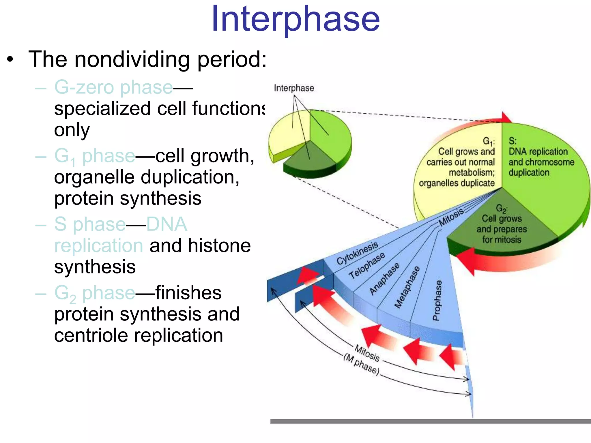
1. The cell cycle consists of interphase and mitosis. Interphase includes the G1, S, and G2 phases where the cell grows and duplicates its DNA.
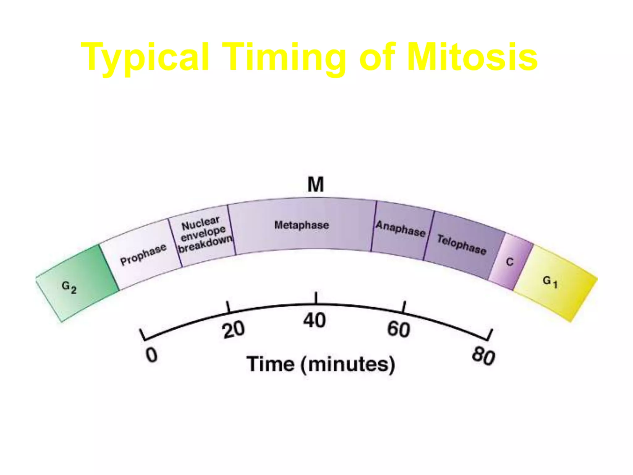
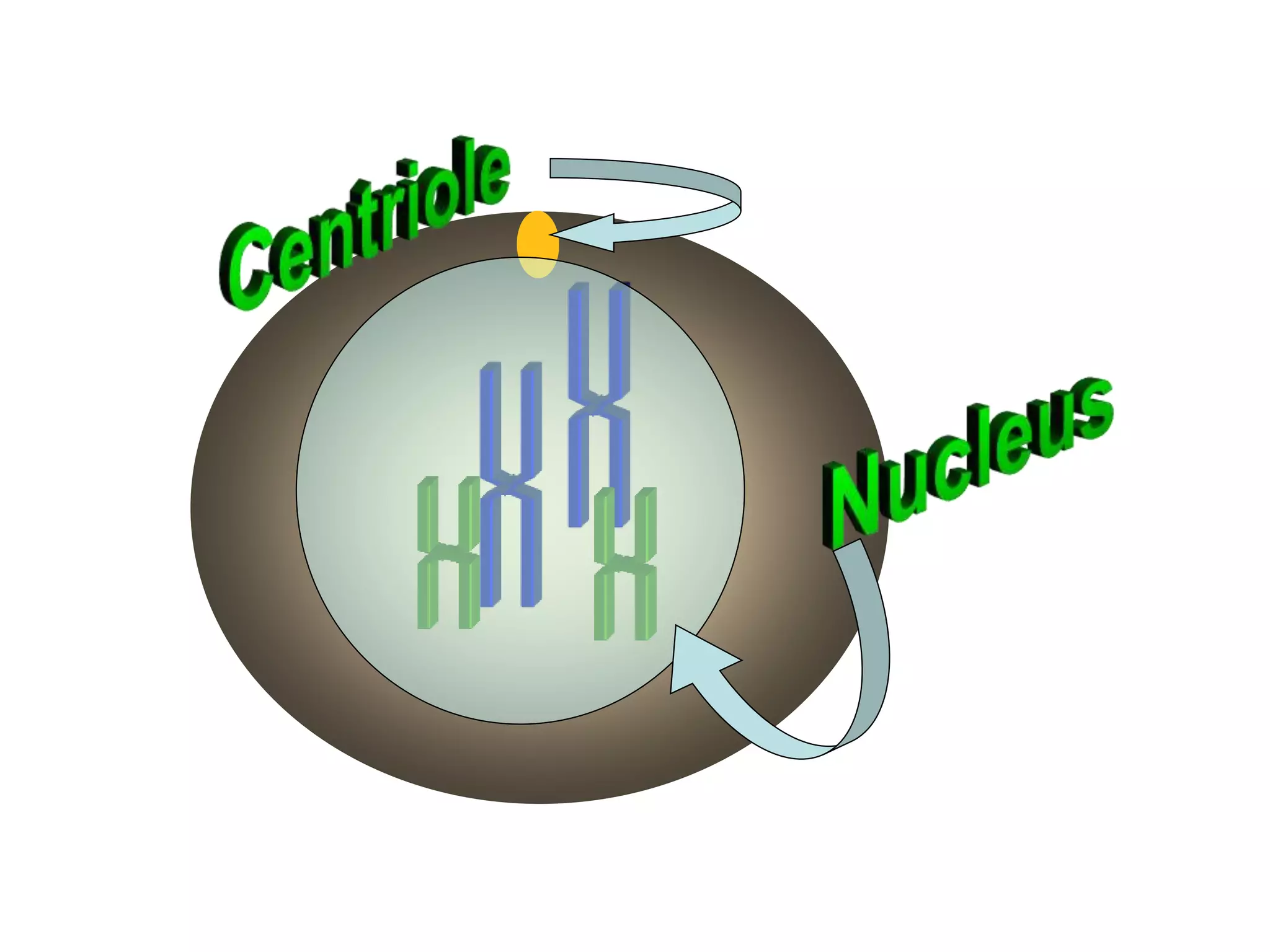
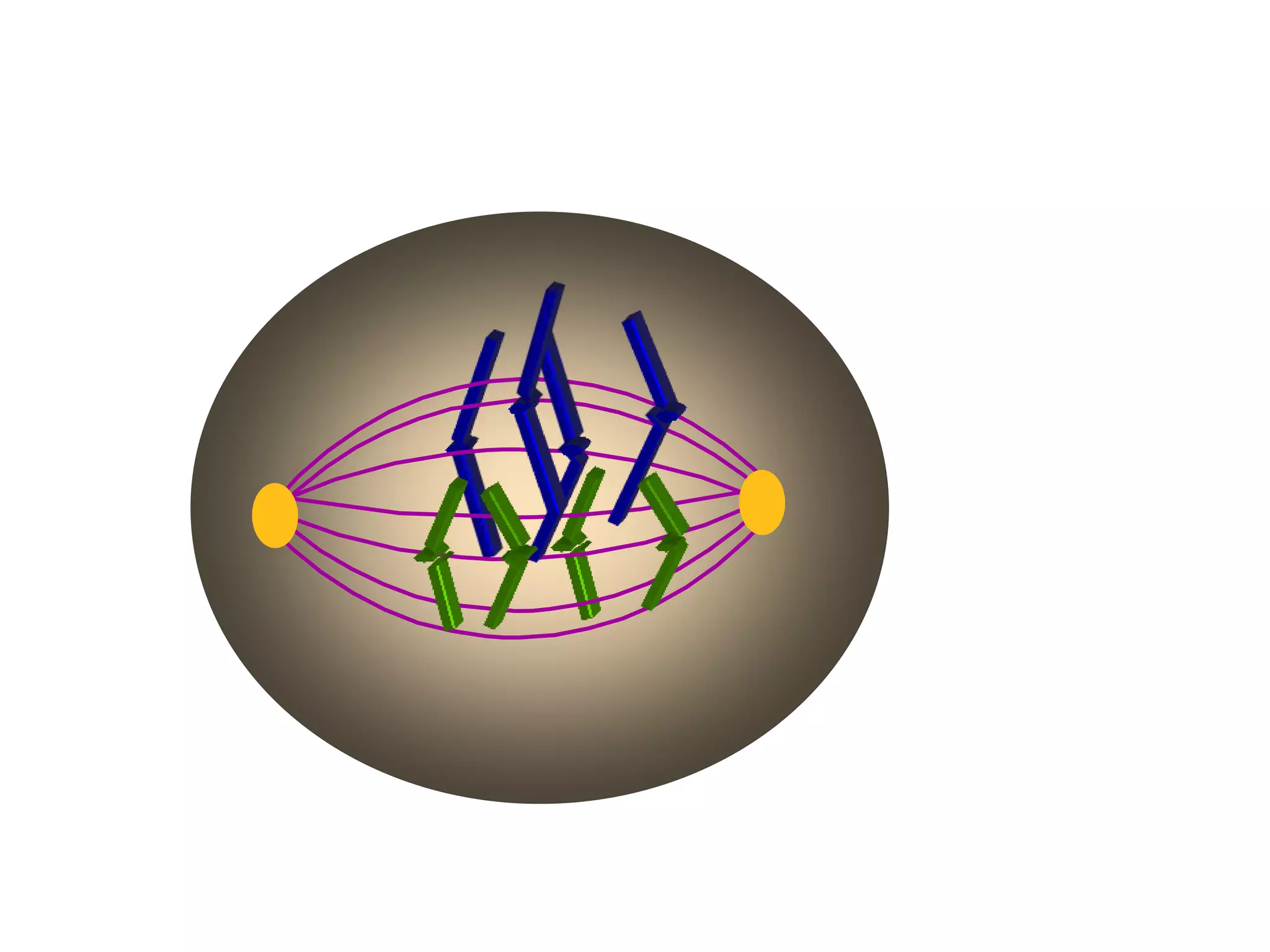
2. Mitosis is divided into prophase, metaphase, anaphase, and telophase where the duplicated chromosomes separate and the cell divides. Cytokinesis then divides the cytoplasm.
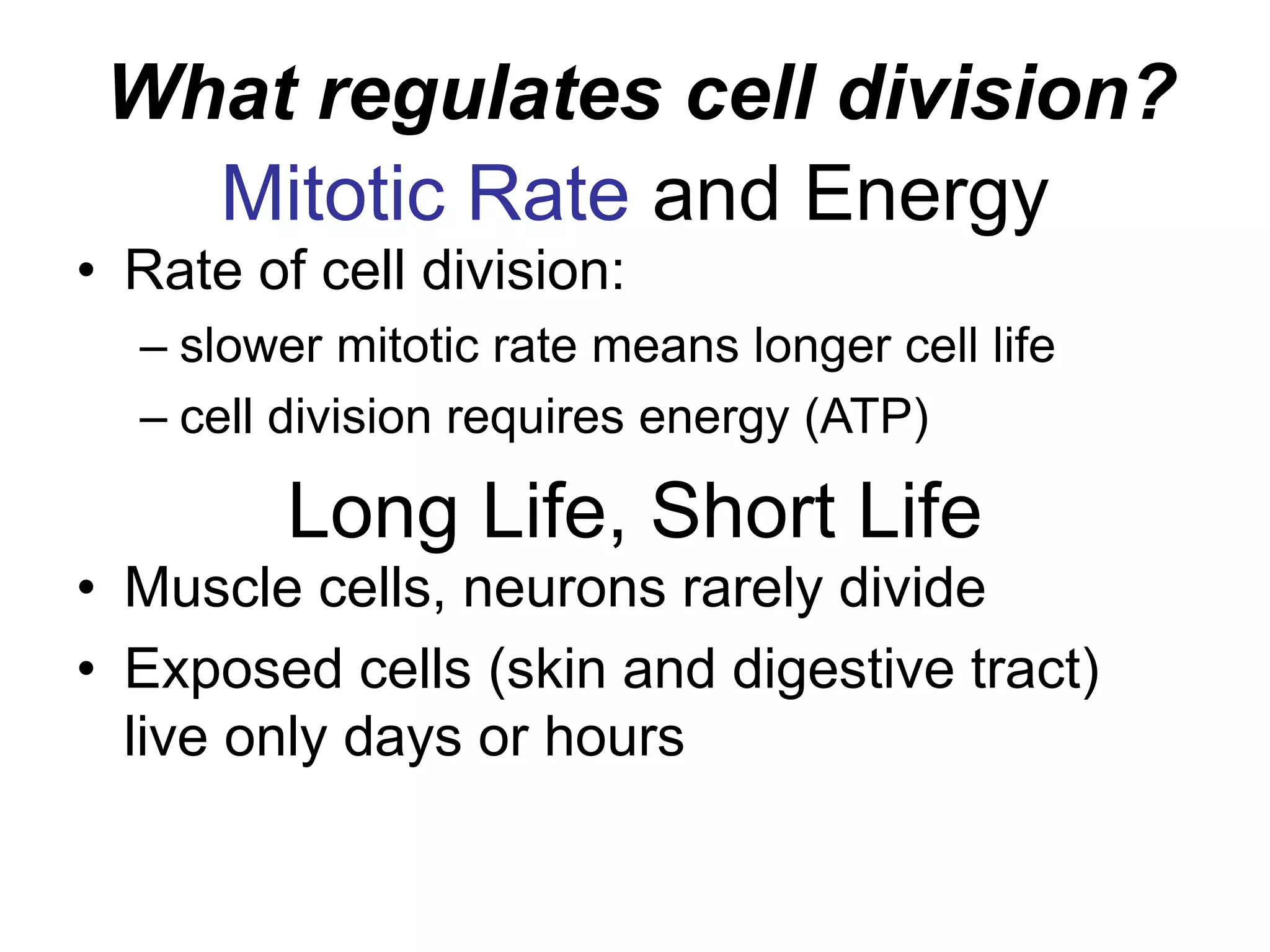
3. Cell division is regulated by both internal and external factors like maturation promoting factors and growth factors that increase division or repressor genes that decrease division. Abnormal cell regulation can lead to tumor formation.