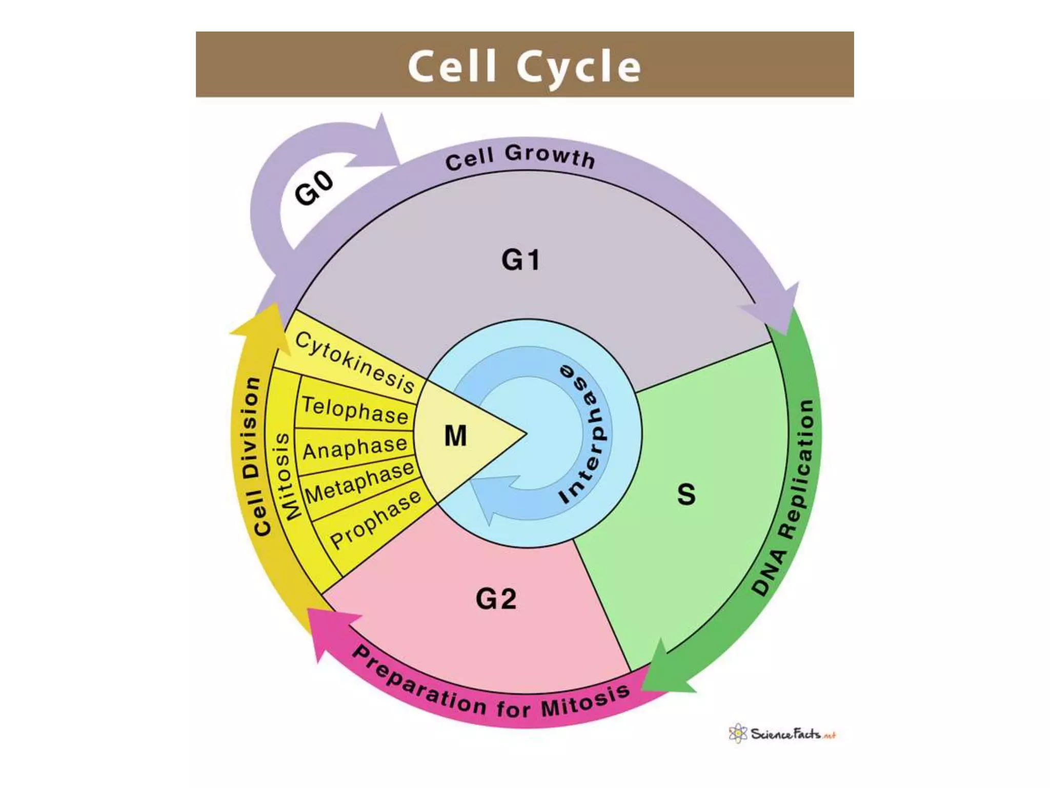
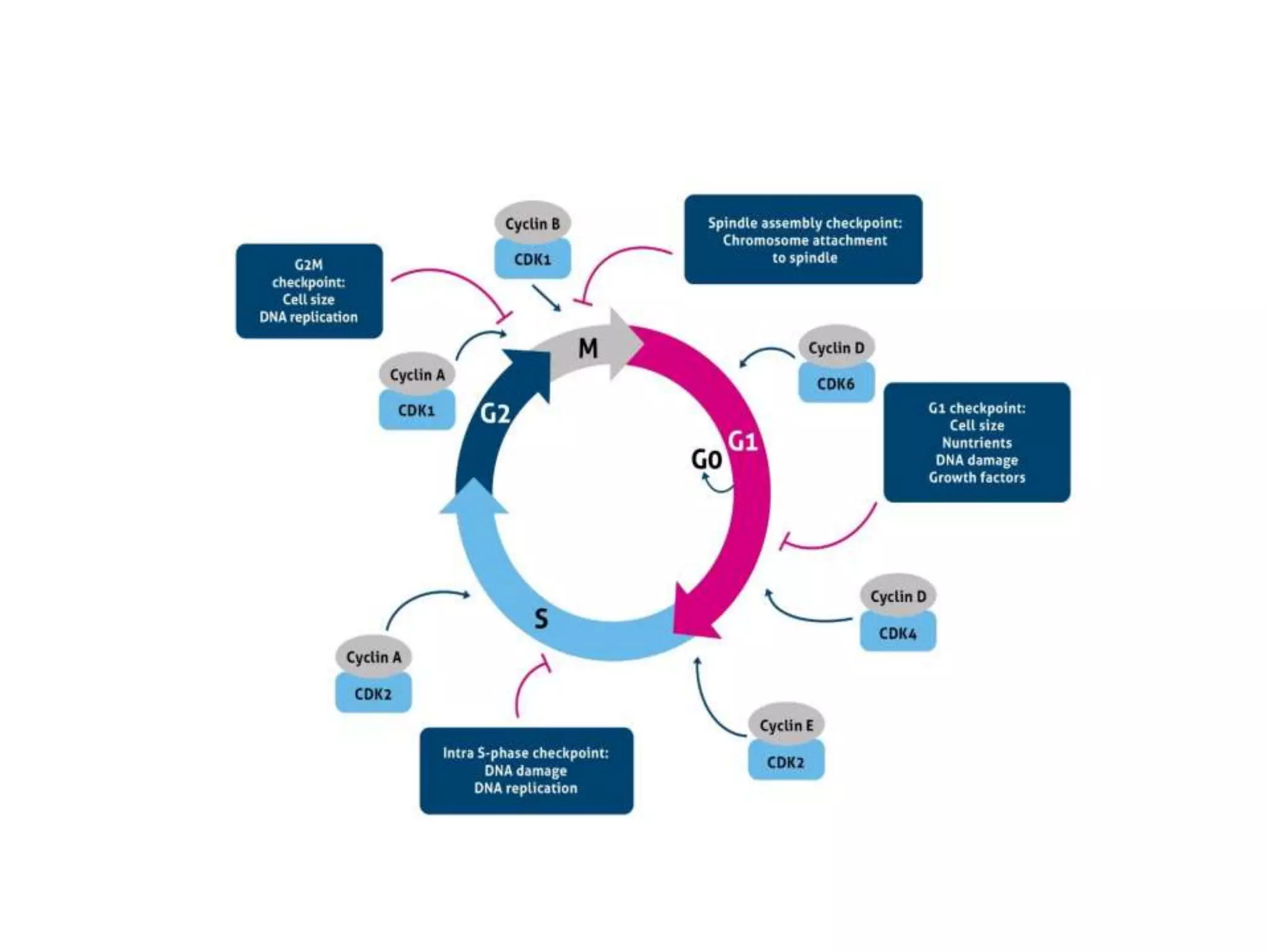
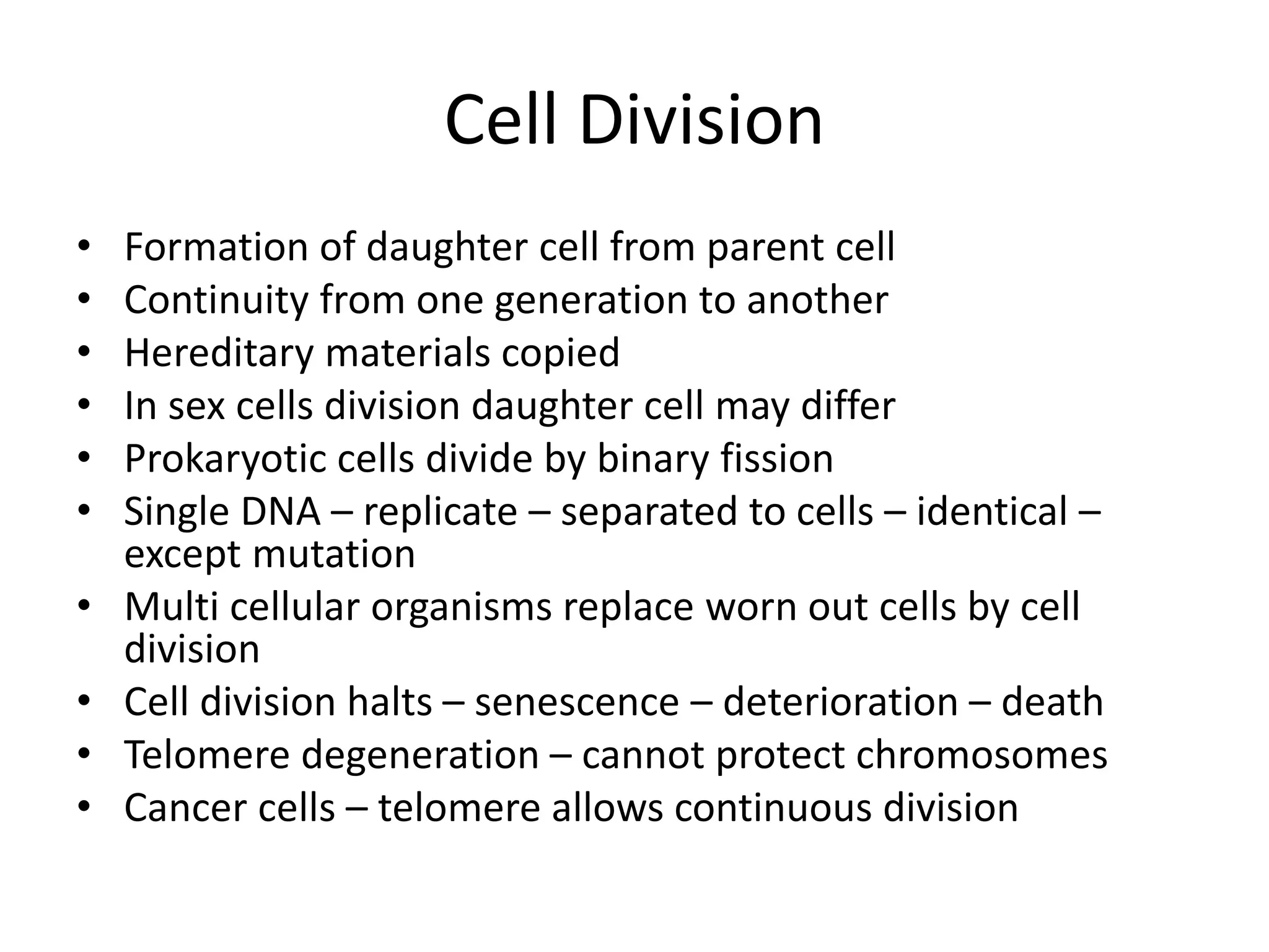
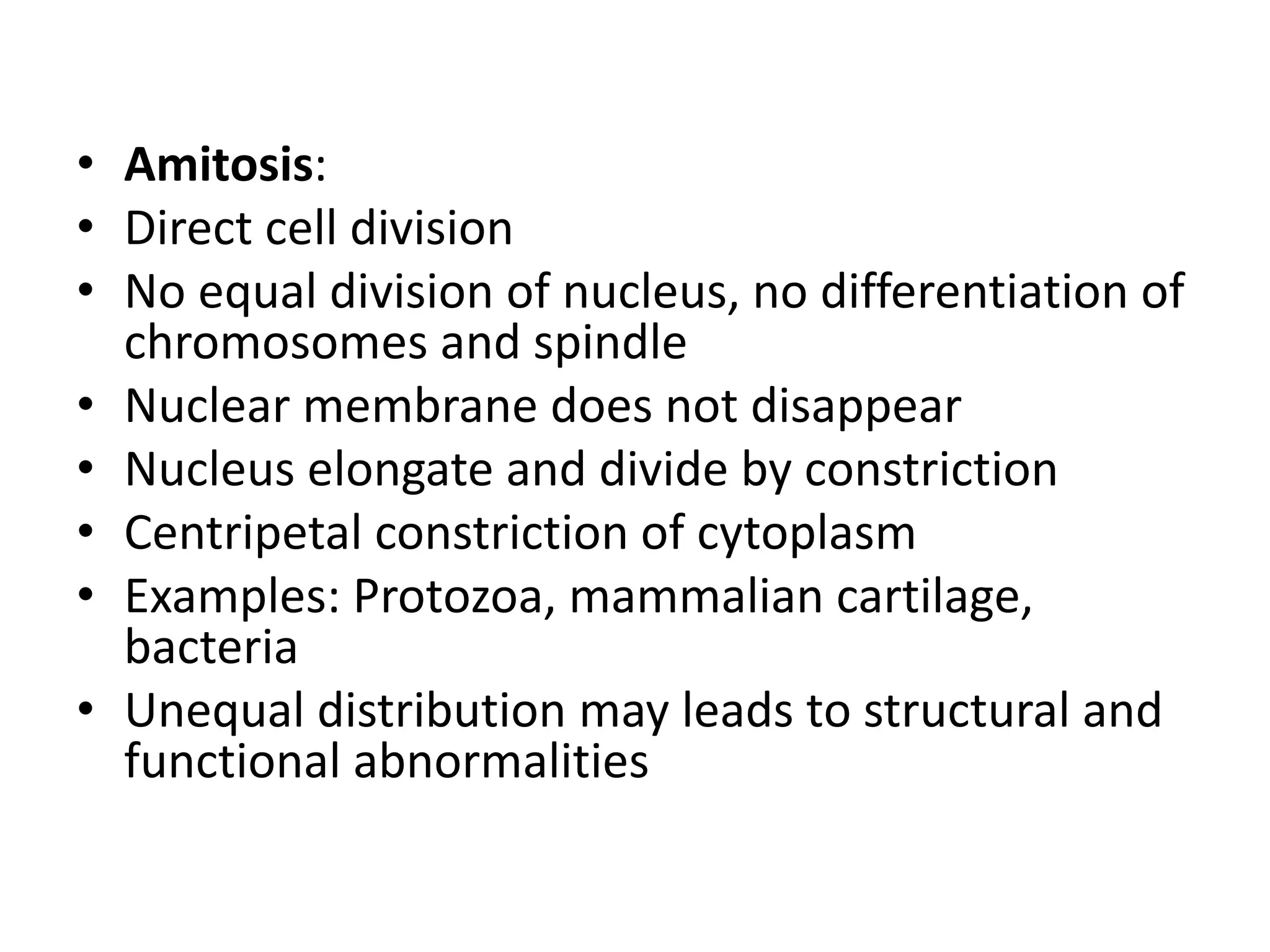
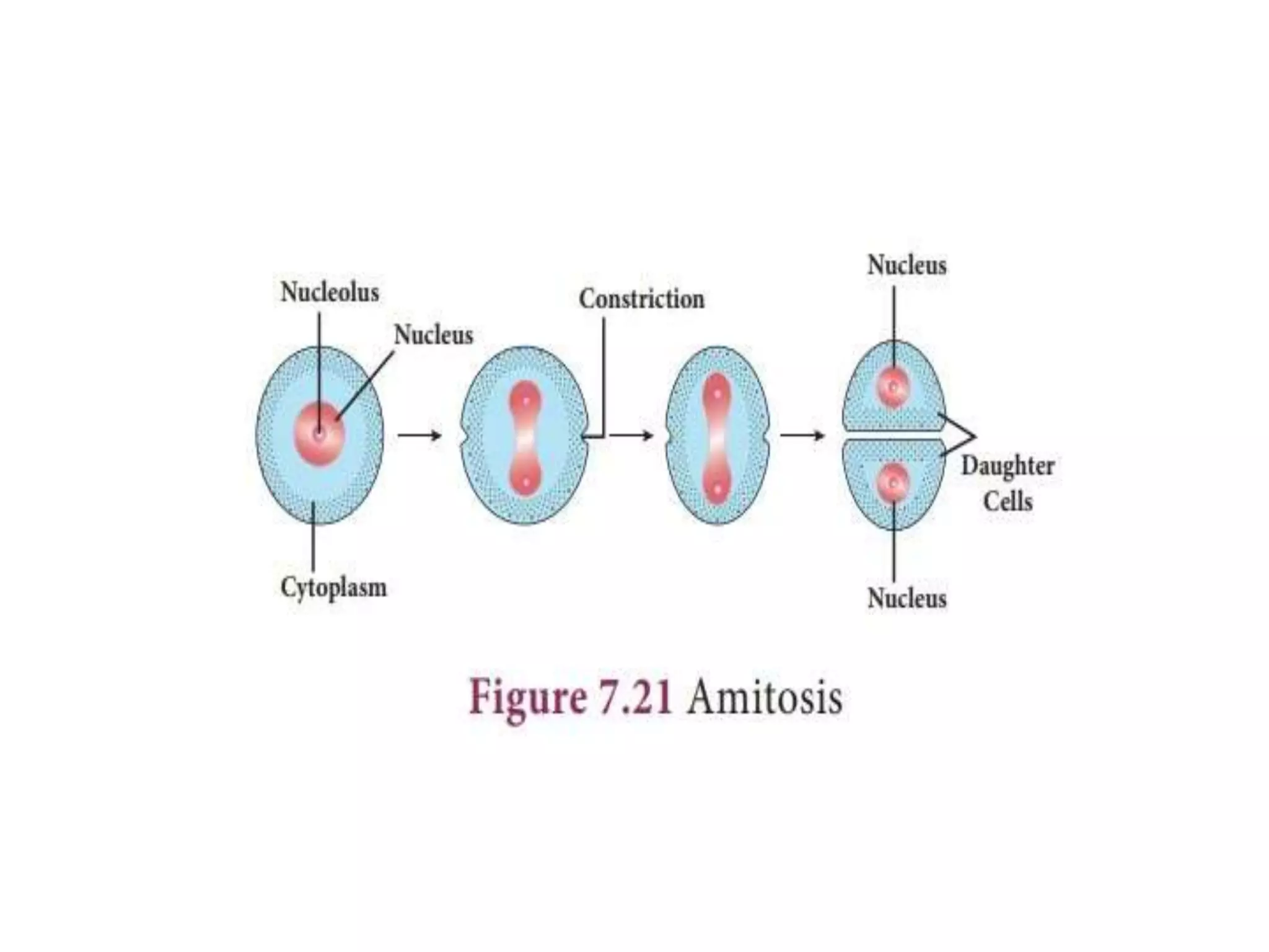
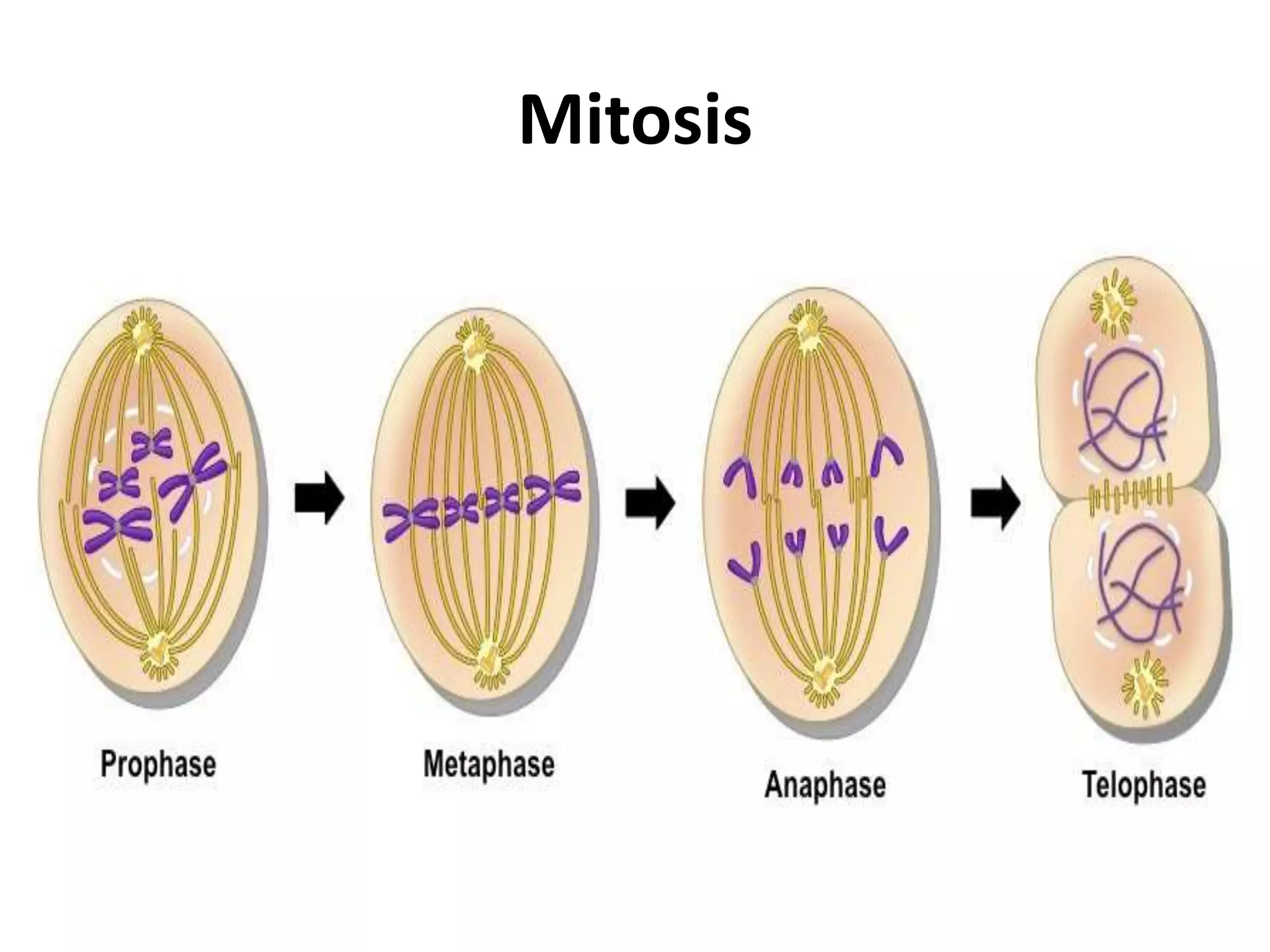
CELL CYCLE AND CELL DIVISION
DR. C. SANKARANARAYANAN
INTRODUCTION
• Living matter – able to reproduce
• Unicellular – duplication – cell division – enlarge
to get original size
• multicellular- - zygote – cell division - growth and
development – adult.
• History:
• Prevost and Dumas – 1824 – cleavage of zygote
• Remak – 1841 - new cells from pre-existing cells
• Rudolf Virchow – 1855, 1859 – cell lineage theory
• Strasburger – 1873 – two nuclei develop from pre-
existing one
• Boveri and Flemming – 1879-80 – somatic cell division
• Flemming – 1882 – coined the term ‘MITOSIS’
• Braur, Sutton, Van-Benden, Strasburger and Winiwater
– 1887-1900 – studied meiosis
• Farmer and Moore – 1905 – coined the term ‘MEIOSIS’
• Gregorie – Meiosis I and Meiosis II
• Howard and Pele – 1953 – Cell Cycle
• Montegomery – coined term ‘synapsis’
• Montose J. Moses – 1955 – Synaptonemal complex
• Cell cycle -> sequence of growth and division
-> formation of cell from parent cell -> division
-> daughter cell -> cell cycle
• Generation time -> period between two
divisions -> depends upon various factors.
• Generation time of E.coli - 20 minutes
• Epithelial cells – 8 to 10 hours
• Onion root tip meristems – 20 hours
• Phases of cell cycle:
• 1. long undividing state – interphase
• 2. short phase of nuclear division,
Multiplication or M phase
• Interphase:
• Preparing for mitosis
• Doubling of DNA, studied using autoradiography
with labelled thymidine
• Synthesis of DNA in synthetic period or S phase
• Followed by two “gaps” – G1 and G2
• Not a resting period
• Intense metabolism
• DNA uncoiled , visible as chromatin
• Volume of nucleus increases
• Nucleolus maximum size
• More ATP form from biomolecules
• Molecules needed stored
• Period 10 – 20 hours – 75 to 90% of
generation time
• Damaged DNA repaired
• FISH can be done – Fluoresccence in situ
hybridization. Fluorophore, sulfoindocyasin is
used to visualize DNA
• Starting point – gap1 (G1) active, make cell
components needed for cell progeny
• Next S phase – synthesis of DNA and proteins
like spindle fibres
• Next gap 2 (G2) – additional proteins, no DNA
synthesis
• No DNA synthesis in G1 and G2.
• M. phase - Multiplication phase – actual cell
division
• Karyokinesis
• Stages – Prophase, metaphase, anaphase,
telophase and cytokinesis
• Then daughter cell enters G1 or Go phase
• G1 phase is also called antephase
• Go phase –discovered by Lajtha – 1963
• If DNA is damaged P53 protein block cell cycle
leads to cell death or apoptosis
Cell Division
• Formation of daughter cell from parent cell
• Continuity from one generation to another
• Hereditary materials copied
• In sex cells division daughter cell may differ
• Prokaryotic cells divide by binary fission
• Single DNA – replicate – separated to cells – identical –
except mutation
• Multi cellular organisms replace worn out cells by cell
division
• Cell division halts – senescence – deterioration – death
• Telomere degeneration – cannot protect chromosomes
• Cancer cells – telomere allows continuous division
• Amitosis:
• Direct cell division
• No equal division of nucleus, no differentiation of
chromosomes and spindle
• Nuclear membrane does not disappear
• Nucleus elongate and divide by constriction
• Centripetal constriction of cytoplasm
• Examples: Protozoa, mammalian cartilage,
bacteria
• Unequal distribution may leads to structural and
functional abnormalities
Mitosis
• Means – ‘thread of fibres’ in Greek
• Formation of identical cells, cellular xerox
• Fleming coined the term
• Phases:
• Interphase: period between two mitosis, resting
and preparatory phase
• Replication of DNA, synthesis of nuclear proteins,
formation of centriole, synthesis of ATP,
formation of spindle
• Duplication of chromosome – monads to dyad
• Prophase:
• Cell become spheroid
• Increase in viscosity, and refractivity
• Chromatin reticulum disappears
• Shortening and thickening of chromosome fibers
• Outside the nucleus two centrosomes formed by
microtubules of tubilin protein
• Due to repulsive interaction of tubules centrosomes
push to the opposite ends of the cell
• In animal cells and fungi fibrils form like a spokes of a
wheel called aster
• Centriole participate – centric mitosis
• Plants no centriole – acentric mitosis
• Aster formation in animals and fungi –astral mitosis
• No aster – an-astral mitosis
• Spindle form between two poles
• Microtubules number 16 in yeast and 5000 in higher
plants
• Two types of centrioles – central spindle for
centrosome division during early prophase and
metaphasic centrioles for anaphasic movement of
chromosomes
• Disappearance of nuclear membrane and no
differentiation of cytoplasm and nucleoplasm
• Each chromosome form two kinetochore at
the centromere, one attached to each
chromatid for attachment of microtubules and
formation of spindle fibres
• Metaphase:
• Chromosomes reaches central equatorial plane
• Smaller chromosomes at the center and larger chromosomes at the
periphery
• Centriole lie at the poles
• Continuous spindle fibres form fro pole to pole, not attached to
chromosomes, form stem-korper or pushing body and help in
separation
• Chromosomal fibres connect kinetochore and pole, pull
chromosomes
• Inter-zonal fibres connect centromere and pole, pull chromosomes
• Bringing chromosomes on the equator of the spindles is called
congression
• Spindle – 90-95% tubulin, 3-5% RNA and traces of actin, myosin and
lipid
• Anaphase:
• Separation of centromere
• Pulling of chromosomes to poles by spindle
• At the end two groups of chromosomes with
equal number form at the pole equal to
parental cell
• Telophase:
• Reversal of prophase
• Two nuclei form and nuclear membrane appear
• Spindle fibres and aster disappear
• Cytokinesis:
• Animal cell cleavage furrow form to become two
cells
• Plant cell - cellplate – cell wall and middle lamella
form to become two cells
Mitosis
MEIOSIS
• “Meio” means to lessen
• Farmer and Moore 1905 coined the term
• First studied by Van Benedin – 1887
• Strasburger – 1888, Sutton - !900,
Winiwater – 1900.
• Gregoire – Meiosis I and Meioisis II
• Diploid cell become haploid cells
• Haploid gametes form from diploid cell
• During sexual reproduction in eukaryotes.
• Male and female gametes fuse to form zygote
• One of the main reasons for variations
• Chromosome number constant
• Variations are the basis of evolution
• Types:
• 1. Zygotic life cycle: Initial Meiosis
• Haploid organism -> male and female gametes fusion -
> Zygote ( diploid) -> meiosis -> four haploid cells ->
new organism. Eg: Bryophytes
• 2. Gametic life cycle: Terminal Meiosis
• Organism diploid -> germ cells -> meiosis -> gametes ->
fusion of gametes -> zygote -> organism Eg: Human
beings
• 3. Sporic Meiosis: Diplohaplontic, intermediate meiosis
• Diploid sporophyte -> meiosis -> micro and
megaspores -> male and female gametes by mitosis ->
Zygote after fusion -> embryo -> that is meiosis
between zygote and gamete
• Phases of meiosis:
• Meiosis I:
• Prophase I:
• Leptotene : slender tread of chromatin become
chromosomes after condensation in nucleus
• It looks like thread of beads – chromomeres
• Irregularly arranged or towards centriole forming
a “ bouquet”. It is due to chromosome
attachment to nuclear membrane.
• In plant cells form tangle of threads – “Synezetic
knot”
• Zygotene: Mating thread
• Chromosome become shorter and thickened
• Pairing of homologous chromosomes “
Synapsis” or “Syndesis” or Synizesis”
• Pairing from centromere to ends – Procentric or
from end to centre – Proterminal pairing
• Combined homologous chromosomes – bivalent.
• It is also called tetrad because of four sister
chromatids.
• Synaptonemal complex formed during
zygotene.
• It is a ribbon like structure formed between
pairing homologous chromosomes.
• It has central and lateral elements – LC fibres
central fibres connects chromosomes and
lateral elements connects nuclear membrane
• Pachytene: Thick thread
• Longest stage
• It begins when synapsis is completed
• Chromosome as bivalent or tetrad, that is each
bivalent with four chromatids
• Each pair of chromatids attached by a centromere
called sister chromatids
• Chromatids of different pair called non-sister
chromatids.
• Breakage and reunion after interchange between
them is called crossing over.
• Diplotene: Double thread
• Beginning of separation of paired homologous
chromosomes
• Held together at one or two points called
chiasmata
• Number of chiasmata varies depends on length
• Terminal or intertidal chiasma
• Chiasma displaced along the length –
terminalization due to despiraling of
chromosomes – expressed as terminalization
coefficent {T}
• Synaptimal complex disappear
• Diakinesis:
• Chromosomes more contracted
• Bivalents move towards periphery
• Homologous remain in contact by terminal
chiasmata.
• Nucleolus detached or disappear
• Metaphase I :
• Chromosomes arranged at the centre
• Spindle formed and attached to centromere of
the two homologous chromosomes
• Two centromeres of each bivalent lie on
opposite sides of the equatorial plate
• It differs from mitosis, that is each bivalent has
two centromeres.
• Anaphase I :
• Homologous chromosomes move towards
poles
• Due to crossing over separating chromosomes
are different.
• Centromere do not divide like mitosis
• Telophase I :
• Centromere arrives pole
• Each cell has half the number of
chromosomes, each with two chromatids
• Spindle fibres disappear
• Nuclear membrane form
• Chromosomes uncoil and become chromatin
• It is followed by cytokinesis.
• Meiosis II :
• Like mitosis, but DNA do not duplicate
• Prophase II :
• Nuclear membrane, nucleolus disappear.
• Shortening and thickening of chromatids.
• Centrioles move to the poles and spindle fibres
arranged
• New equatorial plane rotated by 90o, that is
perpendicular to the previous plane
Metaphase II :
• Like mitosis chromosomes oriented on the
equatorial plate and attached with spindle fibres
• Anaphase II :
• Centromeres cleaved
• Allow the kinetochore to pull the sister
chromatids apart.
• Now become sister chromosomes and pulled
toward opposite poles.
• Telophase II :
• Like telophase I uncoiling, lengthening and
disappearance of chromosomes
• Nuclear membrane form
• Finally four haploid cells formed
• Non-disjunction :
• Normal separation of chromosomes called
disjunction.
• If they are not properly separated leads to non-
disjunction
• Leads to more or less amount of genetic material
• Example – trisomy or monosomy
• Downs syndrome - trisomy of 21 chromosome
• Klinefelder’s syndrome – one extra X cromosome
• Significance of Meiosis:
• 1. Sexual reproduction
• 2. Genetic variation
• 3. Mutation
• 4. Genetic information for gametes or
gametophytes and sporophytes.
THANK YOU

Cell cycle and cell division.pptx

  • 1.
    CELL CYCLE ANDCELL DIVISION DR. C. SANKARANARAYANAN
  • 2.
    INTRODUCTION • Living matter– able to reproduce • Unicellular – duplication – cell division – enlarge to get original size • multicellular- - zygote – cell division - growth and development – adult. • History: • Prevost and Dumas – 1824 – cleavage of zygote • Remak – 1841 - new cells from pre-existing cells • Rudolf Virchow – 1855, 1859 – cell lineage theory
  • 3.
    • Strasburger –1873 – two nuclei develop from pre- existing one • Boveri and Flemming – 1879-80 – somatic cell division • Flemming – 1882 – coined the term ‘MITOSIS’ • Braur, Sutton, Van-Benden, Strasburger and Winiwater – 1887-1900 – studied meiosis • Farmer and Moore – 1905 – coined the term ‘MEIOSIS’ • Gregorie – Meiosis I and Meiosis II • Howard and Pele – 1953 – Cell Cycle • Montegomery – coined term ‘synapsis’ • Montose J. Moses – 1955 – Synaptonemal complex
  • 4.
    • Cell cycle-> sequence of growth and division -> formation of cell from parent cell -> division -> daughter cell -> cell cycle • Generation time -> period between two divisions -> depends upon various factors. • Generation time of E.coli - 20 minutes • Epithelial cells – 8 to 10 hours • Onion root tip meristems – 20 hours
  • 5.
    • Phases ofcell cycle: • 1. long undividing state – interphase • 2. short phase of nuclear division, Multiplication or M phase
  • 6.
    • Interphase: • Preparingfor mitosis • Doubling of DNA, studied using autoradiography with labelled thymidine • Synthesis of DNA in synthetic period or S phase • Followed by two “gaps” – G1 and G2 • Not a resting period • Intense metabolism • DNA uncoiled , visible as chromatin • Volume of nucleus increases • Nucleolus maximum size
  • 7.
    • More ATPform from biomolecules • Molecules needed stored • Period 10 – 20 hours – 75 to 90% of generation time • Damaged DNA repaired • FISH can be done – Fluoresccence in situ hybridization. Fluorophore, sulfoindocyasin is used to visualize DNA
  • 8.
    • Starting point– gap1 (G1) active, make cell components needed for cell progeny • Next S phase – synthesis of DNA and proteins like spindle fibres • Next gap 2 (G2) – additional proteins, no DNA synthesis • No DNA synthesis in G1 and G2.
  • 9.
    • M. phase- Multiplication phase – actual cell division • Karyokinesis • Stages – Prophase, metaphase, anaphase, telophase and cytokinesis • Then daughter cell enters G1 or Go phase • G1 phase is also called antephase • Go phase –discovered by Lajtha – 1963 • If DNA is damaged P53 protein block cell cycle leads to cell death or apoptosis
  • 12.
    Cell Division • Formationof daughter cell from parent cell • Continuity from one generation to another • Hereditary materials copied • In sex cells division daughter cell may differ • Prokaryotic cells divide by binary fission • Single DNA – replicate – separated to cells – identical – except mutation • Multi cellular organisms replace worn out cells by cell division • Cell division halts – senescence – deterioration – death • Telomere degeneration – cannot protect chromosomes • Cancer cells – telomere allows continuous division
  • 13.
    • Amitosis: • Directcell division • No equal division of nucleus, no differentiation of chromosomes and spindle • Nuclear membrane does not disappear • Nucleus elongate and divide by constriction • Centripetal constriction of cytoplasm • Examples: Protozoa, mammalian cartilage, bacteria • Unequal distribution may leads to structural and functional abnormalities
  • 15.
    Mitosis • Means –‘thread of fibres’ in Greek • Formation of identical cells, cellular xerox • Fleming coined the term • Phases: • Interphase: period between two mitosis, resting and preparatory phase • Replication of DNA, synthesis of nuclear proteins, formation of centriole, synthesis of ATP, formation of spindle • Duplication of chromosome – monads to dyad
  • 17.
    • Prophase: • Cellbecome spheroid • Increase in viscosity, and refractivity • Chromatin reticulum disappears • Shortening and thickening of chromosome fibers • Outside the nucleus two centrosomes formed by microtubules of tubilin protein • Due to repulsive interaction of tubules centrosomes push to the opposite ends of the cell • In animal cells and fungi fibrils form like a spokes of a wheel called aster
  • 18.
    • Centriole participate– centric mitosis • Plants no centriole – acentric mitosis • Aster formation in animals and fungi –astral mitosis • No aster – an-astral mitosis • Spindle form between two poles • Microtubules number 16 in yeast and 5000 in higher plants • Two types of centrioles – central spindle for centrosome division during early prophase and metaphasic centrioles for anaphasic movement of chromosomes
  • 19.
    • Disappearance ofnuclear membrane and no differentiation of cytoplasm and nucleoplasm • Each chromosome form two kinetochore at the centromere, one attached to each chromatid for attachment of microtubules and formation of spindle fibres
  • 23.
    • Metaphase: • Chromosomesreaches central equatorial plane • Smaller chromosomes at the center and larger chromosomes at the periphery • Centriole lie at the poles • Continuous spindle fibres form fro pole to pole, not attached to chromosomes, form stem-korper or pushing body and help in separation • Chromosomal fibres connect kinetochore and pole, pull chromosomes • Inter-zonal fibres connect centromere and pole, pull chromosomes • Bringing chromosomes on the equator of the spindles is called congression • Spindle – 90-95% tubulin, 3-5% RNA and traces of actin, myosin and lipid
  • 25.
    • Anaphase: • Separationof centromere • Pulling of chromosomes to poles by spindle • At the end two groups of chromosomes with equal number form at the pole equal to parental cell
  • 27.
    • Telophase: • Reversalof prophase • Two nuclei form and nuclear membrane appear • Spindle fibres and aster disappear • Cytokinesis: • Animal cell cleavage furrow form to become two cells • Plant cell - cellplate – cell wall and middle lamella form to become two cells
  • 29.
  • 30.
    MEIOSIS • “Meio” meansto lessen • Farmer and Moore 1905 coined the term • First studied by Van Benedin – 1887 • Strasburger – 1888, Sutton - !900, Winiwater – 1900. • Gregoire – Meiosis I and Meioisis II • Diploid cell become haploid cells
  • 31.
    • Haploid gametesform from diploid cell • During sexual reproduction in eukaryotes. • Male and female gametes fuse to form zygote • One of the main reasons for variations • Chromosome number constant • Variations are the basis of evolution
  • 32.
    • Types: • 1.Zygotic life cycle: Initial Meiosis • Haploid organism -> male and female gametes fusion - > Zygote ( diploid) -> meiosis -> four haploid cells -> new organism. Eg: Bryophytes • 2. Gametic life cycle: Terminal Meiosis • Organism diploid -> germ cells -> meiosis -> gametes -> fusion of gametes -> zygote -> organism Eg: Human beings • 3. Sporic Meiosis: Diplohaplontic, intermediate meiosis • Diploid sporophyte -> meiosis -> micro and megaspores -> male and female gametes by mitosis -> Zygote after fusion -> embryo -> that is meiosis between zygote and gamete
  • 33.
    • Phases ofmeiosis: • Meiosis I: • Prophase I: • Leptotene : slender tread of chromatin become chromosomes after condensation in nucleus • It looks like thread of beads – chromomeres • Irregularly arranged or towards centriole forming a “ bouquet”. It is due to chromosome attachment to nuclear membrane. • In plant cells form tangle of threads – “Synezetic knot”
  • 34.
    • Zygotene: Matingthread • Chromosome become shorter and thickened • Pairing of homologous chromosomes “ Synapsis” or “Syndesis” or Synizesis” • Pairing from centromere to ends – Procentric or from end to centre – Proterminal pairing • Combined homologous chromosomes – bivalent. • It is also called tetrad because of four sister chromatids.
  • 35.
    • Synaptonemal complexformed during zygotene. • It is a ribbon like structure formed between pairing homologous chromosomes. • It has central and lateral elements – LC fibres central fibres connects chromosomes and lateral elements connects nuclear membrane
  • 36.
    • Pachytene: Thickthread • Longest stage • It begins when synapsis is completed • Chromosome as bivalent or tetrad, that is each bivalent with four chromatids • Each pair of chromatids attached by a centromere called sister chromatids • Chromatids of different pair called non-sister chromatids. • Breakage and reunion after interchange between them is called crossing over.
  • 37.
    • Diplotene: Doublethread • Beginning of separation of paired homologous chromosomes • Held together at one or two points called chiasmata • Number of chiasmata varies depends on length • Terminal or intertidal chiasma • Chiasma displaced along the length – terminalization due to despiraling of chromosomes – expressed as terminalization coefficent {T} • Synaptimal complex disappear
  • 38.
    • Diakinesis: • Chromosomesmore contracted • Bivalents move towards periphery • Homologous remain in contact by terminal chiasmata. • Nucleolus detached or disappear
  • 41.
    • Metaphase I: • Chromosomes arranged at the centre • Spindle formed and attached to centromere of the two homologous chromosomes • Two centromeres of each bivalent lie on opposite sides of the equatorial plate • It differs from mitosis, that is each bivalent has two centromeres.
  • 43.
    • Anaphase I: • Homologous chromosomes move towards poles • Due to crossing over separating chromosomes are different. • Centromere do not divide like mitosis
  • 45.
    • Telophase I: • Centromere arrives pole • Each cell has half the number of chromosomes, each with two chromatids • Spindle fibres disappear • Nuclear membrane form • Chromosomes uncoil and become chromatin • It is followed by cytokinesis.
  • 47.
    • Meiosis II: • Like mitosis, but DNA do not duplicate • Prophase II : • Nuclear membrane, nucleolus disappear. • Shortening and thickening of chromatids. • Centrioles move to the poles and spindle fibres arranged • New equatorial plane rotated by 90o, that is perpendicular to the previous plane
  • 48.
    Metaphase II : •Like mitosis chromosomes oriented on the equatorial plate and attached with spindle fibres • Anaphase II : • Centromeres cleaved • Allow the kinetochore to pull the sister chromatids apart. • Now become sister chromosomes and pulled toward opposite poles.
  • 49.
    • Telophase II: • Like telophase I uncoiling, lengthening and disappearance of chromosomes • Nuclear membrane form • Finally four haploid cells formed
  • 51.
    • Non-disjunction : •Normal separation of chromosomes called disjunction. • If they are not properly separated leads to non- disjunction • Leads to more or less amount of genetic material • Example – trisomy or monosomy • Downs syndrome - trisomy of 21 chromosome • Klinefelder’s syndrome – one extra X cromosome
  • 52.
    • Significance ofMeiosis: • 1. Sexual reproduction • 2. Genetic variation • 3. Mutation • 4. Genetic information for gametes or gametophytes and sporophytes.
  • 53.