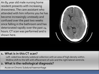
BRAIN CT REVISION
‫رادیولوژی‬ ‫جامع‬ ‫سایت‬
WWW.RADIOLOGYHA.COM
ChanYon Quan
5 Basic Densities
 Air
 Fat
 Soft tss / Fluid
 Calcium
 Metal
HYPODENSE
•Air
•Fat
•CSF
ISODENSE
•Brain tss
•Subacute SDH
HYPERDENSE
•Blood
•Iodine contrast
•Calcium
•Metal
An 84 year old male nursing home
resident presents with increasing
drowsiness.The care assistant who
attended with him informs you he has
become increasingly unsteady and
confused over the past two weeks
since falling in the bathroom and has
deteriorated rapidly over the past 24
hours. CT scan was performed and is
shown here.
1. What is in this CT scan?
Left -sided low density subdural collection with an area of high density within.
Midline shift to the left with effacement of sulci and the right lateral ventricle.
2. What is the radiological diagnosis?
Acute on Chronic Subdural haemorrhage
A 46 year old male with known
Adult Polycystic Kidney Disease
(APKD) presents to his GP with
sudden onset headache
associated with nausea and
vomiting.
1. What is the diagnosis?
SAH
2. What does his CT scan
show?What complication is
he developing?
Ruptured aneurysm
Blood in the subarachnoid spaces
Hydrocephalus – enlarged temporal
horns of the lateral ventricle
3. In which part of circle of Willis his aneurysm arose from?
Anterior communicating artery (ACOM)
A 24 year old female fell from her
horse during a cross country
competition, hitting her head on a
wooden fence on the way to the
ground. She was taken by
ambulance to A&E where on
examination she had a Glasgow
coma scale of 11/15. CT scan is
shown here..
1. Describe the two radiological abnormalities shown.
Soft tissue swelling over the right parietal bone
Right sided hyper-dense eliptical area
2. Give a radiological diagnosis.
Acute EDH
3. What other abnormalities should always be looked for in EDH?
Skull fracture- 80+% of EDH are associated with a skull fracture
Pneumocephalus – Mount Fuji sign

Case Studies - Brain CT scan explained.pptx

  • 1.
    BRAIN CT REVISION ‫رادیولوژی‬‫جامع‬ ‫سایت‬ WWW.RADIOLOGYHA.COM ChanYon Quan
  • 2.
    5 Basic Densities Air  Fat  Soft tss / Fluid  Calcium  Metal HYPODENSE •Air •Fat •CSF ISODENSE •Brain tss •Subacute SDH HYPERDENSE •Blood •Iodine contrast •Calcium •Metal
  • 3.
    An 84 yearold male nursing home resident presents with increasing drowsiness.The care assistant who attended with him informs you he has become increasingly unsteady and confused over the past two weeks since falling in the bathroom and has deteriorated rapidly over the past 24 hours. CT scan was performed and is shown here. 1. What is in this CT scan? Left -sided low density subdural collection with an area of high density within. Midline shift to the left with effacement of sulci and the right lateral ventricle. 2. What is the radiological diagnosis? Acute on Chronic Subdural haemorrhage
  • 4.
    A 46 yearold male with known Adult Polycystic Kidney Disease (APKD) presents to his GP with sudden onset headache associated with nausea and vomiting. 1. What is the diagnosis? SAH 2. What does his CT scan show?What complication is he developing? Ruptured aneurysm Blood in the subarachnoid spaces Hydrocephalus – enlarged temporal horns of the lateral ventricle 3. In which part of circle of Willis his aneurysm arose from? Anterior communicating artery (ACOM)
  • 5.
    A 24 yearold female fell from her horse during a cross country competition, hitting her head on a wooden fence on the way to the ground. She was taken by ambulance to A&E where on examination she had a Glasgow coma scale of 11/15. CT scan is shown here.. 1. Describe the two radiological abnormalities shown. Soft tissue swelling over the right parietal bone Right sided hyper-dense eliptical area 2. Give a radiological diagnosis. Acute EDH 3. What other abnormalities should always be looked for in EDH? Skull fracture- 80+% of EDH are associated with a skull fracture
  • 6.