2. Physiologic thrombosis is
counterbalanced by intrinsic
antithrombotic properties and
fibrinolysis. Under normal
conditions, a thrombus is confined
to the immediate area of injury
and does not obstruct flow to
critical areas, unless the blood
vessel lumen is already
diminished.
3.
4. The principal clinical syndromes
that result from thrombosis are as
follows:
Acute myocardial infarction (AMI)
Deep vein thrombosis (DVT)
Pulmonary embolism (PE)
Acute ischemic stroke (AIS)
Acute peripheral arterial occlusion
Occlusion of indwelling catheters
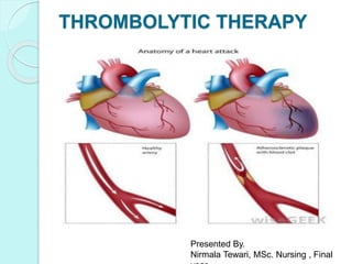
5. Thrombolytic therapy is the use of
drugs to break up or dissolve blood
clots, which are the main cause of
both heart attacks and stroke.
Thrombolytic medications are
approved for the immediate
treatment of stroke and heart attack.
The thrombolytic agents available
today are serine proteases that work
by converting plasminogen to the
natural fibrinolytic agent plasmin.
6. Fibrinolytic agents, sometimes
referred to as plasminogen
activators, are divided into 2
categories:
1) Fibrin-specific agents It
include alteplase (tPA*), reteplase
(recombinant plasminogen
activator [r-PA]), and tenecteplase,
produce limited plasminogen
conversion in the absence of fibrin.
7. *Tissue plasminogen activator (tPA) is a
naturally occurring fibrinolytic agent
found in vascular endothelial cells and
is involved in the balance between
thrombolysis and thrombogenesis. It
exhibits significant fibrin specificity and
affinity. At the site of the thrombus, the
binding of tPA and plasminogen to the
fibrin surface induces a conformational
change that facilitates the conversion
8. Currently available agents
include the following:
Alteplase
Reteplase
Tenecteplase
Urokinase
Prourokinase
Anisoylated purified
streptokinase activator
complex (APSAC;
anistreplase)
Streptokinase
9. Alteplase
Alteplase was the first recombinant
tissue-type plasminogen activator and
is identical to native tPA.
In vivo, tissue-type plasminogen
activator is synthesized and made
available by cells of the vascular
endothelium. It is the physiologic
thrombolytic agent responsible for
most of the body’s natural efforts to
prevent excessive thrombus
propagation
10. Alteplase is fibrin-specific and has a
plasma half-life of 4-6 minutes.
In theory, alteplase should be
effective only at the surface of fibrin
clot but in practice a systemic lytic
state is seen, with moderate
amounts of circulating fibrin
degradation products and a
substantial risk of systemic
bleeding. Alteplase may be
readministered as necessary;
11. Reteplase
Reteplase is a second-generation
recombinant tissue-type plasminogen
activator that seems to work more
rapidly and to have a lower bleeding risk
than the first-generation agent alteplase.
It is a synthetic nonglycosylated deletion
mutein of tPA that contains 355 of the
527 amino acids of native tPA.
As reteplase does not bind fibrin as
tightly as native tPA does, it can diffuse
more freely through the clot rather than
bind only to the surface as tPA does. At
high concentrations, reteplase does not
12. Cont.
The biochemical modifications also
resulted in a molecule with a longer half-
life (approximately 13-16 minutes),
which allows bolus administration.
Reteplase is FDA-approved for AMI and
is administered as 2 boluses of 10 U
given 30 minutes apart, with each bolus
administered over 2 minutes.
It is not antigenic and almost never is
associated with any allergic
13. Tenecteplase
Tenecteplase, the latest thrombolytic
agent approved for use in clinical
practice, was approved by the FDA as a
fibrinolytic agent in 2000.
It is produced by recombinant DNA
technology using Chinese hamster ovary
cells. Its mechanism of action is similar to
that of alteplase, and it is currently
indicated for the management of AMI.
Tenecteplase has a half-life ranging
initially from 20-24 minutes to 130
14. Urokinase
Urokinase is a physiologic thrombolytic
agent that is produced in renal
parenchymal cells. Unlike streptokinase,
urokinase directly cleaves plasminogen to
produce plasmin. (When it is purified from
human urine, approximately 1500 L of
urine are needed to yield enough
urokinase to treat a single patient. )
It is indicated only for massive PE and PE
accompanied by unstable hemodynamics.
15. Cont.
Allergic reactions are rare, and
the agent can be administered
repeatedly without antigenic
problems.
Urokinase is the fibrinolytic
agent that is most familiar to
interventional radiologists and
that has been used most often
for peripheral intravascular
16. Prourokinase
• Prourokinase is a new
fibrinolytic agent that is currently
undergoing clinical trials.
• Prourokinase is relatively fibrin-
specific,It has been studied in
the settings of AMI, AIS, and
peripheral arterial occlusion
17. Streptokinase
Streptokinase is produced by beta-
hemolytic streptococci. By itself, it is not a
plasminogen activator, but it binds with
free circulating plasminogen (or with
plasmin) to form a complex that can
convert additional plasminogen to
plasmin.
Streptokinase activity is not enhanced in
18. Side effects of
streptokinase :
Allergic problems- As Streptokinase is
produced from streptococcal bacteria, it
often causes febrile reactions and other.
Streptokinase usually cannot be
administered safely a second time within
6 months, because it is highly antigenic
and results in high levels of
19. Anisoylated purified
streptokinase activator complex/
Anistreplase
APSAC (anistreplase) is a complex of
streptokinase and plasminogen that
does not require free circulating
plasminogen to be effective.
It has many theoretical benefits over
streptokinase but suffers antigenic
problems similar to those of the parent
compound.
Like streptokinase, anistreplase does
not distinguish between fibrin-bound
20. Contraindication Of
Thrombolytic Therapy
Absolute contraindications for
fibrinolytic use in STEMI include
the following
Prior intracranial hemorrhage (ICH)
Known structural cerebral vascular lesion,
malignant intracranial neoplasm
Ischemic stroke within 3 months
Suspected aortic dissection
Active bleeding or bleeding diathesis
(excluding menses)
Significant closed head trauma or facial
trauma within 3 months
Intracranial or intraspinal surgery within 2
months
21. Relative contraindications for
fibrinolytic use in STEMI include the
following:
History of chronic, severe, poorly controlled
hypertension
Significant hypertension on presentation
(systolic blood pressure > 180 mm Hg or
diastolic blood pressure > 110 mm Hg
Traumatic or prolonged (> 10 minutes)
cardiopulmonary resuscitation (CPR) or major
surgery less than 3 weeks previously
History of prior ischemic stroke not within the
last 3 months
Dementia
Recent (within 2-4 weeks) internal bleeding
Noncompressible vascular punctures
22. Complications Of Thrombolytic
Therapy
OHemorrhage,
OAllergic Reactions
OEmbolism
OStroke, And
OReperfusion Arrhythmia
OThe most feared complication of
fibrinolysis is intracranial
hemorrhage (ICH), but serious
hemorrhagic complications can occur
from bleeding at any site in the body.
23. Risk factors for
hemorrhagic complications
of thrombolytic therapy :
Increasing age
Lower body weight
Elevated pulse pressure
Uncontrolled hypertension
Recent stroke or surgery
Presence of a bleeding diathesis
Severe congestive heart failure
24. Precautions For
Thrombolytic Therapy
Thrombolytic therapy may cause bleeding.
To lower the risk of serious bleeding,
people who are given this drug should
move around as little as possible and
should not try to get up on their own
unless told to do so by a health care
professional.
Special care should be taken with
people-
heart or blood vessel disease
stroke (recent or in the past)
25. Stomach ulcer or colitis
Severe liver disease
Active tuberculosis
Recent falls, injuries, or blows to the
body or head
Recent injections into A blood vessel
Recent surgery, including dental surgery
Tubes recently placed in the body for
any reason
Recent delivery of A baby
Patient with recent streptococcal (strep)
Pregnant and lactating mother
26. Side Effects Of Thrombolytic
Therapy
People who are given thrombolytic therapy
should also be alert to the signs of bleeding
inside the body and should check with a
physician immediately if any of the following
symptoms occur:
blood in the urine
blood or black, tarry stools
constipation
coughing up blood
vomiting blood or material that looks like
coffee grounds
nosebleeds
27. Conts..
sudden, severe, or constant
headaches
Pain or swelling in the abdomen
or stomach
back pain or backache
severe or constant muscle pain or
stiffness
stiff, swollen, or painful joints
Other side effects of thrombolytic
agents are possible. Anyone who
28. Interactions Of Thrombolytic
Therapy
People who take certain medicines may be
at greater risk for severe bleeding when
they receive a thrombolytic agent. Anyone
who is given a thrombolytic agent should
tell the physician about all other
prescription or nonprescription (over-the-
counter) medicines he or she is taking.
Among the medicines that may increase
the chance of bleeding are:
Aspirin and other medicines for pain and
inflammation
Blood thinners (anticoagulants)
Antiseizure medicines, such as depakote
29. Nursing Responsibility In
Thrombolytic Therapy:
PREINFUSION CARE-
History Taking And Perform A Physical
Assessment- Information obtained from the
history and physical exam helps to determine
whether thrombolytic therapy is
appropriate.The goal is to initiate thrombolytic
therapy within 30 minutes of arrival.
Evaluate For Contraindications To
Thrombolytic Therapy:
recent surgery or trauma (including prolonged
CPR), bleeding disorders or active bleeding,
cerebral vascular accident, neurosurgery
30. Consent for treatment and
counselling - Inform the client of
the purpose of the therapy. Discuss
the risk of bleeding and the need to
keep the extremity immobile during
and after the infusion. Minimal
movement of the extremity is
necessary to prevent bleeding from
the infusion site.
31. DURING THE INFUSION
Assess and record vital signs and the
infusion site for hematoma or bleeding
every 15 minutes for the first hour, every
30 minutes for the next 2 hours, and
then hourly until the intravenous
catheter is discontinued.
Assess pulses, color, sensation, and
temperature of both extremities with
each vital sign check. Vital signs and the
site are frequently assessed to detect
possible complications.
Remind the client to keep the extremity
32. Conts..
Hypotension may develop keeping the
bed flat helps maintain cerebral
perfusion.
Maintain continuous cardiac monitoring
during the infusion.
Keep antidysrhythmic drugs and the
emergency cart readily available for
treatment of significant dysrhythmias.
Ventricular dysrhythmias commonly
occur with reperfusion of the ischemic
33. POSTINFUSION CARE-
Assess vital signs, distal pulses, and
infusion site frequently as needed. The
client remains at high risk for bleeding
following thrombolytic therapy.
Evaluate response to therapy:
normalization of ST segment, relief of chest
pain, reperfusion dysrhythmias, early
peaking of the CK and CK-MB band. These
are signs that the clot has been dissolved
and the myocardium is being reperfused.
Maintain bed rest for 6 hours. Keep the
head of the bed at or below 15 degrees.
Reinforce the need to keep the extremity
straight and immobile. Avoid any injections
for 24 hours after catheter removal.
34. Conts.
Perform routine care in a gentle manner
to avoid bruising or injury.
Peripheral bleeding may occur at
puncture sites, and there may not be
sufficient fibrin to form a clot. Direct or
indirect pressure may be needed to
control the bleeding.
Assess body fluids, including urine,
vomitus, and feces, for evidence of
bleeding
35. Conts..
Monitor hemoglobin and hematocrit
levels, prothrombin time (PT), and
partial thromboplastin time (PTT). These
provide additional means of assessing
for bleeding.
Administer platelet-modifying drugs
(e.g., aspirin, dipyridamole) as ordered.
Platelet inhibitors decrease platelet
aggregation and adhesion and are used
to prevent reocclusion of the artery.
Report manifestations of reocclusion,
including changes in the ST segment,
36. Conclusion
For thrombolytic therapy to be effective
in treating stroke or heart attack,
prompt medical attention is very
important. The drugs must be given
within a few hours of the beginning of
a stroke or heart attack. However, this
treatment is not right for every patient
who has a heart attack or a stroke. To
increase the chance of survival and
reduce the risk of serious, permanent
damage, anyone who has signs of a
38. History Of IABP –
The IABP device was pioneered at Grace
Sinai Hospital in Detroit during the early
1960s by Dr . Adrian Kantrowitz and his
team.
The first publication of intra-aortic balloon
counter-pulsation appeared in the
American Heart Journal of May by S.
Moulopoulos, S. Topaz and W. Kolff.
The device and the balloons were then
developed for commercial use between
1967 and 1969 heart surgery by William
Rassman, M.D. at Cornell Medical Center
39. The first clinical implant was
performed at Maimonides Medical
Center , Brooklyn, N.Y . in Oct., 1967.
The patient, a 48-year-old woman, was
in cardiogenic shock and unresponsive
to traditional therapy . An IABP was
inserted by a cut down on the left
femoral artery. Pumping was performed
for approximately 6 hours. Shock
reversed and the patient was
discharged.
The size of the original balloon was 15
French but eventually 9 and 8 French
40. Description Of The Device
The Intra-aortic balloon pump (IABP) is a
mechanical device that increases
myocardial oxygen perfusion while at
the same time increasing cardiac
output. Increasing cardiac output
increases coronary blood flow and
therefore myocardial oxygen delivery .
41.
42. How IABP Works…????
It consists of a cylindrical
polyethylene balloon that sits in the
aorta, approximately 2 centimeters
(0.79 in) from the left subclavian
artery and counterpulsates.
A computer-controlled mechanism
inflates the balloon with helium from
a cylinder during diastole, usually
linked to either an electrocardiogram
(ECG) or a pressure transducer at
43. Cont..
It actively deflates in systole
increasing forward blood flow by
reducing afterload through a vacuum
effect.
It actively inflates in diastole
increasing blood flow to the coronary
arteries via retrograde flow .
These actions combine to decrease
45. Why Helium is used???
Helium is used because its low
viscosity allows it to travel
quickly through the long
connecting tubes, and has a
lower risk than air of causing
an embolism should the
46. Effects of Inflation during diastole:
Increase Systolic Pressure
Increase Pressure in the Aortic Root
During Diastole
Increase Coronary Perfusion
Pressure
Improved Oxygen Delivery To The
Myocardium
Decrease Angina
Haemodynamic effects of
counter pulsation
47. Effects of deflation during systole
Decrease afterload
Decrease peack systolic pressure
Decrease myocardial oxygen
consumption
Increase forward flow decreasing
preload
Decrease PA pressure, including
PAWP
Decrease crackles
Increase SV possybly with
Improve sensorium
Warmed skin
52. Contraindications of IABP
Absolute contraindication
The following conditions will always exclude
patients for treatment:
Severe aortic valve insufficiency
Aortic dissection
Severe aortoiliac occlusive disease and
bilateral carotid stenosis
53. Relative
Contraindication
The following conditions
make IABP therapy
inadvisable except under
special circumstances:
Prosthetic vascular grafts
in the aorta
Aortic aneurysm
Aortofemoral grafts
59. Cardioversion is a medical procedure by
which an abnormally fast heart rate
(tachycardia) or cardiac arrhythmia is
converted to a normal rhythm using
electricity or drugs.
The types of cardioversion are as follows
Synchronized electrical cardioversion
Pharmacological cardioversion
60. Synchronized Electrical
Cardioversion:
Synchronized electrical cardioversion uses a
therapeutic dose of electric current to the heart at a
specific moment in a cardiac cycle. (Defibrilation uses a
therapeutic dose of electric current to the heart at a
random moment in a a cardiac cycle, and is the most
effective resuscitation measure for cardiac arrest
associated with ventricular fibrillation and pulseless
ventricular tachycardia).
When synchronized electrical cardioversion is performed
as an elective procedure, the shocks can be performed
in conjunction with drug therapy until sinus rhythm is
attained. After the procedure, the patient is monitored to
ensure stability of the sinus rhythm.
61. Synchronized electrical cardioversion is
used tSynchronized electrical
cardioversion is used to treat
hemodynamically unstable conditions
like
supraventricular (or narrow complex)
tachycardias,
including atrial fibrillation and atrial
flutter.
It is also used in the emergent treatment
of wide complex tachycardias,
including ventricular tachycardia, when
a pulse is present.
63. Pharmacological Cardioversion:
Pharmacological cardioversion also called chemical
cardioversion , uses antiarrhythmic medication instead
of an electrical shock.
Various antiarrhythmic agents can be used to return the
heart to normal sinus rhythm. Pharmacological
cardioversion is an especially good option in patients
with fibrillation of recent onset. Drugs that are effective
at maintaining normal rhythm after electric
cardioversion, can also be used for pharmacological
cardioversion. Drugs like amiodarone, diltiazem,
verapamil and metoprolol are frequently given before
cardioversion to decrease the heart rate, stabilize the
patient and increase the chance that cardioversion is
successful. There are various classes of agents that are
most effective forpharmacological cardioversion.
64. Class I: These agents are sodium (Na) channel blockers (which slow conduction by blocking
the Na+ channel) and are divided into 3 subclasses a, b and c.
Class Ia - It slows phase 0 depolarization in the ventricles and increases the absolute
refractory period.
Procainamide, quinidine and disopyramide are Class Ia agents.
Class 1b- Drugs lengthen phase 3 repolarization. They include lidocaine, mexiletine and
phenytoin.
Class Ic- Greatly slow phase 0 depolarization in the ventricles (however unlike 1a have no
effect on the
refractory period). Flecainide, moricizine and propafenone are Class Ic agents.
Class II: these agents are beta blockers which inhibit SA and AV node depolarization and slow
heart rate. They also decrease cardiac oxygen demand and can prevent cardiac remodeling.
Not all beta blockers are the same, some are cardio selective (affecting only beta 1 receptors)
while others are non-selective (affecting beta 1 and 2 receptors). Beta blockers that target the
beta-1 receptor are called cardio selective because beta-1 is responsible for increasing heart
rate; hence a beta blocker will slow the heart rate.
Class III: These agents (prolong repolarization by blocking outward K+ current): amiodarone
and sotalol are effective class III agents. Ibutilide is another Class III agent but has a different
mechanism of action (acts to promote influx of sodium through slow-sodium channels). It has
been shown to be effective in acute cardioversion of recent-onset atrial fibrillation and atrial
flutter .
Class IV: These drugs are calcium (Ca) channel blockers. They work by inhibiting the action
potential of the SA and AV nodes. If the patient is stable, adenosine may be administered first,
as the medicine performs a sort of "chemical cardioversion" and may stabilize the heart and let
it resume normal function on its own without using electricity .
65. Defibrillation
A quantity of electrical energy delivered to
depolarize a significant mass of myocardium
over a very short period of time, can lead to
the termination of a tachyarrhythmia to allow
a stable rhythm to be reestablished. This is
known as an electric countershock. The
defibrillator is an electrical device that
delivers a pulse of therapeutic current , an
electric countershock, intended to reverse a
ventricular fibrillation (VF) or a life-threatening
ventricular tachycardia (VT) in the heart of a
patient.
66. When a current is applied to the surface of the body in
excess of 80 milliamps but less than 1 ampere in such a
way that it passes through the heart, the heart
fibrillates. The result is that the cardiac output falls to
less than that required to sustain life. This is
electrocution
In 1956, P. M. Zoll used an AC pulse of current for
defibrillation with some success. However, the reliability
was significantly improved in 1962 when B. Lown
introduced a defibrillator that delivered a short DC pulse
of current to the heart through the chest wall.
Defibrillation occurs because the short and strong
current stimulus causes simultaneous depolarization of
a significant amount of the muscles in the heart. The
first region to repolarize after the depolarization is the
sinoatrial (SA) node. It, therefore, regains control of the
pacing of the heart.
67. Two types of waveforms can be
produced at the electrodes:
Monophasic waveform - Unipolar and
delivers current in one direction through
the heart requiring a higher energy level
to terminate arrhythmias. Found in older
defibrillators.
Biphasic waveform- Bipolar, the
current flows in two directions through
the myocardium with reversal of polarity
during the return phase.
68. Indications for
defibrillation/cardioversion:
Urgent- Haemodynamic instability,
acute respiratory distress, congestive
heart failure, and angina due to
tachyarrhythmias. Sinus tachycardia
often associated with hypotension should
not be mistaken for a shockable rhythm.
Elective- Tachyarrythmias not
precipitating situations as above. Risk,
benefits and other safer alternatives to
be considered. Prior anticoagulation to
be considered
69. Conduct Of Defibrillation/Cardioversion:
Electrodes:
Hand-held paddles- Paddle sizes range from 8 to 13 cm in diameter
for adults (4.5 cm for infants). Transthoracic resistance is decreased
by larger paddle size and increasing the pressure applied, and
adequate conductive gel applied to the paddle. This improves the
efficacy of countershock.
Self-adhesive pads- Equally effective, no gel required, decrease
contact with patient and bed during delivery of shock minimizing
electrocution of the staff. Can be used for external pacing if the
defibrillator has pacing capabilities as well. Easy to use.
Anatomic placement- Optimal placement is controversial. Biphasic
waveform makes positioning less of an issue.
I)Anterior/Lateral- Anterior pad/paddle on right infraclavicular chest
and lateral pad/paddle on the left midaxillary line(5th-6th).
Ii) Anterior/Posterior- Anterior pad/paddle on right infraclavicular
chest and posterior pad/paddle to the left of the spine at the level of
the lower scapula.
70. Patient preparation:
In urgent indications- There is not much
time for preparation. Countershock is
urgently delivered because of the nature
of the underlying conditions.
In elective cases- Nil per os (NPO) for 6
to 8 hours to reduce risk of aspiration. To
obtain informed consent. Recording a 12
lead ECG and heart rhythm monitoring
before and after countershock. To ensure
adequate sedation with agents like
midazolam and/or fentanyl with rapid
onset and offset of action.
71. Cardioversion/Defibrillation Procedure:
If QRS amplitude is low, changing leads may optimize the size -
essential for synchronization for cardioversion. In this case the
“synchronization” function has to be selected.
Initial energy output appropriate for the specific device and
arrhythmia present on the monitor to be selected as follows:
Vfib, pulseless VT: (Asynchronous): monophasic, 360 J(Joules);
biphasic, 120 to 200J
VT with pulse: (Synchronous): monophasic, 100 J; biphasic, J
unknown
Afib: (Synchronous): monophasic, 100 to 200 J(Joules); biphasic,
100 to 120J
Atrial flutter:(Synchronous): monophasic, 50 to 100 J; biphasic, J
unknown
Capacitor is to be charged, area is to be cleared, and then the shock
is to be delivered. One should be aware that some devices default
back to “unsynchronized mode” after the shock is delivered.If there
is no change in rhythm, energy output is to be escalated as
appropriate.
72. Types Of Defibrillator
Manual external defibrillator
Manual external defibrillator & monitor are used in
conjunction with electrocardiogram readers, which the
healthcare provider uses to diagnose a cardiac condition. The
healthcare provider will then decide what charge (in joules) to
use, based on proven guidelines and experience, and will
deliver the shock through paddles or pads on the patient' s
chest. As they require detailed medical knowledge, these
units are generally only found in hospitals and on some
ambulances.
Manual internal defibrillator
This is virtually identical to the external version, except that
the charge is delivered through internal paddles in direct
contact with the heart. These are almost exclusively found in
operating theatres (rooms), where the chest is likely to be
open, or can be opened quickly by a surgeon.
73.
74. Automated external defibrillator (AED)
These simple-to-use units are based on computer technology which is designed to analyze the
heart rhythm itself, and then advise the user whether a shock is required. They are designed to
be used by lay persons, who require little training to operate them correctly. They are usually
limited in their interventions to delivering high joule shocks for VF (ventricular fibrillation) and
VT (ventricular tachycardia) rhythms, making them generally of limited use to health
professionals, who could diagnose and treat a wider range of problems with a manual or semi-
automatic unit. The automatic units also take time (generally 10–20 seconds) to diagnose the
rhythm, where a professional could diagnose and treat the condition far more quickly with a
manual unit.
There are 2 types of AEDs :
Fully Automated and Semi Automated : Most aeds are semi automated. A semi automated
AED automatically diagnoses heart rhythms and determines if a shock is necessary . If a
shock is advised, the user must then push a button to administer the shock. A fully automated
AED automatically diagnoses the heart rhythm and advises the user to stand back while the
shock is automatically given.
Also, some types of aeds come with advanced features, such as a manual override or an
ECG display .In order to make them highly visible, public access aeds often are brightly
coloured, andare mounted in protective cases near the entrance of a building. When these
protective cases are opened, and the defibrillator removed, some will sound a buzzer to alert
nearby staff to their removal but do not necessarily summon emergency services. All trained
AED operators should also know to phone for an ambulance when sending for or using an
AED, as the patient will be unconscious, which always requires ambulance attendance.
75. Implantable cardioverter-defibrillator (ICD)
It is also known as automatic internal cardiac defibrillator (AICD). These devices are implants, similar to pacemakers (and
many can also perform the pacemaking function). They constantly monitor the patient' s heart rhythm, and automatically
administer shocks for various life-threatening arrhythmias, according to the device' s programming. Many modern devices
can distinguish between ventricular fibrillation, ventricular tachycardia, and more benign arrhythmias like supraventricular
tachycardia and atrial fibrillation. Some devices may attempt overdrive pacing prior to synchronised cardioversion. When
the life-threatening arrhythmia is ventricular fibrillation, the device is programmed to proceed immediately to an
unsynchronized shock.
There are cases where the patient' s ICD may fire constantly or inappropriately is considered a medical emergency, as it
depletes the device' s battery life, causes significant discomfort and anxiety to the patient, and in some cases may actually
trigger life-threatening arrhythmias. Some emergency medical services personnel are now equipped with a ring magnet to
place over the device, which effectively disables the shock function of the device while still allowing the pacemaker to
function (if the device is so equipped). If the device is shocking frequently , but appropriately , EMS personnel may
administer sedation.
Wearable cardiac defibrillator
A development of the AICD is a portable external defibrillator that is worn like a vest. The unit monitors the patient 24 hours
a day and will automatically deliver a biphasic shock if needed. This device is mainly indicated in patients awaiting an
implantable defibrillator . As of February 2011 only one company manufactures these portable external defibrillators and
they are of limited availability .
Modelling defibrillation
The efficacy of a cardiac defibrillator is highly dependent on the position of its electrodes. Most internal defibrillators are
implanted in octogenarians, but a few children need the devices. Implanting defibrillators in children is particularly difficult
because children are small, will grow over time, and possess cardiac anatomy that differs from that of adults. Recently ,
researchers were able to create a software modeling system capable of mapping an individual’ s thorax and determining the
optimal position for an external or internal cardiac defibrillator . According to the critical mass hypothesis, defibrillation is
effective only if it produces a threshold voltage radient in a large fraction of the myocardial mass. Usually , a gradient of
three to five volts per centimeter is needed in 95% of the heart. Voltage gradients of over 60 V/cm can damage tissue. The
modeling software seeks to obtain safe voltage gradients above the defibrillation threshold.
76. Special Circumstances:
External cardioversion and defibrillation in patients with
implanted pacemakers and defibrillators – External energy
delivery may alter programming. Energy may also be
conducted down an internal lead causing local myocardial
injury or changing the device’s functional threshold. Never
place paddles/pads directly over the internal device. Perform
interrogation of the implanted device immediately after the
counter shock delivery.
Cardioversion and defibrillation in pregnancy – Procedure
has been performed in all trimesters without obvious foetal
effect or induction of premature labour. Consider foetal heart
rhythm monitoring during cardioversion.
Accidental hypothermia – Arrythmias may be refractory to
the conventional therapy unless patient has been rewarmed
to a core temperature between 300 C and 320 C.