Module of cardiacvascular
system
Physiological properties of
myocardium
2.
Physiological function of
myocardium
•Physiological function of the heart is
rhythmical pumping of blood that it
receives from the veins into arteries.
Contraction of myocardium is known
as their systole, relaxation diastole.
The heart works with out interruptions
and pumps into arterial system about
10 t in 24 hours
3.
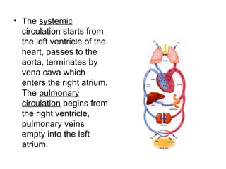
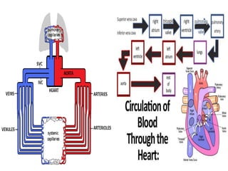
• The systemic
circulationstarts from
the left ventricle of the
heart, passes to the
aorta, terminates by
vena cava which
enters the right atrium.
The pulmonary
circulation begins from
the right ventricle,
pulmonary veins
empty into the left
atrium.
5.
The indicators ofheart
functional statement
• Quantity of blood pumping to the
arterial system in 1 min is called
minute volume of blood. It same for
both ventricles and equal 4,5 -5 liters
at rest condition.
• Systolic volume is quantity of blood
pumping to the arterial system during
one systole equal 65-70 ml.
6.
The indicators ofheart
functional statement
• Minute volume of circulation is
characterized by total amount of
blood , pumping by left and right
parts of heart in 1 min.
• Cardiac index is relation of minute
volume over total body surface in
meters.
7.
Physiology of myocardium
Structural-functionalunit of myocardium is
myocardiocyte connecting in series by discs
and forms muscle fiber.
There are two types of muscle fibers in the heart
depending on morphological structure and
functional accomplishment:
• Muscle fibers of working myocardium.
• Muscle fibers of conducting system of the
heart.
8.
Physiology of myocardium
Meanmass occupies working muscle
fibers, they carry out pumping function.
The second type are atypical, specific
muscle fibers responsible for
conduction of excitation from the
pacemaker to muscle fibers of working
myocardium or contractile muscle
fibers.
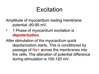
Excitation
Amplitude of myocardiumresting membrane
potential -80-90 mV.
• 1 Phase of myocardium excitation is
depolarization.
After stimulation of the myocardium quick
depolarization starts. This is conditioned by
passage of Na+ across the membranes into
the cells. The alteration of potential difference
during stimulation is 100-120 mV.
11.
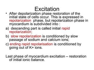
Excitation
• After depolarizationphase restoration of the
initial state of cells occur. This is expressed in
repolarization phase, but repolarization phase in
myocardium is subdivided into :
a) descending part is called initial rapid
repolarization;
b) slow repolarization is conditioned by slow
passage of sodium and calcium ions;
c) ending rapid repolarisation is conditioned by
going out of K+ ions.
Last phase of myocardium excitation – restoration
of initial ionic balance.
12.
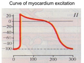
Curve of myocardiumexcitation
• Amplitude of myocardium resting
membrane potential is -80 - -90 mV
• Amplitude of myocardium resting
membrane potential is -80 - -90 mV
• Amplitude of myocardium resting
membrane potential is -80 - -90 mV
13.
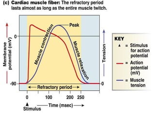
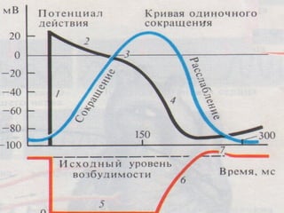
Excitability
Significant peculiarity ofthe myocardial
action potential is prolonged
repolarization phase. This property
prevents the myocardium from
repeated stimulation. Duration of
action potential of ventricle
myocardium 0.3 sec.
14.
Excitability
• Period I– absolute rerefractory state
(0,27) is characterized by total
inexcitability even to suprathreshold
stimuli. This period corresponds to
depolarization, initial rapid and slow
repolarization phases of action
potential and this period extends
throughout the whole period of
contraction.
15.
Excitability
• For thisreason heart muscle
can not be tetanised. This long
refractory period ensures
sufficient time for recovery of
the cardiac muscle. This is the
reason why cardiac muscle
can’t be fatigued.
17.
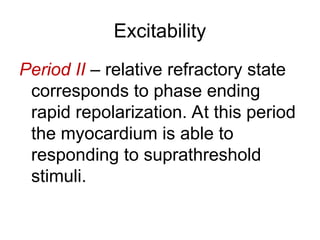
Excitability
Period II –relative refractory state
corresponds to phase ending
rapid repolarization. At this period
the myocardium is able to
responding to suprathreshold
stimuli.
18.
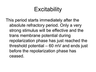
Excitability
This period startsimmediately after the
absolute refractory period. Only a very
strong stimulus will be effective and the
trans membrane potential during
repolarization phase has just reached the
threshold potential – 60 mV and ends just
before the repolarization phase has
ceased.
19.
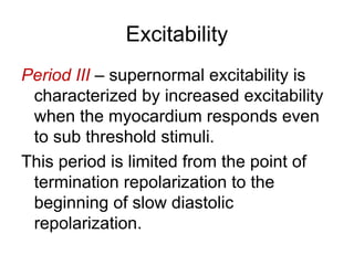
Excitability
Period III –supernormal excitability is
characterized by increased excitability
when the myocardium responds even
to sub threshold stimuli.
This period is limited from the point of
termination repolarization to the
beginning of slow diastolic
repolarization.
21.
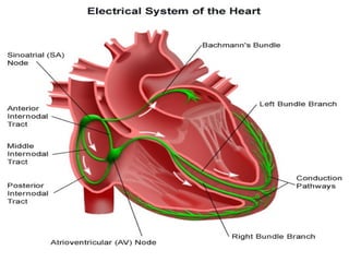
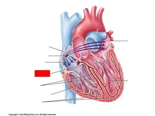
Conductivity
Main condition ofnormal activity of the heart
is anatomical wholeness of conducting
system. In the atrial myocardium excitation
spreads at a rate 0.8-1.0 m/sec. In the
atrio - ventricular region the impulses
transmitted along the bundle branch at a
higher velocity 4,5-5 m/sec. In the working
myocardium excitation is transmitted – 1
m/sec
22.
Conductivity
Atrio-ventricular delay: Adelay in the
conduction of impulses occurs when
excitation is propagated from atrial
muscle fibers to the cells of atrio-
ventricular node. Duration of this delay
0.08-0.09 sec.
Atrio-ventricular delay is responsible for
the fact that ventricular excitation
begins after atrial.
23.
Conductivity
This is importantphysiological property,
which ensures correlating the work of
different parts of the heart and rhythmical
pumping of blood.
The impulse from the bundle of His passes
quickly through the right and left bundle
branches and reaches the Purkinje fibers
and ventricular muscle fibers as well.
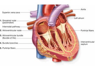
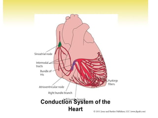
Automatism
Automatism of themyocardium is ability to
excitation under influence of impulses
arising in the heart itself without influences
of other stimulation.
• Nature of automatism is slow spontaneous
depolarization of myocardiocytes in the
pacemakers (automatically centers).
26.
Automatism
• Automatism centerof I order: Sino-atrial
node. This is situated in the right atrium
near the vena cava. Sino-atrial node
generates impulses by frequency 60-80
per min.
• So sino-atrial node regulates rhythm of
cardiac contractions. This is governor of
the heart.
27.
Automatism
Automatism center ofII order: Atrio-ventricular ore
Assof-Tawara node is located in right atrium in
interatrial septum. This center produces
impulses 40-50 per minute. Atrio-ventricular
node gives rises to Gis’s bundle. This is
muscular bridge conducting impulses from atria
to ventricles. Common branch of Gis’s bundle is
divided into 2 peduncles: the right providing the
right ventricle excitation and the left provide the
left ventricle.
28.
Automatism
Automatism center ofIII order: Purkinje’s
fibers. The terminal branches of the
conducting system are represented by a
network of Purkinje’s fibers distributed in
the sub-endocardial tissue that form
anastomoses with muscle fibers of the
myocardium. Frequency of impulses 20
per min.
29.
Automatism
• Law ofautomatism gradient.
According to this law diminution of
frequency of produced impulses
occur from sino-atrial node to
Purkinje’s fibers.
33.
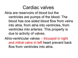
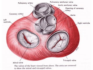
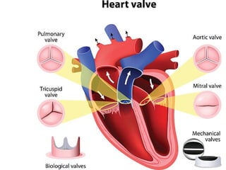
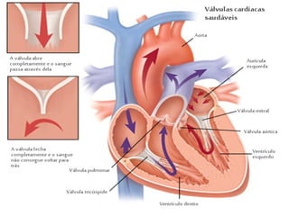
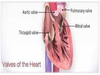
Cardiac valves
Atria arereservoirs of blood but the
ventricles are pumps of the blood. The
blood has one sided blood flow from veins
into atria, from atria into ventricles, from
ventricles into arteries. This property is
due to activity of valves.
Atrio-ventricular valves – tricuspid in right
and mitral valve in left heart prevent back
flow from ventricles into atria.
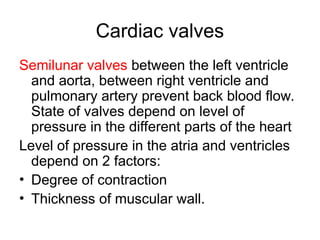
34.
Cardiac valves
Semilunar valvesbetween the left ventricle
and aorta, between right ventricle and
pulmonary artery prevent back blood flow.
State of valves depend on level of
pressure in the different parts of the heart
Level of pressure in the atria and ventricles
depend on 2 factors:
• Degree of contraction
• Thickness of muscular wall.
39.
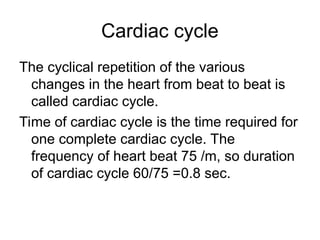
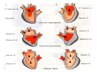
Cardiac cycle
The cyclicalrepetition of the various
changes in the heart from beat to beat is
called cardiac cycle.
Time of cardiac cycle is the time required for
one complete cardiac cycle. The
frequency of heart beat 75 /m, so duration
of cardiac cycle 60/75 =0.8 sec.
40.
Cardiac cycle
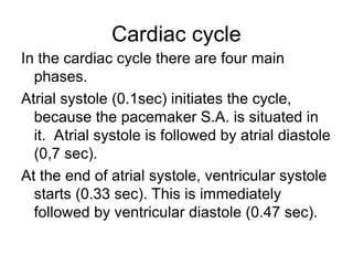
In thecardiac cycle there are four main
phases.
Atrial systole (0.1sec) initiates the cycle,
because the pacemaker S.A. is situated in
it. Atrial systole is followed by atrial diastole
(0,7 sec).
At the end of atrial systole, ventricular systole
starts (0.33 sec). This is immediately
followed by ventricular diastole (0.47 sec).
41.
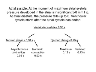
Atrial systole: Atthe moment of maximum atrial systole,
pressure developed in the atria is insignificant 5-8 mm Hg.
At atrial diastole, the pressure falls up to 0. Ventricular
systole starts after the atrial systole has ended.
Ventricular systole - 0.33 s
Tension phase - 0,08 s Ejection phase - 0.25 s
Asynchronous Isometric Maximum Reduced
contraction contraction 0.12 s 0.13 s
0.05 s 0.03 s
42.
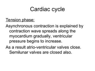
Cardiac cycle
Tension phase:
Asynchronouscontraction is explained by
contraction wave spreads along the
myocardium gradually, ventricular
pressure begins to increase.
As a result atrio-ventricular valves close.
Semilunar valves are closed also.
43.
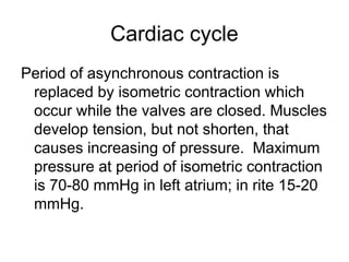
Cardiac cycle
Period ofasynchronous contraction is
replaced by isometric contraction which
occur while the valves are closed. Muscles
develop tension, but not shorten, that
causes increasing of pressure. Maximum
pressure at period of isometric contraction
is 70-80 mmHg in left atrium; in rite 15-20
mmHg.
44.
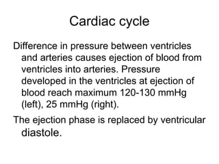
Cardiac cycle
Difference inpressure between ventricles
and arteries causes ejection of blood from
ventricles into arteries. Pressure
developed in the ventricles at ejection of
blood reach maximum 120-130 mmHg
(left), 25 mmHg (right).
The ejection phase is replaced by ventricular
diastole.
45.
Cardiac cycle
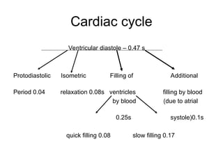
Ventricular diastole– 0.47 s
Protodiastolic Isometric Filling of Additional
Period 0.04 relaxation 0.08s ventricles filling by blood
by blood (due to atrial
0.25s systole)0.1s
quick filling 0.08 slow filling 0.17
46.
Cardiac cycle
Protodiastolic period– interval between relaxation
of the ventricles and closure of the valves.
Relaxation period when myocardium relaxes at
both closed valves, pressure in ventricles falls
below than in the atria, immediately a.v. valves
open, blood from the atria flows into the
ventricles and phase of maximum filling takes
place. The rises of pressure in the ventricles
causes phase of reduced filling.
48.
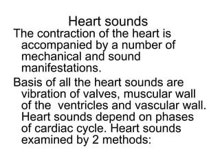
Heart sounds
The contractionof the heart is
accompanied by a number of
mechanical and sound
manifestations.
Basis of all the heart sounds are
vibration of valves, muscular wall
of the ventricles and vascular wall.
Heart sounds depend on phases
of cardiac cycle. Heart sounds
examined by 2 methods:
49.
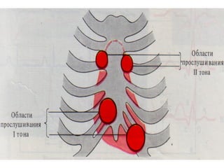
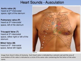
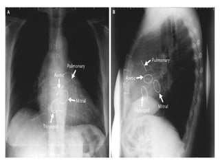
Heart sounds
1) Auscultation:By applying of phonendoscope on
external surface of chest.
2) Phonocardiography: This is method of graphical
recording of the heart sounds by means of
microphone connected to ECG machine. And
recording of heart sounds on the moving band of
paper. There are four heart sounds in human:
1st and 2nd heart sounds are examined by
auscultation: 3rd and 4th by
phonocardiography.
50.
Origin of HeartSounds
1st heart sound / Systolic is conditioned by
vibration of atrio-ventricular valves also
ventricular wall at closing. This
corresponds to period of isometric
contraction of ventricles. 1st systolic heart
sound is prolonged, dull and low, duration
0.12 sec.
51.
Origin of HeartSounds
2nd heart sound / Diastolic: Short,
high pitched, duration 0.08 sec.
2nd diastolic heart sound is
conditioned by closing of
semilunar valves. At ending of
ejection period.
52.
Origin of HeartSounds
3rd heart sound is due to vibration of
cardiac wall during maximum filling of the
ventricles.
4th heart sound occurs at atrial systole.
This is caused by vibration of ventricular
wall at additional filling of ventricles by
blood.
53.
Origin of HeartSounds
Apex beat:
This is short time protrusion of wall of the chest in
the 5th intercostal space along midclavicular line
in the left. Apex beat corresponds to tension
phase of ventricular systole of cardiac cycle.
Heart Murmurs:
They are pathological sounds , conditioned by
backflow of the blood in the parts of the heart.
At congenital and acquired valvular diseases.