
Recommended
Recommended
More Related Content
Similar to Cardiorenal Syndrome Classification, Pathophysiology, Diagnosis, and Treatment Strategies A Scientific Statement From the American Heart Association.pdf
Similar to Cardiorenal Syndrome Classification, Pathophysiology, Diagnosis, and Treatment Strategies A Scientific Statement From the American Heart Association.pdf (20)
Recently uploaded
Recently uploaded (20)
Cardiorenal Syndrome Classification, Pathophysiology, Diagnosis, and Treatment Strategies A Scientific Statement From the American Heart Association.pdf
- 1. Circulation. 2019;139:00–00. DOI: 10.1161/CIR.0000000000000664 TBD TBD, 2019 e1 ABSTRACT: Cardiorenal syndrome encompasses a spectrum of disorders involving both the heart and kidneys in which acute or chronic dysfunction in 1 organ may induce acute or chronic dysfunction in the other organ. It represents the confluence of heart-kidney interactions across several interfaces. These include the hemodynamic cross-talk between the failing heart and the response of the kidneys and vice versa, as well as alterations in neurohormonal markers and inflammatory molecular signatures characteristic of its clinical phenotypes. The mission of this scientific statement is to describe the epidemiology and pathogenesis of cardiorenal syndrome in the context of the continuously evolving nature of its clinicopathological description over the past decade. It also describes diagnostic and therapeutic strategies applicable to cardiorenal syndrome, summarizes cardiac- kidney interactions in special populations such as patients with diabetes mellitus and kidney transplant recipients, and emphasizes the role of palliative care in patients with cardiorenal syndrome. Finally, it outlines the need for a cardiorenal education track that will guide future cardiorenal trials and integrate the clinical and research needs of this important field in the future. Janani Rangaswami, MD, Vice Chair Vivek Bhalla, MD, FAHA John E.A. Blair, MD Tara I. Chang, MD, MS Salvatore Costa, MD Krista L. Lentine, MD, PhD Edgar V. Lerma, MD, FAHA Kenechukwu Mezue, MD, MSc Mark Molitch, MD Wilfried Mullens, MD, PhD Claudio Ronco, MD W.H. Wilson Tang, MD, FAHA Peter A. McCullough, MD, MPH, FAHA, Chair On behalf of the American Heart Association Council on the Kidney in Cardiovascular Disease and Council on Clinical Cardiology © 2019 American Heart Association, Inc. AHA SCIENTIFIC STATEMENT Cardiorenal Syndrome: Classification, Pathophysiology, Diagnosis, and Treatment Strategies A Scientific Statement From the American Heart Association Circulation https://www.ahajournals.org/journal/circ Key Words: AHA Scientific Statements ◼ acute kidney injury ◼ biomarkers ◼ cardio-renal syndrome ◼ chronic kidney disease ◼ dialysis ◼ diuretics ◼ heart failure ◼ hospitalization ◼ kidney transplantation ◼ mortality ◼ ultrafiltration T he nuanced and highly interdependent relationship between the kidney and the heart was described as early as 1836 by Robert Bright, who outlined the significant cardiac structural changes seen in patients with advanced kidney disease.1 Since then, numerous advances have been made in summarizing the car- diorenal link in terms of hemodynamic phenotypes, pathophysiology, therapeutic options, and clinical outcomes. The overlap of cardiovascular and kidney disease ex- tends across several interfaces. These include the hemodynamic interactions of the heart and kidney in heart failure, the impact of atherosclerotic disease across both organ systems, neurohormonal activation, cytokines, the biochemical perturbations across the anemia–inflammation–bone mineral axis in chronic kidney disease (CKD), and structural changes in the heart unique to kidney disease progression. However, the term cardiorenal syndrome (CRS) encompasses a spectrum of disorders involv- ing both the heart and kidneys in which acute or chronic dysfunction in 1 organ may induce acute or chronic dysfunction in the other organ. This scientific state- ment focuses primarily on the definition of, pathophysiology of, and diagnostic and therapeutic strategies in CRS. It also summarizes cardiorenal interactions in special populations such as patients with diabetes mellitus and kidney transplant (KT) recipients. Finally, it outlines the need for comprehensive cardiorenal trial end Downloaded from http://ahajournals.org by on March 17, 2019
- 2. Rangaswami et al Cardiorenal Syndrome TBD TBD, 2019 Circulation. 2019;139:00–00. DOI: 10.1161/CIR.0000000000000664 e2 CLINICAL STATEMENTS AND GUIDELINES points and the scope of a dedicated cardiorenal educa- tion track that will encapsulate the clinical and research needs of this important field for the future. METHODOLOGY The need for a comprehensive overview of the epide- miology of, pathophysiology of, diagnostic tools in, and therapeutic options for CRS was identified by the Council on the Kidney in Cardiovascular Disease of the American Heart Association (AHA). A writing group was commis- sioned to review the current literature and to develop an expert-based consensus summary on CRS. Members of the writing group were chosen for their expertise in heart failure, kidney disease, metabolic factors, and therapeutic strategies in the management of CRS. The writing group held a series of teleconferences and web-based com- munications from October 2017 tor2018. A manuscript outline was developed on the initial conference call, with individual section reviews being assigned to authors on the basis of their expertise. All authors had continuous access to the working document to provide input, and each section editor provided critical review and revisions. The writing group used MEDLINE (1966–pres- ent) and the Cochrane Central Register of Controlled Trials as the primary sources for the literature search, which was limited to human subjects and the English language. Related article searches were conducted in MEDLINE to find additional relevant articles. In addition, writing group members recommended articles outside the scope of the formal searches. Key relevant search words and Medical Subject Heading descriptors included kidney disease, renal insuf- ficiency, chronic renal/chronic kidney, acute kidney injury, end-stage renal or end-stage kidney disease, albu- minuria, congestive/myocardial/heart failure, cardiomy- opathy, cardiorenal, predialysism, and ultrafiltration. Key search abbreviations included CRS, CKD, CRF, CRD, AKI, RI, WRF, KT, CRT, ICD, CRT-D, ACEi/ARB, MRA, BB, ARNI, DM, T1DM, T2DM, SGLT-2 inhibitors, GLP-1 agonists, DPP-4 inhibitors, HF, HFrEF, HFpEF, and UF. (A full list of abbreviations, including search terms used in the manu- script, is available as an Online Appendix.) Finally, find- ings from conference proceedings, medical textbooks, and relevant online data sources were also reviewed. Certain topics within this statement may have been reviewed in other clinical practice guidelines and sci- entific statements published by other working groups, including AHA/American College of Cardiology task forces. When appropriate, these relevant guidelines have been referenced without the need to reiterate recommendations contained in those guidelines or statements. Suggestions/considerations agreed on by consensus within the writing group are included in spe- cific areas when there is a desire to provide some guid- ance to the cardionephrology community. CONFLICT OF INTEREST The AHA has a strict conflict-of-interest policy for all writing groups Each writing group member declared all relevant current conflicts, and 50% of the writing group were free of relevant conflicts. The chair and vice chair did not have any relevant industry-related con- flicts. The writing group members updated an electron- ic file of conflict-of-interest data from the beginning of the work until the article was published, and each member reported any new relevant conflicts at the be- ginning of each teleconference. See the Writing Group Disclosures table for details on individual conflict-of- interest reporting. DEFINITION AND PHENOTYPES OF CRS The first attempt at formally defining CRS came from the Working Group of the National Heart, Lung, and Blood Institute in 2004, which defined CRS as the re- sult of interactions between the kidneys and other cir- culatory compartments that increase circulating volume, which exacerbates the symptoms of heart failure (HF) and disease progression.2 The National Heart, Lung, and Blood Institute’s definition also stated that at its ex- treme, cardiorenal dysregulation leads to CRS, in which therapy to relieve congestive symptoms of HF is limited by further decline in renal function. This cardiocentric definition remains the cornerstone of CRS as commonly observed in the setting of acute decompensated HF, now called acute HF (AHF). Recognizing a wider clinical spectrum that may represent cardiorenal dysregulation, the Acute Dialysis Quality Initiative outlined a consensus approach in 2008 that phenotyped CRS into 2 major groups, cardiorenal and renocardiac syndromes, based on the primum movens of the disease process.3,4 This was further grouped into 5 subtypes based on disease acuity and sequential organ involvement, which are out- lined in Table 1. The goals of this consensus definition of CRS were to facilitate reliable characterization of the clinical presentation of cardiorenal dysregulation for di- agnostic and therapeutic purposes, to streamline inclu- sion criteria in epidemiological studies, to identify target treatment populations, and to develop novel diagnostic tools for the diagnosis and management of CRS. The Acute Dialysis Quality Initiative classification of CRS overcame some of the initial ambiguity in de- fining CRS and helped clinicians deliver phenotype- based goal-directed therapies for CRS at the bedside. Although simplifying the clinical approach to CRS, it also recognized the inevitability of overlap between dif- ferent phenotypes and the evolution of 1 subtype to the other during disease progression. However, in clini- cal practice, identifying the initial insult and subsequent events that result in decompensated acute or chronic CRS/renocardiac syndrome can be challenging. Several Downloaded from http://ahajournals.org by on March 17, 2019
- 3. Rangaswami et al Cardiorenal Syndrome Circulation. 2019;139:00–00. DOI: 10.1161/CIR.0000000000000664 TBD TBD, 2019 e3 CLINICAL STATEMENTS AND GUIDELINES complex interconnected pathways culminate in CRS, including diabetes mellitus, hypertension, HF, athero- sclerosis, endothelial cell dysfunction, anemia and dis- orders of iron metabolism, and chronic inflammation, many of which do not have well-defined temporal progression patterns. To this end, an alternative clas- sification of CRS based on the various clinical manifes- tations of CRS regardless of the initial organ of injury was proposed by Hatamizadeh et al5 that encompasses manifestations of hemodynamic compromise, uremic or vascular manifestations, neurohumoral disturbances, anemia/iron and bone mineral metabolism perturba- tions, and the malnutrition inflammation complex. Determining the significance of fluctuations in kidney function that meet the criteria for acute kidney injury (AKI) in the context of CRS represents a core challenge in standardizing its definition and phenotypes, particu- larly in the setting of AHF, in which decongestive thera- pies may complicate the assessment of biomarkers of renal function (especially for serum creatinine and urine output). Historically, the description of an acute decline in kidney function in the CRS literature has included the use of inconsistent terms such as kidney impairment and renal insufficiency, thus limiting accurate quantifi- cation of kidney injury and its clinical significance in a consistent fashion. Initial efforts toward standardizing the definition of AKI through the use of the RIFLE (risk, injury, failure, loss of kidney function, and end-stage kidney disease [ESKD]) criteria came from the Acute Di- alysis Quality Initiative in 20026 and were subsequently modified by the Acute Kidney Injury Network.7 The 2012 Kidney Disease: Improving Global Outcomes guideline on the evaluation and management of AKI harmonized these 2 sets of criteria to allow early AKI detection, to permit epidemiological comparisons, and to standardize entry criteria and end points in clinical trials.8 The standardized criteria for the diagnosis of AKI greatly improved the sensitivity of detection of AKI with emphasis on small fluctuations in serum creatinine and urine output; however, they may not represent true renal tubular injury when observed in the context of diuresis in the setting of AHF. Ahmad et al9 demonstrated that tubular injury quantified by validated urine biomarkers was not associated with worsening renal function esti- mated with cystatin C (CysC) with aggressive diuresis in patients with AHF. These findings suggest that small to moderate fluctuations in measurements of renal func- tion with clinically available biomarkers (such as serum creatinine) in the context of aggressive diuresis in AHF may be dissimilar from other causes of AKI such as sep- sis or drug-induced nephrotoxicity. Thus, underpinning the difference between true AKI with evidence of tubu- lar injury and pseudo-AKI or worsened renal function from functional changes in estimated glomerular filtra- tion rate (eGFR) is critical in preventing suboptimal de- livery of appropriate goal-directed therapies such as de- congestion and renin-angiotensin-aldosterone system (RAAS) inhibition in CRS.10 The cornerstone in making this distinction between AKI and worsened renal func- tion (without injury) in the setting of AHF, azotemia, and declining urine output rests on a combination of clinical assessment of perfusion status, relevant hemo- dynamic parameters (invasive and noninvasive), detec- tion of bedside markers of intrinsic renal injury evident on urine microscopy, and a thorough investigation of alternative explanations for worsening renal function. In the absence of evidence for intrinsic causes of kidney injury, small fluctuations in serum creatinine in the con- text of delivering appropriate goal-directed therapies in AHF may not have the same negative prognostic impact of AKI as seen with alternative causes9 and may rep- resent the effect of relative plasma underfilling or the therapeutic intended target effects of medical therapies for AHF, which are outlined in subsequent sections. To this end, the incorporation of novel biomarkers of car- diac and kidney injury to delineate the presence (or ab- sence) of organ damage and to guide therapeutic strat- egies in CRS represents a new dimension in improving the accuracy of the definition of CRS and its treatment targets for the future. Table 1. Classification of CRS Based on the Consensus Conference of the Acute Dialysis Quality Initiative Phenotype Nomenclature Description Clinical Examples Type 1 CRS Acute CRS HF resulting in AKI ACS resulting in cardiogenic shock and AKI, AHF resulting in AKI Type 2 CRS Chronic CRS Chronic HF resulting in CKD Chronic HF Type 3 CRS Acute renocardiac syndrome AKI resulting in AHF HF in the setting of AKI from volume overload, inflammatory surge, and metabolic disturbances in uremia Type 4 CRS Chronic renocardiac syndrome CKD resulting in chronic HF LVH and HF from CKD-associated cardiomyopathy Type 5 CRS Secondary CRS Systemic process resulting in HF and kidney failure Amyloidosis, sepsis, cirrhosis ACS indicates acute coronary syndrome; AHF, acute heart failure; AKI, acute kidney injury; CKD, chronic kidney disease; CRS, cardiorenal syndrome; HF, heart failure; and LVH, left ventricular hypertrophy. Downloaded from http://ahajournals.org by on March 17, 2019
- 4. Rangaswami et al Cardiorenal Syndrome TBD TBD, 2019 Circulation. 2019;139:00–00. DOI: 10.1161/CIR.0000000000000664 e4 CLINICAL STATEMENTS AND GUIDELINES PATHOPHYSIOLOGICAL MECHANISMS IN CRS The conventional explanation for the development of CRS in the setting of a cardiocentric primum movens focuses on the inability of the failing heart to generate forward flow, thus resulting in prerenal hypoperfusion. Inadequate renal afferent flow activates the RAAS axis, the sympathetic nervous system, and arginine vaso- pressin secretion, leading to fluid retention, increased preload, and worsening pump failure.11 However, the presence of a low-flow state only partly explains the pathophysiology of CRS. The ADHERE registry (Acute Decompensated Heart Failure National Registry) noted that the incidence of rising serum creatinine was similar among patients with AHF and reduced versus preserved systolic function.12 In addition, many patients hospital- ized with evidence of acute CRS have preserved or even elevated blood pressure and normal left ventricular (LV) ejection fraction (EF).13 The kidneys are not first in line for delivery of oxygenated blood, yet they receive a dis- proportionately large fraction (25%) of cardiac output (CO) because they are a low-resistance circuit. The dif- ference between arterial driving pressure and venous outflow pressures must remain sufficiently large for adequate renal blood flow and glomerular filtration. In this context, the concept of elevated central venous pressures (CVPs) resulting in renal venous hypertension, increased renal resistance, and ultimately impaired in- trarenal blood flow has been shown in early experimen- tal models14 and in more contemporary experiences in patients with AHF using invasive hemodynamic moni- toring.15,16 Merrill17 elegantly demonstrated large reduc- tions in renal blood flow in subjects with decompensat- ed HF with relative preservation of glomerular filtration rate (GFR). This was explained by a concomitant in- crease in filtration fraction derived from elevated intra- glomerular pressures from efferent arteriolar constric- tion in the setting of elevated renin levels. However, in severe decompensated HF with markedly elevated renal venous pressures and decreased renal blood flow, the compensatory increase in filtration fraction is lost and results in declining GFR.18 In this setting, the decrease in intraglomerular pressures and reduced GFR are driven by preglomerular vasoconstriction from extreme levels of RAAS and neurohumoral activation. In addition, the enhanced activation of the neurohumoral axis results in increased proximal tubular sodium and water reabsorp- tion to maintain effective plasma volumes, eventually resulting in oliguria and worsening congestion.19 These renal hemodynamic regulatory mechanisms are also the rationale behind the elevations in serum creatinine from decreased glomerular hydraulic pressures seen with the administration of RAAS inhibitors, with little changes in renal blood flow per se, and translate into true worsen- ing of renal function only when reductions in mean ar- terial pressure exceed renal autoregulatory capacity.18,20 This is the basis for the elevations in serum creatinine seen with RAAS inhibition in trials such as CONSEN- SUS (Cooperative North Scandinavian Enalapril Survival Study), which is discussed further in the RAAS Inhibi- tion in Chronic CRS section on pharmacotherapies.21 Finally, the low-resistance nature of the renal vascula- ture and parenchyma and the very low oxygen tension in the outer medulla also explain the unique sensitiv- ity of the kidneys to hypotension-induced injury. Thus, both hemodynamic instability and antecedent hypoten- sion should be considered in the consultative evaluation of a patient with developing CRS. In a post hoc analysis of the ESCAPE trial (Evaluation Study of Congestive Heart Failure and Pulmonary Artery Catheterization Effectiveness), right atrial (RA) pres- sure was the only hemodynamic parameter associated with baseline renal dysfunction.22 This observation was also confirmed in a broad spectrum of cardiovascular patients undergoing right-sided heart catheterization, in whom increased CVP was associated with reduced GFR and all-cause mortality.23 Along the same lines, elevated intra-abdominal pressures (IAPs) in the set- ting of AHF may contribute to renal dysfunction by causing renal compression and reduced perfusion.24 Hemodynamic metrics reflective of right ventricular (RV) function such as the RV stroke work index may have prognostic impact on kidney function in HF (including in patients with HF with preserved ejection fraction [HF- pEF]), thus underscoring the influence of RV function on renal hemodynamics.25 However, data on the neu- rohumoral perturbations and sodium and water reten- tion in isolated RV failure models in humans are scarce. Early experimental models inducing RV failure by grad- ed valvular damage showed a decrease in renal blood flow, preserved GFR, and intense salt and water reten- tion.26 Other investigators have shown that despite the presence of pulmonary baroreceptors, when CO is kept constant, pulmonary arterial (PA) distension did not have a direct effect on renal hemodynamics.27 The renal hemodynamic changes and the retention of sodium and water observed in patients with PA hyper- tension therefore may be mediated by systemic rather than PA baroreceptors, as has been shown in other edematous states.28 Thus, in the clinical context of CRS, the relative effects of declining RV function and elevated RV afterload on renal hemodynamics are less clear. The cardiorenal neural reflexes initiating from the PA circula- tion or the RV have not been well delineated, and the elevated levels of natriuretic peptides seen with PA hyper- tension/RV dysfunction do not account for the sodium avid state seen in RV failure, albeit their negative prog- nostic significance.29,30 Other mechanisms of the direct effect of RV dysfunction on renal hemodynamics include interventricular asynchrony and pericardium-mediated RV-LV interactions. This is a consequence of prolonged Downloaded from http://ahajournals.org by on March 17, 2019
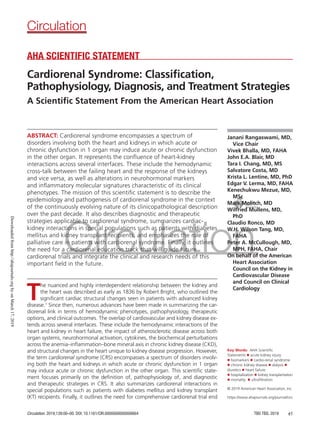
- 5. Rangaswami et al Cardiorenal Syndrome Circulation. 2019;139:00–00. DOI: 10.1161/CIR.0000000000000664 TBD TBD, 2019 e5 CLINICAL STATEMENTS AND GUIDELINES contraction of the RV free wall seen with RV pressure overload exceeding LV pressures in early diastole, re- sulting in paradoxical septal movement, which causes reduced LV end-diastolic filling.31,32 Finally, although RV function is a central determinant of CRS hemody- namics, surgical models such as the Fontan procedure demonstrate the ability to maintain CO and functional capacity by bypassing the RV in the presence of nor- mal LV function and the absence of pulmonary vascular disease.33,34 Several nonhemodynamic pathways that exacerbate cardiac or kidney injury are operative in CRS, central to which are activation of the sympathetic nervous sys- tem, chronic inflammation, imbalance in the proportion of reactive oxygen species/nitric oxide production, and persistent RAAS activation.35 Circulating levels of TNF- α (tumor necrosis factor-α), IL-1 (interleukin-1), and IL-6 (interleukin-6), which are elevated in experimen- tal models of AKI, have direct cardiodepressant effects such as a reduction in LVEF. Uremic cardiomyopathy (type 4 CRS) is characterized by significant burden of LV hypertrophy on which FGF-23 (fibroblast growth fac- tor-23) has recently been shown to have an indepen- dent causal effect.36 Because the hypertrophy of the LV is associated with a reduction in capillary density, partic- ularly in the central endocardium, it is conceivable that microvascular ischemia plays a role in the progression of uremic cardiopathy. Endothelial stretch from peripheral venous congestion causes conversion of vascular endo- thelium from a quiescent to a proinflammatory pheno- type, highlighting the importance of decongestion in CRS beyond its hemodynamic effects.37 Finally, data are emerging on the cross-talk between cardiac and kidney dendritic cells, which play a central role in innate and Figure 1. Pathophysiology of neurohumoral and inflammatory pathways involved in cardiorenal syndrome. α/π GST indicates α/π glutathione S-transferase; Alk Phos, alkaline phosphatase; ANP, atrial natriuretic peptide; AVP, arginine vasopressin; BNP, B-type natriuretic peptide; Cr, creatinine; GFR, glomerular filtration rate; GGT, γ-glutamyl transferase; IL-18, interleukin-18; KIM-1, kidney injury molecule-1; LDH, lactate dehydroge- nase; L-FAP, L-type fatty acid protein; NAG, N-acetyl-β-d-glucosaminidase; and NGAL, neutrophil gelatinase-associated lipocalin. Reprinted from Ismail et al39 with permission from Elsevier. Copyright © 2012, Elsevier. Downloaded from http://ahajournals.org by on March 17, 2019
- 6. Rangaswami et al Cardiorenal Syndrome TBD TBD, 2019 Circulation. 2019;139:00–00. DOI: 10.1161/CIR.0000000000000664 e6 CLINICAL STATEMENTS AND GUIDELINES adaptive immune responses in the context of CRS.38 The key pathophysiological pathways involved in CRS are outlined in Figure 1.39 DIAGNOSTIC STRATEGIES IN CRS HF is a complex mechanical and neurohumoral syn- drome resulting in stasis of blood in the lungs and periphery, causing the cardinal features of effort intol- erance and edema. Diagnosis of HF requires the pres- ence of signs and symptoms, along with evidence of a structural or functional cardiac abnormality,40 and in CRS, this requirement extends to the heart and kidneys. Several diagnostic tools help establish the structural and functional derangements characteristic of CRS, including biomarkers, noninvasive imaging modalities, invasive hemodynamic monitoring, and adjuvant vol- ume measurement techniques, which are summarized in the following sections. Biomarkers Biomarkers of cardiac and kidney injury may provide valu- able information when applied to the clinical context of CRS and can serve to indicate early cardiac or renal injury, the repair process, and long-term sequelae.41 They repre- sent an opportunity to prognosticate CRS, to discriminate between CRS phenotypes, and to serve as markers for tar- geted therapeutic interventions. Although biomarkers of myocardial injury (troponin) and wall tension (BNP [B-type natriuretic peptide]/NT-proBNP [N-terminal pro-BNP]) are routinely used in clinical practice, biomarkers of AKI are emerging as an additional dimension in diagnostic algo- rithms.ThedefinitionsofAKIusedtodayarelinkedtochang- es in creatinine or urine output, resulting in a significant time lag of 24 to 48 hours to institute corrective measures. Table 2 summarizes key biomarkers of CRS based on site of origin and diagnostic and prognostic value in AKI, HF, and, when applicable, CRS. Renal Biomarkers in CRS Markers of Glomerular Filtration and Integrity CysC and albuminuria represent biomarkers of glomer- ular filtration and integrity in CRS. CysC is a 13-kDa cys- teine protease, ubiquitous in all nucleated cells, that is produced at a constant rate, freely filtered, completely reabsorbed, and not secreted in renal tubules. In a sub- set of patients with chronic HF in the Cardiovascular Health Study, the highest quartile of serum CysC (1.55 mg/L) was associated with twice the risk of cardiovas- cular mortality adjusted for baseline characteristics.42 In patients presenting with AHF, serum CysC was a strong indicator of rehospitalization and short- and long-term mortality43,44 and had additive prognostic value when combined with other CRS biomarkers such as NT-proB- NP and cardiac troponin T.45 Similarly, albuminuria had a strong prognostic value for all-cause mortality, cardio- vascular death, and readmission in patients with HF in substudies of 3 major HF trials: CHARM (Candesartan in Heart Failure Assessment of Reduction in Mortality and Morbidity), GISSI-HF (Gruppo Italiano per lo Studio della Sopravvivenza nella Insufficienza Cardiaca–Heart Failure), and Val-HeFT (Valsartan in Heart Failure).46–49 It is important to note that biomarkers of glomerular integrity such as serum creatinine and CysC have differ- ing sources of bias when estimating GFR, particularly in advanced CRS.50,51 To this end, measurement of tubu- lar secretory clearance may provide different metabolic profiles of retained solutes eliminated by tubular secre- tion and filtration (eg, indoxyl sulfate and p-cresyl sul- fate) and thus refine the approach to quantification of kidney function and drug dosing and improve predic- tion of cardiovascular disease and kidney outcomes.52 Markers of Renal Tubular Injury Urine microscopy is a readily available clinical biomarker that has diagnostic value in distinguishing an intrinsic cause of AKI from functional changes in serum creati- nine in the setting of AHF. In addition, a urine sediment severity score based on the number of renal tubular epithelial cells and granular casts was shown to have prognostic value in the prediction of worsening AKI during hospitalization.53 Several novel urinary biomark- ers have shown promise in identifying tubular injury in AKI; some assays are available for in vitro use and are briefly described below. NGAL (neutrophil gelatinase-associated lipocalin), a 25-kDa protein found in neutrophil granules that is se- creted by renal tubular epithelium, myocardial cells, and other specific organ sites, has been extensively studied in CRS and has diagnostic and prognostic value in AHF and chronic HF. NGAL is the most upregulated protein produced by the kidneys in the setting of AKI. A meta- analysis of 10 studies involving ≈2000 patients with predominantly CRS identified early serum and urine NGAL measurements as predictors of dialysis and death with a pooled area under the curve of 0.78 and 0.75, respectively.54 Serial measurements of NGAL in AHF increase its predictive value for AKI, with the change in NGAL from baseline to peak producing an area under the curve of 0.91 compared with 0.69 for NGAL at ad- mission only.55 NGAL assays are available for clinical use outside but not within the United States. The combination of TIMP-2 (tissue inhibitor of metal- loproteinase-2) and IGFBP7 (insulin-like growth factor– binding protein 7), both tubular biomarkers involved in G1 cell cycle arrest during the early phase of cell injury, is available for clinical use in the United States. Kashani et al56 compared the performance of TIMP-2 and IGFBP7 in combination with other biomarkers of AKI in the SAPPHIRE validation cohort (Systolic and Pulse Pressure Hemodynamic Improvement by Restoring Elasticity) in Downloaded from http://ahajournals.org by on March 17, 2019
- 7. Rangaswami et al Cardiorenal Syndrome Circulation. 2019;139:00–00. DOI: 10.1161/CIR.0000000000000664 TBD TBD, 2019 e7 CLINICAL STATEMENTS AND GUIDELINES 728 critically ill patients without evidence of AKI at en- rollment. In this study, the combination of urine TIMP-2 and IGFBP7 was superior to previously described mark- ers of AKI (P0.002). Although the performance of TIMP-2 and IGFBP7 has been validated in several set- tings of AKI, the relationship between cell cycle arrest markers and CRS has not yet been described, and there are no reported studies of this biomarker combination measured serially in AHF. The promising markers of tu- bular injury in AKI and their specific role in CRS (if avail- able) are summarized in Table 2. Urinary biomarkers that correlate with measures of congestion such as BNP or NT-proBNP may play a role in phenotyping CRS in AHF and guide decongestive therapies.57 Perhaps the most critical role that novel AKI markers can have is in their negative predictive value in distinguishing functional serum creatinine fluctuations from true AKI. This distinction at the bedside may influ- ence or even guide the delivery of goal-directed therapy in CRS in the future; however, tubular biomarkers are influenced by the degree of baseline functioning renal tissue and thus may be inaccurate at low filtration rates, representing an important limitation of these markers. Finally, biomarkers that represent the transition to chro- nicity on the AKI-CKD continuum may help phenotype the shift from acute to chronic CRS and assist with ap- propriate clinical therapies and prognostication. Cardiac Biomarkers in CRS The “2017 ACC/AHA/HFSA Focused Update of the 2013 ACCF/AHA Guideline for the Management of Heart Failure” reiterated the existing Class 1A recom- mendation for the use of BNP and its inactive cleav- age proBNP in the diagnosis/exclusion of HF, as well as establishing prognosis and quantifying severity in AHF and chronic HF.58 Patients with CKD have higher Table 2. Biomarkers of Renal and Cardiac Injury Based on Site of Origin and Diagnostic and Prognostic Roles in AKI, HF, and CRS Biomarkers Characteristics/Site of Origin Diagnostic Value Prognostic Value Cardiac biomarkers cTn Marker of myocardial injury ACS ACS, HF, CKD BNP Marker of myocardial stretch HF, ACS, CRS HF, CRS sST2 Member of IL-1 family of receptors … HF, CRS Galectin-3 β-Galactoside binding lectin (intracellular and extracellular) … HF, CRS Kidney biomarkers Biomarkers of glomerular integrity Serum creatinine Skeletal muscle AKI, CRS HF, CRS CysC All nucleated cells CRS CRS Albuminuria Marker of glomerular integrity/PCT disruption CRS CRS Biomarkers of tubular injury TIMP*IGFBP7 Involved in G1 cell cycle arrest; may stimulate renal epithelium in an autocrine and paracrine fashion and sensitize for upcoming insults AKI AKI recovery Serum NGAL 25-kDa protein found in neutrophil granules; secreted by myocardium, renal tubules, activated immune cells, hepatocytes, lung, and colon AKI CRS Urine NGAL Loop of Henle, collecting ducts AKI, CRS CRS NAG PCT CRS, AKI CRS KIM-1 Type 1 cell membrane glycoprotein expressed in regenerating PCT epithelium AKI CRS IL-18 Cytokine mediating inflammation and AKI through the nuclear factor-κB pathway AKI CRS L-FABP Renal PCT AKI … H-FABP Cardiomyocytes, distal tubule HF, CRS … Urine angiotensinogen … AKI, CRS CRS α-1 Microglobulin Synthesized in liver; freely filtered through glomerular capillaries and reabsorbed by PCT AKI AKI recovery ACS indicates acute coronary syndrome; AKI, acute kidney injury; BNP, B-type natriuretic peptide; CKD, chronic kidney disease; CRS, cardiorenal syndrome; cTn, cardiac troponin; CysC, cystatin C; ellipses (...), data not available or reported.; HF, heart failure; H-FABP, heart-type fatty acid–binding protein; IGFBP7, insulin-like growth factor protein 7; IL, interleukin; KIM-1, kidney injury molecule-1; L-FABP, liver-type fatty acid–binding protein; NAG, N-acetyl-κ-d-glucosaminidase; NGAL, neutrophil gelatinase-associated lipocalin; PCT, proximal convoluted tubule; sST2, soluble suppressor of tumorigenicity; and TIMP, tissue inhibitor of metalloproteinase. Downloaded from http://ahajournals.org by on March 17, 2019
- 8. Rangaswami et al Cardiorenal Syndrome TBD TBD, 2019 Circulation. 2019;139:00–00. DOI: 10.1161/CIR.0000000000000664 e8 CLINICAL STATEMENTS AND GUIDELINES baseline BNP levels compared with matched patients with normal renal function because of impaired renal clearance (more notably with NT-proBNP), as well as chronic pressure/volume overload and CKD-associated cardiomyopathy.59,60 BNP levels are also significantly elevated in patients with evidence of CRS compared with patients with AHF without renal impairment.61 Future studies are necessary to determine the inter- pretation of fluctuations in natriuretic peptide levels in the context of administration of angiotensin receptor blocker (ARB)/neprilysin inhibitor therapy, especially in patients with CRS.62 ST2 (suppressor of tumorigenicity 2) is a decoy pro- tein produced by the endothelial cells lining the LV and aortic outflow tract in response to biomechanical strain. ST2 binds to the IL-33 (interleukin-33) receptor on car- diomyocytes and satellite cells in the heart, and instead of receiving favorable signal transduction, the ST2 effect results in myocyte dysfunction and tissue fibrosis. ST2 measurements offer incremental value to natriuretic peptides levels in predicting HF-related deaths and hospi- talizations and notably are not affected by renal function.58 Galectin-3 is a member of the β-galactoside– binding lectin family that is synthesized by cardiac macrophages and known to interact with specific ex- tracellular matrix proteins, including laminin, synexin, and integrins. In a recent study of 232 patients with New York Heart Association (NYHA) class III or IV HF, Lok et al63 used NT-proBNP and eGFR to adjust for severity of heart disease and degree of renal dysfunc- tion and demonstrated that serum galectin-3 levels were independent predictors of cardiovascular mor- tality.64 In a secondary analysis of the CORONA trial (Controlled Rosuvastatin Multinational Trial in Heart Failure) and COACH trial (Coordinating Study Evalu- ating Outcomes of Advising and Counseling Failure), patients whose galectin-3 levels increased by 15% over 3 to 6 months had a significantly increased adjusted risk for all-cause mortality and hospitalization for HF (HHF).65 Tang et al66 reported in a single-center study of subjects with chronic HF that higher galec- tin-3 levels were associated with worse renal function and poorer survival and that galectin-3 remained an independent predictor of all-cause mortality in a multi variate analysis of several factors, including eGFR. High-sensitivity cardiac troponins I and T are estab- lished diagnostic and prognostic markers in acute myo cardial infarction (MI). In addition to their diagnostic value, cardiac troponins have prognostic implications when elevated in acute decompensated HF even in the absence of myocardial ischemia or underlying coro- nary artery disease, and elevated levels are associated with a higher risk of death.58 The prevalence of el- evated cardiac troponins increases with declining GFR, and a sustained elevation is associated with a higher mortality risk.67 Imaging Modalities Up to 40% of patients hospitalized for AHF present with a type 1 CRS phenotype.68 Reduction in renal per- fusion pressure from elevated CVP plays a critical role, along with reduced CO in the pathogenesis of AKI in CRS.15 Noninvasive imaging modalities play an impor- tant role in establishing markers of venous conges- tion and impaired forward flow in CRS and are readily accessible clinical tools at the bedside. Echocardiog- raphy may help in diagnosing the congestive state by hemodynamic parameters, including CVP, systolic PA pressure, pulmonary capillary wedge pressure/left atrial pressure, and CO.69 Besides CVP, other useful echocar- diographic measurements include lateral and septal wall longitudinal motion (E′) in relation to the mitral inflow velocity (E). The E/E′ ratio directly correlates with pulmonary capillary wedge pressure, with an E/E′ 15 correlating to a pulmonary capillary wedge pressure of ≥18 mmHg.70,71 In addition, echocardiography carries prognostic value specific to phenotypes in CRS. In a retrospective cohort study in a large healthcare sys- tem, acute CRS (types 1 and 3) was associated with the highest risk of death compared with CKD without CRS (hazard ratio [HR], 3.13 [95% CI, 2.72–3.61]).72 Patients with CRS type 4 had better survival than patients with acute CRS (HR, 0.48 [95% CI, 0.37– 0.61]). Sixteen percent of patients with type 2 CRS and 20% of patients with type 4 CRS developed acute CRS, whereas 14% of patients with acute CRS progressed to CKD or chronic HF. Decreasing LVEF, increasing PA pressure, and higher RV diameter were independently associated with higher incidence of CRS. Renal ultrasonography and intrarenal venous flow patterns are emerging tools in identifying renal venous congestion and its clinical significance in CRS. Iida et al73 examined intrarenal venous flow patterns mea- sured by intrarenal Doppler ultrasound that were as- sociated with RA pressures and correlated strongly with clinical outcomes. In their study cohort of 217 patients hospitalized with AHF, 54% of subjects exhibited a continuous intrarenal venous flow pattern that invari- ably had low RA pressures (estimated 10 mmHg) and favorable prognosis (95% survival at 1 year). In contrast, about one-quarter of patients with discontinuous intra- renal venous flow, with either increased RA pressures (26%) or monophasic patterns (23%), had the poorest prognosis (40% survival at 1 year).73 In subjects with HF, intravascular expansion results in significant blunt- ing of renal venous flow before a significant increase in cardiac filling pressures is demonstrated and correlates with less diuretic efficiency.74 Other renal hemodynamic parameters such as renal arterial resistive index and re- nal perfusion index, although showing correlation with CVP, mean arterial pressures, and effective renal plasma flow, have not extended to being predictors of clinical Downloaded from http://ahajournals.org by on March 17, 2019
- 9. Rangaswami et al Cardiorenal Syndrome Circulation. 2019;139:00–00. DOI: 10.1161/CIR.0000000000000664 TBD TBD, 2019 e9 CLINICAL STATEMENTS AND GUIDELINES outcomes in CRS.73 Renal ultrasonography provides information on chronicity of disease using renal size, echogenicity, cortical thickness, and abnormal cortico- medullary ratios, which are helpful in identifying pro- gression from type 1 CRS to a more indolent type 2 CRS phenotype or establishing AKI or CKD as the primary perturbation in the clinical presentation of CRS.75 Uremic cardiomyopathy evolves through the course of progression of CKD, with subtle alterations in cardiac structure occurring even before a clinically significant decline in renal function.76 Speckle echocardiography with strain analysis allows a more detailed analysis of myocardial systolic function in the setting of normal LVEF and may have additive value over echocardio- graphic assessment of EF, including in uremic cardiomy- opathy (type 4 CRS).77 In a study of 40 control subjects and 90 patients with CKD across a range of eGFR, LV longitudinal systolic strain and early and late diastolic strain rates were significantly reduced in patients with CKD(−16.9±3.8%,1.6±0.5%,and1.3±0.4%inpatients with CKD versus −22.5±0.6%, 2.3±0.2%, and 1.9±0.1% in control subjects; P0.001 for all), despite overall pres- ervation of EF.78 Krishnasamy et al79 demonstrated that global longitudinal strain was a significant predictor of all-cause mortality in CKD (HR, 1.08 [95% CI, 1.01– 1.15]) in a single-center experience with 447 subjects. Cardiac magnetic resonance imaging is the standard noninvasive method of assessing ventricular dimen sions and function and fibrosis. Myocardial fibrosis in patients with uremic cardiomyopathy (type 4 CRS) occurs through multiple mechanisms not uniquely related to coronary artery disease. Early attempts to characterize and quantify myocardial fibrosis in ESKD with gadolinium-enhanced cardiac magnetic resonance imaging described a high prevalence of late gadolin- ium enhancement characteristic of coronary artery disease but also described a noninfarct pattern typical of more diffuse fibrosis.76 The limitations in the use of gadolinium in advanced CKD resulting from the risk of nephrogenic systemic fibrosis were overcome in 2 recent studies that described prolonged native T1 relaxation time and abnormal global longitudinal strain in patients with prevalent HFpEF undergoing hemodial- ysis compared with control subjects.80,81 The validation of non–gadolinium-based cardiac magnetic resonance in advanced CKD opens new possibilities in identifying subclinical LV dysfunction and has high potential as a tool for future studies in characterizing cardiac struc- ture in future cardiorenal studies. Volume Status Determination Strategies in CRS Fluid overload represents a core target for treatment in the process of optimizing the vicious cycle of CRS. However, the optimal method to assess fluid status and to determine dry weight and appropriate decongestion in decompensated HF or kidney disease remains an un- resolved issue. This section describes the role of several modalities available in conjunction with clinical assess- ment of volume status. Bioimpedance Vector Analysis Bioimpedance vector analysis (BIVA) is a noninvasive bedside volume assessment technique based on the electric principle that the body is a circuit with a given resistance (opposition of current flow through intra- cellular and extracellular solutions) and reactance (the capacitance of cells to store energy). With BIVA, total body water may be measured by placing a pair of elec- trodes on the dorsum of the wrist and ipsilateral ankle and then applying a 50-kHz current to the body. BIVA is displayed graphically so that relative hydration is de- picted as vector length. Shorter vectors are associated with volume overload, whereas longer vectors equate to volume depletion (Figure 2). BIVA has shown promis- ing results in distinguishing dyspnea caused by HF from other causes in patients presenting to the emergency department.82,83 BIVA has also been combined with BNP to guide discharge timing in patients with AHF,84 preventing AKI in the setting of high-dose diuretics for HF,85 and prognosticating patients with high risk of rehospitalization and cardiovascular mortality.86,87 In a recent study using a body composition analysis based on bioimpedance, a derived measure of fluid overload was found to be a key management parameter associ- ated with mortality on both the low and high ends of the measurement.88 Measurement of IAP In advanced HF, inefficient natriuresis with progressive volume overload may ultimately lead to a state of sys- temic congestion with increased IAP if the capacitance function of the splanchnic vasculature is insufficient.24 In 60% of patients admitted with AHF, measurements of IAP are elevated beyond the baseline value range of 5 to 7 mmHg.24 Bedside noninvasive measurements of IAP can be obtained with a urinary bladder catheter connected to a transducer. Reversing increased IAP by decongestive therapy ameliorates serum creatinine in this setting, presumably by alleviating abdominal congestion.89 Relative Blood Volume Monitoring Devices Devices that monitor relative blood volume have gener- ated interest in optimizing volume status in decompen- sated HF. Radiolabeled albumin tracer injections (BVA- 100, Daxor) are commercially available as a measuring tool for intravascular blood volume. A wide range of total blood volume values were reported in a small co- hort of patients hospitalized with AHF, with margin- ally reduced intravascular volume after diuretic therapy despite large reductions in body weight.90 It is unknown Downloaded from http://ahajournals.org by on March 17, 2019
- 10. TBD TBD, 2019 Circulation. 2019;139:00–00. DOI: 10.1161/CIR.0000000000000664 e10 CLINICAL STATEMENTS AND GUIDELINES Rangaswami et al Cardiorenal Syndrome whether the addition of blood volume measurement devices will affect clinical outcomes in patients with AHF in the context of CRS. Implantable Hemodynamic Monitoring Devices The CHAMPION trial (CardioMEMS Heart Sensor Allows Monitoring of Pressure to Improve Outcomes in NYHA Class III Heart Failure Patients) demonstrated a lower hospitalization rate (HR, 0.72 [95% CI, 0.59–0.88]) and a trend toward lower mortality (HR, 0.68 [95% CI, 0.45–1.02]) in 456 patients with HF with reduced ejection fraction (HFrEF) in the group who received PA pressure–guided HF management versus control sub- jects.91 Mean baseline eGFR in this study was 61.1±22.8 mL/min per 1.73 m2 for the study group and 62.3±23.4 mL/min per 1.73 m2 for the control group (P=0.69). The hospitalization reduction and survival benefit were am- plified by increasing the application of guideline-directed medical therapy. Currently, data on the efficacy of this device in patients with CRS or HF with advanced CKD are lacking. An implantable device (Optivol, Medtronic) has been used to assess transthoracic impedance as a measure of pulmonary fluid status.92 Direct measurements of in- trathoracic impedance with an implanted device have been shown to have prognostic value in HF.93 Howev- er, a reduction in outpatient visits for HF symptoms or hospital admissions with the use of device alerts has not been demonstrated.94,95 Specific data on outcomes with CRS using implantable intrathoracic impedance measurements are currently lacking. Invasive Hemodynamic Monitoring in CRS Routine evaluation of invasive hemodynamics has not been recommended in AHF because the ESCAPE trial did not show a reduction in either mortality or rehos- pitalizations with such a strategy in patients with equi- poise for right-sided heart catheterization.96 A post hoc Figure 2. Bioimpedance vector analysis (BIVA) in a patient undergoing ultrafiltration (UF). Relative hydration status is determined by the net vector of resistance to an applied current and reactance. Results from BIVA are compared with measurements made in healthy reference populations and are plotted as ellipses corresponding to the 50th, 75th, and 90th percentiles. Phase angle corresponds to the portion of electric current that is stored and subsequently released in a different phase and depends on cell integrity, cell membrane permeability, and total body water. BNP indicates B-type natriuretic peptide; and ECFV, extracellular fluid volume. Downloaded from http://ahajournals.org by on March 17, 2019
- 11. Rangaswami et al Cardiorenal Syndrome Circulation. 2019;139:00–00. DOI: 10.1161/CIR.0000000000000664 TBD TBD, 2019 e11 CLINICAL STATEMENTS AND GUIDELINES analysis of the ESCAPE trial showed that a PA cathe- ter–guided strategy was associated with less average increase in creatinine but did not decrease the inci- dence of defined worsening renal impairment during hospitalization or affect renal function after discharge relative to clinical assessment alone.22 Nevertheless, PA catheterization might still be warranted in patients with CRS who are difficult to treat, aiming to identify and treat subclinical congestion while avoiding intra- vascular underfilling and modulating hemodynamics to improve dual organ function. Common relevant sce- narios include underdiagnosis of culprit hemodynamic contributors such as pulmonary hypertension (PH) or cardiogenic shock, underestimation of valvular dysfunc- tion such as mitral regurgitation or tricuspid regurgita- tion, and accurate assessment of volume overload or RV failure. The RA/pulmonary capillary wedge pressure ratio, reflecting a disproportionate increase in RV to LV pressures, is inversely associated with eGFR in patients with AHF.97 Notably, cardiorenal hemodynamic mea- surements as assessed by invasive catheterization are confounded by the presence of elevated IAP or ascites, which represents a clinical caveat when PA catheteriza- tion is used in the context of CRS.24 The relative successes and failures of adjuvant meth- ods in assessing volume status and guiding diuresis or ultrafiltration goals depend on the degree of plasma re- fill in response to decongestive therapies. Sodium in the subcuticular and interstitial tissues, venous pressure, oncotic pressure, and several other poorly understood factors affect plasma refill rates with diuresis and ultra- filtration.98,99 23 Na-labeled magnetic resonance imaging has demonstrated Na+ in muscle and skin in patients with HF, and diuretic and ultrafiltration treatments can mobilize this Na+ deposition in varying rates.99,100 Thus, attempts at optimizing congestion in CRS with adjunct volume measurement techniques must factor in the limitations with predicting plasma refill rate with these devices, as well as the practical constraints of imple- menting clinically driven protocols based on theoretical extrapolations of volume assessment. TREATMENT STRATEGIES IN CRS Decongestive Therapies Diuretics Fluid retention and congestion are hallmarks of AHF, and diuretics are a cornerstone of the management in patients with or without CRS. Diuretics are com- monly prescribed (≈90% of patients with AHF),101 but unlike many other pharmacological therapies for HF that are supported by data from large clinical trials, evidence-based best clinical practices for diuretic use in HF remain uncertain, affording immediate relief of HF symptoms but no benefit in short- or long-term mor- tality or rehospitalization.102,103 The AHA and others recently endorsed diuretic use in HF with a Class I rec- ommendation based on expert opinion alone.58 Diuretic therapy is also standard of care for subjects enrolled in interventional clinical trials for HF. Loop diuretics (furosemide, bumetanide, torsemide, ethacrynic acid), named for their site of action in the loop of Henle of the nephron, represent the primary class of diuretics in HF. This section focuses on the effects of loop diuretics on renal hemodynamics and the physiology of diuretic resistance with relevance to CRS. Kidney Injury (Type 1 CRS) and RAAS Activation in Association With Loop Diuretics Loop diuretics inhibit the Na+ K+ 2Cl− cotransporter in the thick ascending limb of the loop of Henle, and Na+ K+ 2Cl− inhibition leads primarily to natriuresis and volume loss in edematous states such as HF. Loop di- uretics have a short duration of action, lasting 2 to 3 hours and up to 6 hours for an intravenous bolus and oral administration, respectively. Oral furosemide has ≈50% bioavailability with a wide range of values,104 explaining the variation in response to oral doses. In- travenous administration and novel subcutaneous infu- sions of furosemide ensure 100% bioavailability.105,106 Torsemide has a longer half-life and thus requires less frequent dosing.107 Given the more predictable oral bioavailability and longer half-life in patients with HF, torsemide may be more effective as a decongestive therapy compared with furosemide, as suggested by several small studies and a recent meta-analysis.108–110 Loop diuretics have multiple effects on neurohor- monal activation and renal and systemic hemodynam- ics that can predispose to kidney injury. Worsening kidney function in AHF (type 1 CRS) is associated with higher rehospitalization rates and mortality,111,112 and several studies have assessed the clinical benefit of dif- ferent dosing protocols for loop diuretics in AHF and their effect on kidney function. The DOSE-AHF trial (Diuretic Optimization Strategies Evaluation in Acute Heart Failure) randomized 308 patients with AHF to bolus versus continuous infusions of furosemide and a low-dose (intravenous equivalent of patient’s home diuretic dose) versus high-dose regimen (2.5 times the patient’s home loop diuretic dose intravenously) in a 2-by-2 factorial design model.113 In continuous ver- sus intermittent diuretic dosing, no significant differ- ences were observed in patients’ symptoms (P=0.47) or change in renal function (P=0.45); that is, no sig- nificant differences in the incidence of type 1 CRS were seen. However there was a trend in favor of the high-dose strategy compared with the standard dose in symptom improvement (P=0.06), without a signifi- cant difference change in renal function (P=0.21). The DIUR-AHF trial (Loop Diuretic Therapy in Acutely De- compensated Heart Failure) randomized 92 patients Downloaded from http://ahajournals.org by on March 17, 2019
- 12. Rangaswami et al Cardiorenal Syndrome TBD TBD, 2019 Circulation. 2019;139:00–00. DOI: 10.1161/CIR.0000000000000664 e12 CLINICAL STATEMENTS AND GUIDELINES with AHF to a bolus or continuous infusion strategy. Like the DOSE-AHF trial, there was no difference in mortality; however, the continuous infusion was as- sociated with greater rates of hyponatremia and the need for vasopressor infusion, and at 6 months, there were higher rates in the composite of rehospitalization or death.114 A post hoc analysis of 198 patients who developed type 1 CRS, pooled from 3 randomized clin- ical trials, DOSE-AHF, CARRESS-HF (Cardiorenal Rescue Study in Acute Decompensated Heart Failure), and ROSE-AHF (Renal Optimization Strategies Evaluation in Acute Heart Failure), compared a urine volume goal-di- rected stepwise diuretic algorithm and standard diuret- ic therapy. The stepwise algorithm aimed for a 24-hour urine volume between 3 and 4 L with furosemide with or without metolazone (a thiazide-type diuretic that inhibits sodium uptake in the downstream nephron segment) and showed more weight loss (−1.5±2.4 kg versus −0.4±1.5 kg; P0.001) and higher net fluid loss (1.705±1.417 L versus 0.892±1.395 L; P0.001) with an improvement in renal function (Δ serum creatinine, −0.1±0.3 mg/dL versus 0.0±0.03 mg/dL; P=0.03)115 compared with standard diuretic therapy. ROSE-AHF specifically compared the effect of low-dose dopamine, nesiritide, or placebo on decongestion and renal func- tion.116 In an ancillary study of ROSE-AHF, investigators measured biomarkers of kidney injury in individuals taking high-dose furosemide. In this analysis, kidney tubular injury detected by biomarkers did not appear to have an association with worsening renal function in the context of aggressive diuresis of individuals with AHF. Of note, the mean baseline eGFR was 44 mL/min per 1.73 m2 , providing relevance for individuals with type 1 and 2 CRS.9 Increases in NGAL, NAG (N-acetyl- β-d-glucosaminidase), and KIM-1 (kidney injury mol- ecule-1) were paradoxically associated with improved survival (HR, 0.80 per 10-percentile increase [95% CI, 0.69–0.91]). These studies in AHF would suggest that loop diuretics per se may not contribute to biomarker- associated renal injury, and a decrease in the eGFR may be a surrogate for severity of cardiac disease. On the basis of the analyses highlighted above, high-dose in- termittent furosemide appears to be safe and effective in AHF. Whether diuretics promote renal injury in indi- viduals with more severe baseline kidney function, for example, stage 4 or 5 CKD, is uncertain. Furthermore, without guidance from assessment of blood volume, rate of plasma refill, or measures of acute tubular in- jury, it is apparent that the use of diuretics in HF is empirical without a proven strategy associated with favorable outcomes from either observational studies or randomized trials. This raises the hope for future trials guided by these parameters to improve outcomes compared with usual care. The potentially deleterious effects of RAAS activa- tion by loop diuretics could theoretically limit the abil- ity to break the neurohormonal vicious cycle with AHF. However, in a follow-up analysis of DOSE-AHF and CARRESS-HF, high-dose loop diuretic therapy did not result in RAAS activation greater than that with low- dose diuretic therapy. In fact, ultrafiltration resulted in a greater increase in plasma renin activity than stepwise pharmacological care. Neither plasma renin activity nor aldosterone was significantly associated with short- term outcomes in AHF and CRS.117 This emphasizes the key concept that blood volume represents a small com- ponent of extracellular volume from which fluid losses are mobilized in the short term by diuretics or ultra- filtration. Reductions in extracellular fluid volume are further limited by the degree of plasma refill from the extracellular fluid into the intravascular space, the im- pairment of which further triggers endogenous produc- tion of hormones such as angiotensin II and vasopres- sin. Thus, a careful clinical assessment of the degree of plasma refill is critical in minimizing triggering of the adaptive neurohormonal responses to impaired plasma refill when decongestive therapies are administered. Diuretic Resistance Diuretic resistance is defined as the attenuation of the maximal diuretic effect that ultimately limits sodium and chloride excretion and is a well-characterized phenom- enon of diuretic use. In contrast to the lack of kidney injury associated with diuretic use,9 diuretic resistance is associated with renal impairment, increased risk of rehospitalization after HF, and mortality.118,119 Several factors contribute to diuretic resistance, in- cluding drug pharmacokinetics and pharmacodynam- ics, the braking phenomenon, and tubular remodeling (Figure 3). Free, unbound loop diuretics must reach the urinary lumen of the thick ascending limb and bind to the site of chloride entry to inhibit Na+ K+ 2Cl− . There- fore, for outpatient therapy, oral bioavailability is the first line of resistance. All loop diuretics are not created equal. Bumetanide and torsemide have higher bioavail- ability than furosemide.120 HF and food intake can pro- long time to peak concentration and the peak drug lev- els.121 Because loop diuretics are 95% protein bound, hypoalbuminemia increases the volume of distribution and reduces the availability of loop diuretics for facili- tated diffusion. Nonsteroidal anti-inflammatory drugs and uremic toxins can also competitively inhibit drug transport across proximal tubular epithelial cells. Specific factors related to CRS promote diuretic re- sistance. The bioavailability of loop diuretics is similar, but CKD reduces excretion of diuretic into the tubu- lar lumen. CKD does not limit the peak effect of drug delivered to the lumen. Overall diuretic-induced sodi- um excretion is reduced in CKD by the reduced and diminished filtered load of sodium. Thus, administra- tion of effective doses multiple times per day can cir- cumvent the above constraints.122,123 HF also reduces Downloaded from http://ahajournals.org by on March 17, 2019
- 13. Rangaswami et al Cardiorenal Syndrome Circulation. 2019;139:00–00. DOI: 10.1161/CIR.0000000000000664 TBD TBD, 2019 e13 CLINICAL STATEMENTS AND GUIDELINES the peak effect of the drug, which may be caused by increased proximal reabsorption of sodium (eg, result- ing from RAAS activation) or increased expression of Na+ K+ 2Cl− .124 These changes necessitate more frequent dosing rather than dose escalation to achieve maximal sodium excretion. Diuretic use (eg, in chronic HF and in type 1 or 2 CRS) can induce the braking phenomenon in the short term and distal tubular hypertrophy in the long term. The braking phenomenon refers to diminished diuretic ef- ficacy with each successive dose. The effect is observed within hours, but the mechanism is unclear. Sodium loss is thought to play a role in the upregulation of proximal and distal sodium transporters, and sodium repletion can attenuate this compensation125 and, in turn, the braking phenomenon. A recent study including indexes of proximal versus sodium reabsorption in subjects with HF treated with furosemide indicates that enhanced distal sodium transport, more than proximal transport, attenuates the maximal efficacy of furosemide.126 This nephron-specific element of diuretic resistance is also more consequential than delivery of the loop diuretic to the site of action127 and forms the rationale for use of thiazide-type diuretics to augment furosemide-induced sodium excretion. Whether the concept of diuretic syn- ergy can be transferred to HF and to CRS is uncertain. A large-scale randomized clinical trial of thiazide-type di- uretics as an adjunct to furosemide in HF or CRS is lack- ing. However, the ATHENA-HF trial (Efficacy and Safety of Spironolactone in Acute Heart Failure) tested spi- ronolactone, a potassium-sparing diuretic that targets another hypertrophied downstream nephron segment, versus placebo and did not demonstrate significant clin- ical benefit.128 Recent data suggest that hypochloremia plays a critical role in neurohormonal activation in pa- tients with HF on high-dose loop diuretics, which may contribute to diuretic resistance in these subjects.129 Diuretic Efficiency The concept of diuretic efficiency focuses on quantify- ing the renal response to a fixed dose of a loop diuretic using net fluid output in milliliters or weight change in kilogram per 40 mg furosemide equivalent130 or natri- uretic response to continuous intravenous furosemide defined as urine sodium to urine furosemide ratio.131 Diuretic efficiency may serve as a prognostic marker in CRS. Patients with diuretic efficiency below the median in the ESCAPE trial experienced nearly 3 times the risk of death compared with those patients with diuretic efficiency above the median, despite adjustment for baseline and in-hospital characteristics (HR, 2.86 [95% CI, 1.53–5.36]).130 As another measure of diuretic ef- ficiency, Singh et al131 measured the ratios of urine so- dium to urine furosemide in 52 patients hospitalized with AHF on continuous furosemide infusions. Patients with a ratio of urine sodium to urine furosemide 2 mmol/mg (indicative of low diuretic efficiency) experi- enced less weight loss and fluid removal in the first 24 hours and were at significantly increased risk for death, HF rehospitalization, and cardiac transplantation in an adjusted multivariate analysis (HR, 2.2 [95% CI, 1.08– 4.49]). In addition, these patients were more likely to experience worsening renal function in the context of decongestive therapies. Thus, measurements of diuretic efficiency may help to identify individuals who develop diuretic resistance and to identify a higher-risk subset of patients with CRS with worse outcomes. Further stud- Figure 3. Mechanisms of diuretic resistance in cardiorenal syndrome. Several extrarenal and renal factors impede the delivery of diuretic to the site of action in the nephron. After initial efficacy, diuretics become less effective because of the braking phenomenon and distal tubular remodeling. Potential strategies to overcome diuretic resistance include increased dose, frequency, and combination diuretic therapy. CCD indicates cortical collecting duct; CNT, connecting tubule; cTAL, cortical thick ascending limb; DCT, distal convoluted tubule; mTAL, medullary thick ascending limb; OMCD, outer medullary collecting duct; and PT, proximal tubule. Downloaded from http://ahajournals.org by on March 17, 2019
- 14. Rangaswami et al Cardiorenal Syndrome TBD TBD, 2019 Circulation. 2019;139:00–00. DOI: 10.1161/CIR.0000000000000664 e14 CLINICAL STATEMENTS AND GUIDELINES ies on the utility of diuretic efficiency in guiding tar- geted treatment strategies in CRS are necessary. Ultrafiltration Ultrafiltration, achieved by passing blood through hol- low fibers made of semipermeable material while ap- plying a negative pressure to the space surrounding the fibers, causes isotonic fluid to be removed from the in- travascular space. The composition of ultrafiltrate con- trasts with the much lower sodium content in the urine produced by loop diuretics132 and allows decongestion without the use of loop diuretics, with potential ben- efits including less potassium wasting, less renin and aldosterone release, and increased sodium loss. Thus, the optimal mode of decongestion in AHF using diure- sis versus ultrafiltration has been the subject of clinical trials, and key aspects of the randomized trials in this field are summarized in Table 3. The UNLOAD trial (Ultrafiltration Versus Intravenous Diuretics for Patients Hospitalized for Acute Decompen- sated Heart Failure) randomized 200 patients within 24 hours of hospitalization for AHF to either loop diuret- ics or ultrafiltration.134 The primary end of weight loss at 48 hours was significantly higher in the ultrafiltra- tion group (5.0±0.68 kg versus 3.1±0.75 kg; P=0.001), whereas dyspnea scores between the groups were not significantly different. There was a significant reduc- tion in 90-day rehospitalization rates in the ultrafiltra- tion arm, a secondary end point. Although UNLOAD demonstrated no differences in episodes of hypoten- sion within the first 48 hours or serum creatinine at 90 days between the 2 groups, it was unclear whether the secondary outcome of reduced readmissions at 90 days could have been achieved in the diuretic arm with more aggressive dose escalation. CARRESS-HF was a landmark study that enrolled 188 patients admitted with AHF and worsening renal function.135 Of all randomized trials for ultrafiltration in AHF, CARRESS-HF represents the only study that in- cluded patients with type 1 CRS. The primary end point was a bivariate change in weight and creatinine at 96 hours after randomization. No significant differences in weight loss were noted between the 2 groups (5.5±5.1 kg in the diuretic group versus 5.7±3.9 kg in the ultra- filtration group; P=0.58). The ultrafiltration group had an increase in serum creatinine of 0.23 mg/dL versus a decrease of 0.04±0.53 mg/dL in the diuretic group (P=0.003). In addition, the patients in the ultrafiltra- tion group experienced a higher rate of adverse events (72% versus 53%; P=0.03). The contrasting results between CARRESS-HF and UNLOAD highlight the nuances in study design, patient selection, and therapeutic algorithms unique to each study. Patients in CARRESS-HF had to demonstrate worsening renal function (CRS) to qualify for inclusion, signifying a sicker group of patients. In addition, ultra- filtration protocols were at fixed rates in CARRESS-HF, which physiologically contrast the documented de- crease in plasma refill rates with continuous ultrafiltra- tion.138 The glomerular filtration and tubular secretion of creatinine with diuresis differ from removal of creati- nine with ultrafiltration with a sieving coefficient of 1 and may not represent the actual effects of either ther- apy on renal function. Despite these issues, CARRESS- HF provided a strong argument against the use of ultra- filtration as primary treatment in patients with type 1 CRS. The AVOID-HF trial (Aquapheresis Versus Intrave- nous Diuretics Hospitalizations for Heart Failure), which sought to address these criticisms with a stepped-up diuretic algorithm and a detailed ultrafiltration proto- col, was terminated before completion because of slow enrollment.137 In the 224 patients who completed the protocol, nonsignificant trends toward reduced HF re- admissions at 90 days were achieved, but an increase in adverse events was also reported in the ultrafiltration group (14.6% versus 5.4%; P=0.026). Future studies that address the utility of ultrafiltration in patients with functional diuretic resistance and frequent readmis- sion for AHF are necessary to see whether clinically and economically meaningful outcomes can be achieved in these high-risk populations. Neurohormonal Modulation and Vasodilator and Inotropic Therapy The maladaptive neurohumoral responses in AHF resulting from type 1 CRS involve key vasoactive pep- tides such as vasopressin, endothelin, and adenosine and a diminished response to endogenous natriuretic peptides. In addition, the hemodynamic compromise that often accompanies HF may contribute to type 1 CRS. This section reviews pharmacological agents that affect neurohormones or improve hemodynamics that have been studied in the treatment of CRS. Arginine vasopressin is a nonapeptide hormone released by posterior pituitary and in conditions of elevated serum osmolarity, reduced cardiac index, or hypovolemia.139 Tolvaptan, a selective V2 receptor an- tagonist, causes aquaresis without loss of sodium. The EVEREST program (Efficacy of Vasopressin Antagonist in Heart Failure Outcome Study With Tolvaptan) evalu- ated the use of tolvaptan in AHF and LVEF 40% and showed similar rates of adverse events in the tolvap- tan and placebo groups with greater degrees of weight reduction in the tolvaptan arm in 2 short-term trials.140 No benefits in reduction in death or the composite of cardiovascular death and HHF were noted in the long- term trial.141 In TACTICS-HF (Targeting Acute Conges- tion With Tolvaptan in Congestive Heart Failure), the addition of tolvaptan to a standardized furosemide regimen did not improve the number of responders at 24 hours despite greater weight loss.142 Similarly, the Downloaded from http://ahajournals.org by on March 17, 2019
- 15. Rangaswami et al Cardiorenal Syndrome Circulation. 2019;139:00–00. DOI: 10.1161/CIR.0000000000000664 TBD TBD, 2019 e15 CLINICAL STATEMENTS AND GUIDELINES SECRET of CHF trial (Short Term Clinical Effects of Tolvaptan in Patients Hospitalized for Worsening Heart Failure With Challenging Volume Management) trial did not show significant improvement in dyspnea in patients with AHF who were selected for greater poten- tial benefit from tolvaptan.143 Although patients with AHF have elevated natriuret- ic peptides, the vasodilatory and natriuretic effects of the endogenous release of these substances are often not enough to overcome the hemodynamic effects of the other neurohormones mentioned. Nesiritide is a recombinant BNP with venous, arterial, and coronary vasodilatory properties that reduce afterload and in- crease CO without inotropic effects. It also causes na- triuresis, improves the GFR, and suppresses the RAAS axis.144,145 The ASCEND-HF trial (Acute Study of Clinical Effectiveness of Nesiritide and Decompensated Heart Failure) randomized 7141 patients with AHF to 1 to 7 days of intravenous nesiritide or placebo. The primary end point of dyspnea improvement, rehospitalization, or death was not statistically different between groups. The coprimary end point of dyspnea improvement at 6 and 24 hours was statistically higher in the nesirit- ide group, but this group also had more hypotension, and there were no differences in renal function.146 The ROSE-AHF trial randomized 360 patients with AHF in- dependent of LVEF and eGFR of 15 to 60 mL/min per 1.73 m2 at 1:1 to low-dose nesiritide or dopamine and, within each randomization, randomized them further at 2:1 into either active treatment or placebo infusions for 72 hours. Low-dose nesiritide had no significant effect on the coprimary end points of cumulative urine volume and change in serum CysC at 72 hours and no effect on the secondary end points reflective of decon- gestion, renal function, or clinical outcomes.116 Although theoretically attractive, neurohormonal modulation in the AHF setting has failed to improve hard clinical and renal end points in large randomized studies. Because of this, only tolvaptan and nesiritide have been approved for use by the US Food and Drug Administra- tion, and their use is limited to specific clinical situations. Inotropes have the potential to improve type 1 CRS by improving CO and reducing venous congestion. Specific inotropes such as dopamine have direct renal effects that may additionally result in improvement of type 1 CRS, but clinical data are mixed. A common theme in studies of inotropic therapy for AHF and reduced EF is that although favorable acute hemodynamic effects are achieved, long-term cardiovascular outcomes are not affected because of the presence of arrhythmias, isch- emia, and worsening long-term myocardial function.147 Dopamine is a catecholamine with effects on the β- and α-adrenergic receptors, as well as the renal dopa- minergic receptors, resulting in cardiac inotropy, systemic vasoconstriction, and improved renal blood flow.148 Early studies supported the renal protective effects of low-dose Table 3. Evidence Table of RCTs Comparing Pharmacological Therapy for Fluid Overload and Ultrafiltration in Patients With Acute Decompensated HF Study Subjects, n Primary End Point UF Protocol Diuretics Protocol Effect on Renal Function Effect on Weight Loss Adverse Events RAPID-CHF133 40 Weight loss at 24 h Single 8-h UF session to maximum rate of 500 mL/min per 1.73 m2 Clinician based NS Similar in both groups; trend toward higher weight loss in UF arm … UNLOAD134 200 Weight loss and dyspnea at 48 h Time and rate of UF flexible; maximum rate of 500 mL/min per 1.73 m2 Clinician based NS UFDT … CARRESS-HF135 188 Change in SCr and weight at 96 h Fixed UF rate of 200 mL/min per 1.73 m2 Prespecified stepped-up algorithm Significant increase in SCr with UF Similar in both groups Higher SAEs in UF arm CUORE136 56 Hospitalization for HF at 1 y Time and rate of UF flexible; maximum rate of 500 mL/min per 1.73 m2 Clinician based Significant increase in SCr with DT at 6 mo Similar in both groups … AVOID-HF*137 224 Time to HF 90 d after discharge Time and rate of UF flexible; maximum rate of 500 mL/min per 1.73 m2 Prespecified algorithm NS Similar in both groups Higher SAEs in UF arm AVOID-HF indicates Aquapheresis Versus Intravenous Diuretics Hospitalizations for Heart Failure; CARRESS-HF, Cardiorenal Rescue Study in Acute Decompensated Heart Failure; CUORE, Continuous Ultrafiltration for Congestive Heart Failure; DT, diuretic therapy; ellipses (...), data not available or reported.; HF, heart failure; NS, not significant; RAPID-CHF, Relief for Acutely Fluid Overloaded Patients With Decompensated Congestive Heart Failure; RCT, randomized controlled trial; SAE, serious adverse event; SCr, serum creatinine; UF, ultrafiltration; and UNLOAD, Ultrafiltration Versus Intravenous Diuretics for Patients Hospitalized for Acute Decompensated Heart Failure. *Trial terminated early. Data as reported on subjects enrolled until trial termination. Downloaded from http://ahajournals.org by on March 17, 2019
- 16. Rangaswami et al Cardiorenal Syndrome TBD TBD, 2019 Circulation. 2019;139:00–00. DOI: 10.1161/CIR.0000000000000664 e16 CLINICAL STATEMENTS AND GUIDELINES dopamine; however, subsequent studies demonstrated a lack of long-term clinical improvement in the treatment of AHF. Meta-analysis data have demonstrated improved urine output but no significant difference in change in cre- atinine, rehospitalization, or mortality with low-dose do- butamineusedinvariousclinicalscenarios.149 Asdiscussed, the ROSE-AHF trial showed no difference in the coprima- ry end points of cumulative urine volume and change in serum CysC at 72 hours or any effect on the secondary end points reflective of decongestion, renal function, or clinical outcomes when a 72-hour infusion of low-dose dopamine was compared with placebo in patients with AHF.116 Post hoc analysis demonstrated a differential ef- fect on 72-hour cumulative urine volume in favor of do- pamine in patients with LVEF ≤40% (P=0.029) compared with nesiritide in patients with LVEF 40% (P=0.001) but no differential effect in change in CysC (P=0.66), sug- gesting a worse clinical effect of low-dose dopamine in patients with HFpEF.150 Other novel inotropes such as levosimendan (calcium-sensitizing agent and potassium channel modulator) and omecamptiv mecarbil (cardiac myosin activators) have limited data in the context of CRS. Although progress has been made in the field of inotrope and vasodilator therapy, its long-term efficacy in the treatment of AHF and type 1 CRS is yet to be demonstrated. RAAS Inhibition in Chronic CRS Angiotensin-Converting Enzyme Inhibitors/ARBs Although the importance of RAAS inhibition in slowing CKD progression is well established, there is a paucity of data on clinically relevant long-term renal end points in trials on RAAS inhibition in HF. Given the known hemo- dynamic (and potentially reversible) effects of angiotensin blockade, interpreting fluctuations in serum creatinine as meaningful renal end points in the context of the use of angiotensin-converting enzyme (ACE) inhibitors and ARBs poses challenges in clinical practice. The benefits of ACE inhibitors in patients with HF and renal impairment have been demonstrated in observational data151,152 and post hoc analyses of randomized controlled trials (RCTs). These studies pertain specifically to the presence of preexisting renal impairment (type 2 or 4 CRS) in outpatient studies with HF, not to acutely decompensated subjects with CRS. CONSENSUS demonstrated a marked reduction in HF-associated mortality and symptom burden and was characterized by a doubling of serum creatinine in 11% of subjects taking enalapril compared with those taking placebo153 However, trends in serum creatinine rise were predominantly early and returned to within 30% of baseline values in most subjects, consistent with the known hemodynamic effects of ACE inhibi- tors, with the effect of concomitant diuretic use and hypotension being independent predictors of doubling of serum creatinine.21 SOLVD (Study of Left Ventricular Dysfunction) reiterated the benefits of enalapril for HF symptoms and hospitalization reduction (LVEF 35%, serum creatinine 2.5 mg/dL) in a much larger popula- tion compared with CONSENSUS (2569 versus 253 sub- jects).154 The enalapril group in SOLVD showed a 33% higher likelihood of a serum creatinine rise of 0.5 mg/ dL, but no data on progression of CKD, ESKD, or dou- bling of creatinine were reported. A post hoc analysis of SOLVD with HF and CKD demonstrated the mortality benefits even in subjects with higher degrees of CKD.155 The overall incidence of hyperkalemia was 6% overall with enalapril, correlating with the severity of renal dysfunction.156 However, in a meta-analysis of 5 place- bo-controlled RCTs of ACE inhibitors in HF by Flather et al,157 drug discontinuation was rarely necessary de- spite higher rates of AKI in the treatment arms versus placebo in most cases. A meta-analysis of 8 trials look- ing at the use of RAAS inhibition in KT demonstrated a higher risk of hyperkalemia (relative risk [RR], 2.44 [95% CI, 1.53–3.9]).158 The strength of evidence of ACE inhibitors in HF with predialytic CKD is not established given the lack of inclusion of these patients in RCTs for HF. Hospitalization and safety reporting data from the ongoing multicenter randomized controlled STOP-ACEi trial (Trial of Angiotensin-Converting Enzyme Inhibitor/ Angiotensin Receptor Blocker Withdrawal in Advanced Renal Disease; ISRCTN62869767) will shed light on the consequences of ACE inhibitors in advanced CKD and related cardiorenal outcomes. Although data on ARBs in CKD and HF specifically are sparse, in a propensity score analysis of 1665 patients with HF (EF 45%) and eGFR 60 mL/min per 1.73 m2 , treatment with an ACE inhibitor or ARB was associated with significant reduc- tions in all-cause mortality (HR, 0.68 [95% CI, 0.74– 0.996]; P=0.04)159 (Tables 4 and 5). The addition of ARBs to ACE inhibitors has been discouraged because of the increased risk of adverse events.176 Neprilysin/Renin-Angiotensin Inhibitors Trials that looked at outcomes with the combination of renin angiotensin system blocker/neprilysin inhibi- tion (sacubitril/valsartan and omapatrilat) provided an excellent opportunity to study the combined approach to RAAS blockade and vasodilator versus RAAS block- ade alone. A recent meta-analysis analyzed data from 3 trials in HFrEF that compared combined neprilysin/ RAAS inhibition with RAAS inhibition alone and in- cluded the following: IMPRESS (Inhibition of Metallo Protease by Omapatrilat in a Randomized Exercise and Symptoms Study of Heart Failure; n=573), OVERTURE (Omapatrilat Versus Enalapril Randomized Trial of Utility in Reducing Events trial; n=5770), and PARADIGM-HF (Prospective Comparison of ARNI With ACEI to Deter- mine Impact on Global Mortality and Morbidity in Heart Failure; n=8399).177 The composite outcome of death or HHF was reduced numerically in patients receiving Downloaded from http://ahajournals.org by on March 17, 2019
- 17. Rangaswami et al Cardiorenal Syndrome Circulation. 2019;139:00–00. DOI: 10.1161/CIR.0000000000000664 TBD TBD, 2019 e17 CLINICAL STATEMENTS AND GUIDELINES Table 4. Evidence Table of Outcomes in HF in Subjects With CKD Treated With ACE Inhibitors Study n Study Design Population CKD Concomitant Therapy Baseline Renal Function Outcome in CKD Group CONSENSUS160–162 235 RCT, enalapril vs placebo Patients with NYHA class IV HF Excluded: GFR 30 mL/min per 1.73 m2 CKD: 55% have Cr 1.58 mg/dL MRA 42% Digoxin 93% β-Blocker 3% Cr 1.45 mg/ dL GFR ≈47 mL/ min per 1.73 m2 Mortality: NS SOLVD Treatment154,163 2569 RCT, enalapril vs placebo HFrEF, EF ≤35%, symptomatic HF Excluded: Cr 2.5mg/dL CKD: CKD ≥3A (41%) CKD ≥3B (10%) MRA 9% Digoxin 67% β-Blocker 8% Cr 1.2 mg/dL Mortality: CKD ≥2: NS HR, 0.88 (95% CI, 0.73–1.06) CKD ≥3B: NS HR, 0.76 (95% CI, 0.54–1.08) HHF: CKD ≥3A: HR, 0.59 (95% CI, 0.48–0.73) CKD ≥3B: HR, 0.69 (95% CI, 0.46–1.02) SOLVD Prevention164 4228 RCT, enalapril vs placebo LV dysfunction EF ≤35%, NYHA class I/II Excluded: Cr 2.0 mg/dL MRA 4% Digoxin 12% β-Blocker 35% Cr 1.2 mg/dL No CKD analysis SAVE165,166 2183 RCT, captopril vs placebo MI with LV dysfunction EF 31% Excluded: Cr ≥2.5 mg/dL CKD: GFR ≥75 mL/min per 1.73 m2 : 37% GFR 75–60 mL/ min per 1.73 m2 : 30% CKD3A: 24% CKD ≥3B: 9% β-Blocker 35% Cr 1.3 mg/dL Mortality: HR, 0.79 (95% CI, 0.65–0.95) HF: HR, 0.69 (95% CI, 0.57–0.84) No subgroup HR in CKD NNT for MI, cardiovascular death, or HF: CKD vs non-CKD=9 vs 19 ATLAS167 3164; 405 not previously on ACE inhibitor RCT, lisinopril high dose vs low dose Symptomatic HF, EF ≤30% Excluded: Cr 2.5 mg/dL CKD: Cr 1.5 31% β-Blocker 11% Digoxin 67% Cr 1.3 mg/dL Adverse event in CKD: high dose vs low dose Hypotension: 31% vs 21.4% Renal dysfunction/ hyperkalemia: 15.7% vs 10% DIG Database168 1707 patients with CKD from DIG data set, 208 after match Propensity score analysis of DIG trial data, ACE inhibitor vs no ACE inhibitor Chronic HF with sinus rhythm, mean EF 28% Excluded: Cr ≥2.5 mg/dL CKD: Cr ≥1.5mg/dL for men and ≥1.3 mg/dL for women Digoxin 47% MRA 12% Cr 1.8 mg/dL GFR 40 mL/ min per 1.73 m2 All-cause mortality Not matched, adjusted: HR, 0.66 (95% CI, 0.49–0.90) Matched, adjusted: 0.58 (95% CI, 0.35–0.96) Berger et al169 4573 Retrospective, ACE inhibitor or ARB vs no ACE inhibitor or ARB Patients with CHF (Framingham criteria) with CKD CKD: CKD1: 22% CKD2: 25% CKD3: 37% CKD4: 11% CKD5: 7% β-Blocker 50% MRA 20% NA All-cause mortality: ACE inhibitor/ARB vs no ACE inhibitor Nondialysis CKD: 11% vs 41%, P=0.05 CKD2: 6.3% vs 8.6% CKD3: 5.4% vs 14% CKD4: 9.4% vs 18.5% Ahmed et al170 1340 Retrospective, propensity- matched analysis, ACE inhibitor/ ARB vs no ACE inhibitor/ARB HFpEF with CKD CKD: CKD ≥3 100% β-Blocker 20% MRA 10% Cr 1.7 mg/dL GFR 40 mL/ min per 1.73 m2 All-cause mortality: Not matched, adjusted: HR, 0.83 (95% CI, 0.72–0.96) Matched: HR, 0.82 (95% CI, 0.70–0.97) (Continued) Downloaded from http://ahajournals.org by on March 17, 2019
- 18. TBD TBD, 2019 Circulation. 2019;139:00–00. DOI: 10.1161/CIR.0000000000000664 e18 CLINICAL STATEMENTS AND GUIDELINES Rangaswami et al Cardiorenal Syndrome combined neprilysin/RAAS inhibition in all 3 trials, with a pooled HR of 0.86 (95% CI, 0.76–0.97; P=0.013). Combined neprilysin/RAAS inhibition compared with ACE inhibitor was associated with more hypotension but less renal dysfunction and hyperkalemia in all 3 tri- als. In the PARAMOUNT trial (Prospective Comparison of ARNI Versus ARB on Management of Heart Failure With Preserved Ejection Fraction), LCZ696 reduced NT-proBNP, blood pressure, and atrial size to a greater extent while preserving eGFR to a greater extent (36- week decline of GFR, 1.6 mL/min per 1.73 m2 in the LCZ696 group versus 5.2 mL/min per 1.73 m2 in the val- sartan group; P=0.007).178 In a subset analysis of PARA- DIGM-HF, treatment with sacubitril/valsartan resulted a slower rate of decrease in eGFR compared with enala- pril, including in patients with CKD, despite a modest increased in albuminuria.179 The HARP-III trial (UK Heart and Renal Protection III), which is a multicenter double- blind RCT comparing 97/103 mg of sacubitril/valsartan (2 times daily) with 300 mg of irbesartan (1 time daily) among 414 patients with CKD, will be the first test of an angiotensin receptor neprilysin inhibitor in patients with CKD with or without proteinuria.180 Mineralocorticoid Receptor Antagonists The long-term efficacy of achieving complete suppression of RAAS with an ACE inhibitor/ARB is limited by the phe- nomenon of aldosterone escape, resulting in an increased level of serum aldosterone. Mineralocorticoid receptor an- tagonists (MRAs), when added to an ACE inhibitor/ARB, can provide more suppression of RAAS with potential long-term cardiorenal benefits. The reduction in mortality and cardiovascular events with HFrEF was demonstrated in RALES (Randomized Aldactone Evaluation Study)181 and EPHESUS (Eplerenone in Post-Acute Myocardial Infarction Heart Failure Efficacy and Survival).182 In the EMPHASIS-HF trial (Eplerenone in Mild Patients Hospitalization and Sur- vival Study in Heart Failure), in which 33% of patients had an eGFR 60 mL/min per 1.73 m2 , the effect of eplerenone on the primary composite end point on HHF or cardiovas- cular death was consistent in patients dichotomized at an eGFR 60 mL/min per 1.73 m2 .183 Data on the safety and efficacy of MRAs in HF with advanced CKD (stage 4 and 5) are limited. However, in appropriately selected patients with symptomatic HFpEF, elevated BNP level, HF admis- sion within 1 year, eGFR 30 mL/min per 1.73 m2 , creati- nine 2.5 mg/dL, and potassium 5.0 mEq/L, particularly in those with elevated BNP levels, use of spironolactone might be considered with close monitoring of potassium and renal function58 (Table 6). Given the universal exclusion of moderate to severe CKD in HF outcomes trials and the lack of reporting on long-term renal outcomes, the true burden of hyper- kalemia in the management of chronic CRS is unclear. Collins and coauthors187 have recently demonstrated in a nationwide electronic medical record (n=1716141 with ≥2 potassium values) that the presence of HF in- creases the fatal risks of hyperkalemia in patients treated with RAAS inhibitors. In this analysis, the overall death rate was 35.7% with hyperkalemia in those subjects with HF, CKD, and DM compared with a death rate of 2.7% in control subjects. In a meta-analysis of clinical trials (n=16065 subjects), the rates of MRA-associated hyperkalemia (9.5%) were ≈2-fold that of control sub- Edner et al171 2410 Prospective, propensity- matched analysis, ACE inhibitor (67%)/ARB (31%)/ both 2% vs no ACE inhibitor/ARB HFrEF, EF ≤39% with CKD4 CKD ≥4: 100% β-Blocker 87% MRA 25% Digoxin 11% GFR 23 mL/ min per 1.73 m2 All-cause mortality: Matched adjusted: HR, 0.83 (95% CI, 0.73–0.94) Overall adjusted: HR, 0.81 (95% CI, 0.73–0.91) Gurwitz et al172 2414 HFrEF and HFpEF with chronic lung disease and CKD HFrEF 32% HFpEF 68% GFR 60 mL/min per 1.73 m2 NA NA HFrEF: All-cause mortality: HR, 0.6 (95% CI, 0.4–0.9) HHF: HR, 0.43 (95% CI, 0.28–0.67) HFpEF: All-cause mortality: HR, 0.5 (95% CI, 0.3–0.8) HHF: HR, 0.35 (95% CI, 0.18–0.68) ACE indicates angiotensin-converting enzyme; ARB, angiotensin receptor blocker; ATLAS, Assessment of Treatment With Lisinopril and Survival; CHF, congestive heart failure; CKD, chronic kidney disease; CONSENSUS, Cooperative North Scandinavian Enalapril Survival Study; Cr, creatinine; DIG, Digitalis Investigation Group; EF, ejection fraction; GFR, glomerular filtration rate; HF, heart failure; HFpEF, heart failure with preserved ejection fraction; HFrEF, heart failure with reserved ejection fraction; HHF, hospitalization for heart failure; HR, hazard ratio; LV, left ventricular; MI, myocardial infarction; MRA, mineralocorticoid receptor antagonist; NA, not applicable; NNT, number needed to treat; NS, not significant; NYHA, New York Heart Association; RCT, randomized controlled trial; SAVE, Survival and Ventricular Enlargement; and SOLVD, Study of Left Ventricular Dysfunction. Table 4. Continued Study n Study Design Population CKD Concomitant Therapy Baseline Renal Function Outcome in CKD Group Downloaded from http://ahajournals.org by on March 17, 2019
- 19. Rangaswami et al Cardiorenal Syndrome Circulation. 2019;139:00–00. DOI: 10.1161/CIR.0000000000000664 TBD TBD, 2019 e19 CLINICAL STATEMENTS AND GUIDELINES jects, and among hyperkalemic subjects, 54% were truly caused by the MRA agent.188 Incorporating the novel oral antihyperkalemic agents (patiromer acetate, sodium zirconium cyclosilicate) into the therapeutic armamentarium of chronic CRS may maximize the additive benefits of MRAs to ACE inhibitors/ARBs.189 β-Adrenergic Blockers β-Adrenergic blockers have been evaluated in numer- ous RCTs and shown to improve NYHA class and LVEF, to alleviate symptoms, to reduce hospitalization bur- den, and to prolong survival. β-Blockers that have been shown to reduce mortality in HF include metoprolol and bisoprolol (β-1 receptor blockers), and carvedilol (α-1, β-1, and β-2 receptor blockers) and are recommended as Class 1A evidence for HFrEF by the 2013 American College of Cardiology Foundation/AHA guidelines on the management of HF.190 Given the paucity of data on β-blockers specific to patients with CKD, the risk/ben- efit profiles of these drugs in CKD depend on post hoc analyses of major RCTs and observational data. The MERIT-HF study (Metoprolol CR/XL Controlled Randomized Intervention Trial in Chronic HF) randomized 3991 patients with NYHA class II to IV HF and EF 40% to metoprolol versus placebo. A secondary analysis that looked at the effects of metoprolol across eGFR ranges of 60, 45 to 60, and 45 mL/min per 1.73 m2 showed significant benefits across all subgroups.191 The benefits were more pronounced in the group with eGFR 45 mL/ min per 1.73 m2 , with a nearly 60% reduction in HHF and mortality. In the SENIORS study (Study of the Effects of Nebivolol Intervention on Outcomes and Rehospitaliza- tion in Seniors With Heart Failure), the composite of all- cause mortality and cardiovascular hospital admissions was significantly reduced in 2112 patients 70 years of age with HF who were randomized to nebivolol versus placebo.192 Although the benefits of nebivolol were ob- served across tertiles of eGFR, the benefit seen in the lowest eGFR group (55 mL/min per 1.73 m2 ) was not as robust as with MERIT-HF. The CIBIS-II study (Cardiac Insufficiency Bisoprolol Study) randomized 2647 patients with NHYA class III to IV HF with EF 35% to bisoprolol versus placebo.193 A serum creatinine of 3.4 mg/dL was a prespecified exclusion criterion. The beneficial effects of bisoprolol with significant reductions in all-cause mor- tality were observed across baseline GFR quartiles. Final- ly, a meta-analysis of 6 RCTs with β-blockers in patients with CKD and HF showed that β-blockers significantly reduced the risk of all-cause mortality (relative risk reduc- tion [RRR], 28%) and cardiovascular mortality (RRR, 34%) compared with placebo.194 Tolerability of β-blockers is limited by fluid retention, which may complicate the management of HF, bradycardia, hypotension, and fatigue. MERIT-HF showed similar rates of tolerance across eGFR ranges. However, in the post hoc analyses of CIBIS-II and SENIORS, rates of β-blocker discontinuation were higher in subgroups with eGFR 45 and 55 mL/ min per 1.73 m2 , respectively. Table 5. Evidence Table of Outcomes in HF in Subjects With CKD Treated With ARBs Study n Study Design Population CKD Concomitant Therapy Baseline Renal Function Outcome in CKD Group Val-HeFT48 5010 RCT, valsartan vs placebo Symptomatic HF, EF 40% Exclude: Cr 2.5 mg/dL CKD ≥2: 58% Proteinuria without CKD: 52% β-Blocker 35% Digoxin 67% GFR 58 mL/min per 1.73 m2 All-cause mortality: HR, 1.01 (95% CI, 0.85–1.20) CHARM- Overall173 7599 RCT, candesartan vs placebo Symptomatic HF, EF 40% Exclude: Cr 3 mg/dL CKD: Cr 2 mg/dL β-Blocker 55% MRA 17% NA Hyperkalemia: Cr 2 vs 2: HR, 4.1 (95% CI, 2.4–7.3) Serious hyperkalemia: Cr 2 vs 2: HR, 3.5 (95% CI, 1.5–7.9) HEAAL174 3846 High- vs low- dose losartan Symptomatic HF, EF 40%, intolerance of ACE inhibitor Exclude: Cr 2.5 mg/dL ACE inhibitor 100% β-Blocker 72% MRA 38% Cr 1.1 mg/dL Death and HF admission GFR: 60 mL/min per 1.73 m2 : HR, 0.98 (95% CI, 0.85–1.13) 60–74: HR, 0.94 (95% CI, 0.78–1.14) 75: HR, 0.72 (95% CI, 0.60–0.86) ELITE175 722 Captopril vs losartan Symptomatic HF, EF 40% Exclude: Cr 2.5 mg/dL ACE inhibitor 100% β -Blocker72% Cr 1.2 mg/dL Worsening renal function in all groups: 2% (−51% to 36%) ACE indicates angiotensin-converting enzyme inhibitor; ARB, angiotensin receptor blocker; CHARM, Candesartan in Heart Failure Assessment of Reduction in Mortality and Morbidity; CKD, chronic kidney disease; Cr, creatinine; EF, ejection fraction; ELITE, Evaluation of Losartan in the Elderly; GFR, glomerular filtration rate; HEAAL, Heart failure Endpoint Evaluation of Angiotensin II Antagonist Losartan; HF, heart failure; HR, hazard ratio; MRA, mineralocorticoid receptor antagonist; NA, not applicable; RCT, randomized controlled trial; and Val-HeFT, Valsartan in Heart Failure. Downloaded from http://ahajournals.org by on March 17, 2019
- 20. TBD TBD, 2019 Circulation. 2019;139:00–00. DOI: 10.1161/CIR.0000000000000664 e20 CLINICAL STATEMENTS AND GUIDELINES Rangaswami et al Cardiorenal Syndrome In summary, there are varying levels of evidence for goal-directed therapies for HF in the CKD population, with a relative paucity of data in patients with advanced CKD.195 Figure 4 provides a summary of the relative strengths of evidence in the use of goal-directed medi- cal therapies for HF across the spectrum of GFR ranges for nondialytic CKD. CARDIORENAL OUTCOMES IN TYPE 2 DIABETES MELLITUS Cardiovascular disease is a major cause of mortality in patients with type 2 diabetes mellitus (T2DM).196 Met- formin is highly effective, has a very low risk of hypo- glycemia, does not cause weight gain, and may reduce cardiovascular events and mortality. Therefore, it is generally recommended as first-line medical therapy for most patients with T2DM when added to lifestyle modification.197–199 However, many patients do not achieve adequate control with metformin alone, and second and even third medications are often neces- sary,198,199 Given the impact of glycemic control on car- diovascular outcomes and the increased cardiovascular risk that was associated with certain glucose-lowering medications, the US Food and Drug Administration outlined the need for cardiovascular safety studies for new glucose-lowering therapies in 2008.200,201 Sub- sequently, several trials have reported cardiovascular safety data across multiple classes of glucose-lowering drugs, including GLP-1 (glucagon like peptide-1) recep- tor agonists, DPP-4 (dipeptidyl peptidase-4) inhibitors, and SGLT-2 (sodium-glucose cotransporter 2) inhibitors, and other trials are ongoing at the time this statement was written. In this section, we highlight key aspects of recently reported safety and cardiovascular outcomes data of the major novel classes of antidiabetic therapy. SGLT-2 Inhibitors SGLT-2 inhibitors are one of the latest classes of glu- cose-lowering therapies available. One SGLT-2 inhibi- tor, empagliflozin, demonstrated impressive results in the multicenter randomized cardiovascular safety trial EMPA-REG OUTCOME (Empagliflozin Cardiovascular Outcome Event Trial in Type 2 Diabetes Mellitus).202 The EMPA-REG OUTCOME Trial randomized 7020 patients with T2DM at high risk for cardiovascular events to receive empagliflozin versus placebo. The trial showed a 14% RRR for the primary composite 3-point major adverse cardiovascular event outcome of cardiovascular death, nonfatal MI, and nonfatal Table 6. Evidence Table of Outcomes in HF in Subjects With CKD Treated With MRAs Study n Study Design Population CKD Concomitant Therapy Baseline Renal Function Outcome in CKD Group RALES184 1663 RCT, spironolactone vs placebo HF, EF 35% Exclude: Cr 2.5 mg/dL CKD: GFR 60 mL/min per 1.73 m2 (48%) ACE inhibitor 94% Digoxin 78% Cr 1.2 mg/dL All-cause mortality: HR, 0.68 (95% CI, 0.56–0.84) Worsening renal function: spironolactone vs placebo 17% vs 7% EMPHASIS-HF185 2737 Eplerenone vs placebo HF, EF35% Exclude: GFR 30 mL/min per 1.73 m2 CKD: CKD 3a: 33% ACE inhibitor 93% β-blocker 87% GFR 71 mL/min per 1.73 m2 HR, 0.66 (95% CI, 0.56–0.78) No difference between subgroups with and without CKD ARTS-HF186 1066 RCT, finerenone with dosage uptitrated vs eplerenone HFrEF with EF 40%, DM with CKD (GFR 30 cc/min per 1.73 m2 ), CKD without DM (GFR 30-60 cc/ min per 1.73 m2 ) Exclude: GFR 30 mL/min per 1.73 m2 CKD: CKD 3a: 71% NA GFR 53 mL/min per 1.73 m2 Decrease in BNP 30%: same in both groups Any adverse event: finerenone less than eplerenone (76.9%) except finerenone 15–20 mg (78.5%) Death, cardiovascular hospitalization, worsening CHF: finerinone better than eplerenone except finerenone 2.5–5mg Hyperkalemia: finerinone better than eplerenone except finerenone 15–20 mg ACE indicates angiotensin-converting enzyme inhibitor; ARTS-HF, Mineralocorticoid Receptor Antagonist Tolerability Study–Heart Failure; BNP, B-type natriuretic peptide; CHF, congestive heart failure; CKD, chronic kidney disease; Cr, creatinine; DM, diabetes mellitus; EF, ejection fraction; EMPHASIS-HF, Eplerenone in Mild Patients Hospitalization and Survival Study in Heart Failure; GFR, glomerular filtration rate; HF, heart failure; HFrEF, heart failure with reduced ejection fraction; HR, hazard ratio; MRA, mineralocorticoid receptor antagonist; NA, not applicable; RALES, Randomized Aldactone Evaluation Study; and RCT, randomized controlled trial. Downloaded from http://ahajournals.org by on March 17, 2019