This document provides an updated summary of the 2011 Society of Thoracic Surgeons guidelines for blood conservation. It outlines changes made to the previous 2007 guidelines, including new recommendations regarding dual anti-platelet therapy management before surgery, drugs to augment red blood cell volume or limit blood loss, use of blood derivatives, blood salvage management, minimally invasive procedures, extracorporeal membrane oxygenation, hemostatic agents, and emphasis on multidisciplinary blood management teams. The methods used to survey the literature for this update differed from previous guidelines by using standardized search terms in the PUBMED database. Major areas of revision from the 2007 guidelines are discussed.
![Ann Thorac Surg FERRARIS ET AL 945
2011;91:944–82 STS BLOOD CONSERVATION REVISION 2011
recombinant Factor VII, antithrombin III, and Factor IX
concentrates, 4) changes in management of blood sal-vage,
5) use of minimally invasive procedures to limit
perioperative bleeding and blood transfusion, 6) recom-mendations
for blood conservation related to extracorpo-real
membrane oxygenation and cardiopulmonary perfu-sion,
7) use of topical hemostatic agents, and 8) new
insights into the value of team interventions in blood
management.
Conclusions. Much has changed since the previously
published 2007 STS blood management guidelines and this
document contains new and revised recommendations.
(Ann Thorac Surg 2011;91:944–82)
© 2011 by The Society of Thoracic Surgeons
1) Executive Summary
Introduction—Statement of the Problem
In the United States, surgical procedures account for
transfusion of almost 15 million units of packed red
blood cells (PRBC) every year. Despite intense interest in
blood conservation and minimizing blood transfusion,
the number of yearly transfusions is increasing [1]. At the
same time, the blood donor pool is stable or slightly
decreased [1, 2]. Donor blood is viewed as a scarce
resource that is associated with increased cost of health
care and significant risk to patients (http://www.hhs.
gov/ophs/bloodsafety/2007nbcus_survey.pdf).
Perioperative bleeding requiring blood transfusion is
common during cardiac operations, especially those pro-cedures
that require cardiopulmonary bypass (CPB).
Cardiac operations consume as much as 10% to 15% of
the nation’s blood supply, and evidence suggests that
this fraction is increasing, largely because of increasing
complexity of cardiac surgical procedures. The majority
of patients who have cardiac procedures using CPB have
sufficient wound clotting after reversal of heparin and do
not require transfusion. Nevertheless, CPB increases the
need for blood transfusion compared with cardiac pro-cedures
done “off-pump” (OPCABG) [3]. Real-world ex-perience
based on a large sample of patients entered into
The Society of Thoracic Surgeons Adult Cardiac Surgery
Database suggests that 50% of patients undergoing car-diac
procedures receive blood transfusion [4]. Complex
cardiac operations like redo procedures, aortic opera-tions,
and implantation of ventricular assist devices re-quire
blood transfusion with much greater frequency
[4–6]. Increasing evidence suggests that blood transfu-sion
during cardiac procedures portends worse short-and
long-term outcomes [7, 8]. Interventions aimed at
reducing bleeding and blood transfusion during cardiac
procedures are an increasingly important part of quality
improvement and are likely to provide benefit to the
increasingly complex cohort of patients undergoing these
operations.
The Society of Thoracic Surgeons Workforce on Evi-dence
Based Surgery provides recommendations for
practicing thoracic surgeons based on available medical
evidence. Part of the responsibility of the Workforce on
Evidence Based Surgery is to continually monitor pub-lished
literature and to periodically update recommen-dations
when new information becomes available. This
document represents the first revision of a guideline by
the Workforce and deals with recent new information on
blood conservation associated with cardiac operations.
This revision contains new evidence that alters or adds to
the 61 previous recommendations that appeared in the
2007 Guideline [9].
2) Methods Used to Survey Published Literature
The search methods used to survey the published liter-ature
changed in the current version compared with the
previously published guideline. In the interest of trans-parency,
literature searches were conducted using stan-dardized
MeSH terms from the National Library of
Medicine PUBMED database list of search terms. The
following terms comprised the standard baseline search
terms for all topics and were connected with the logical
“OR” connector: extracorporeal circulation (MeSH
number E04.292 includes extracorporeal membrane
oxygenation [ECMO], left heart bypass, hemofiltration,
hemoperfusion, and cardiopulmonary bypass), cardio-vascular
surgical procedures (MeSH number E04.100
includes OPCABG, CABG, myocardial revasculariza-tion,
all valve operations, and all other operations on the
heart), and vascular diseases (MeSH number C14.907
includes dissections, aneurysms of all types including left
ventricular aneurysms, and all vascular diseases). Use of
Abbreviations and Acronyms
ACS acute coronary syndrome
AT antithrombin
CABG coronary artery bypass graft surgery
CI confidence interval
CPB cardiopulmonary bypass
ECC extracorporeal circuit
ECMO extracorporeal membrane
oxygenation
EPO erythropoietin
FDA Food and Drug Administration
FFP fresh-frozen plasma
ICU intensive care unit
MUF modified ultrafiltration
OPCABG off-pump coronary artery bypass
graft surgery
PCC prothrombin complex concentrate
PRBC packed red blood cells
PRP platelet-rich plasma
r-FVIIa recombinant activated factor VII
RR relative risk
TEVAR thoracic endovascular aortic repair
VAVD vacuum-assisted venous drainage
ZBUF zero-balanced ultrafiltration
SPECIAL REPORT](https://image.slidesharecdn.com/crxclgf4szi52kwjal69-signature-a407141102b4d26dab61dec9d826bc3f976f09084ffbd565106a4e626689d9ba-poli-141208100715-conversion-gate01/85/2011-blood-conservationupdateguidelines-citazione-2-320.jpg)

![948 FERRARIS ET AL Ann Thorac Surg
STS BLOOD CONSERVATION REVISION 2011 2011;91:944–82
help identify patients with incomplete drug response
who can safely undergo urgent operations.
There is no substitute for good operative technique,
but new evidence suggests that adjunctive topical inter-ventions
that supplement local hemostasis are reason-able.
An emerging body of literature suggests that topical
agents, especially topical antifibrinolytic agents, limit
bleeding in the surgical wound. These agents are espe-cially
important since abnormalities in postoperative
hemostasis start with activation of tissue factor and factor
VII in the surgical wound [10]. Topical agents can poten-tially
interrupt the cascade of hemostatic abnormalities
closer to the source as opposed to replacement therapy
added after the hemostatic insult has occurred.
Table 1. Continued
Blood Conservation Intervention
Class of
Recommendation
(Level of Evidence)
Use of factor IX concentrates, or combinations of clotting factor complexes that include factor IX, may be
considered in patients with hemophilia B or who refuse primary blood component transfusion for
religious reasons (eg, Jehovah’s Witness) and who require cardiac operations.
IIb (C)
Blood salvage interventions
In high-risk patients with known malignancy who require CPB, blood salvage using centrifugation of
salvaged blood from the operative field may be considered since substantial data supports benefit in
patients without malignancy and new evidence suggests worsened outcome when allogeneic transfusion
is required in patients with malignancy.
IIb (B)
Consensus suggests that some form of pump salvage and reinfusion of residual pump blood at the end of
CPB is reasonable as part of a blood management program to minimize blood transfusion.
IIa (C)
Centrifugation of pump-salvaged blood, instead of direct infusion, is reasonable for minimizing post-CPB
allogeneic red blood cell (RBC) transfusion.
IIa (A)
Minimally invasive procedures
Thoracic endovascular aortic repair (TEVAR) of descending aortic pathology reduces bleeding and blood
transfusion compared with open procedures and is indicated in selected patients.
I (B)
Off-pump operative coronary revascularization (OPCABG) is a reasonable means of blood conservation,
provided that emergent conversion to on-pump CABG is unlikely and the increased risk of graft closure
is considered in weighing risks and benefits.
IIa (A)
Perfusion interventions
Routine use of a microplegia technique may be considered to minimize the volume of crystalloid
cardioplegia administered as part of a multimodality blood conservation program, especially in fluid
overload conditions like congestive heart failure. However, compared with 4:1 conventional blood
cardioplegia, microplegia does not significantly impact RBC exposure.
IIb (B)
Extracorporeal membrane oxygenation (ECMO) patients with heparin-induced thrombocytopenia should be
anticoagulated using alternate nonheparin anticoagulant therapies such as danaparoid or direct thrombin
inhibitors (eg, lepirudin, bivalirudin or argatroban).
I (C)
Minicircuits (reduced priming volume in the minimized CPB circuit) reduce hemodilution and are indicated
for blood conservation, especially in patients at high risk for adverse effects of hemodilution (eg, pediatric
patients and Jehovah’s Witness patients).
I (A)
Vacuum-assisted venous drainage in conjunction with minicircuits may prove useful in limiting bleeding
and blood transfusion as part of a multimodality blood conservation program.
IIb (C)
Use of biocompatible CPB circuits may be considered as part of a multimodality program for blood
conservation.
IIb (A)
Use of modified ultrafiltration is indicated for blood conservation and reducing postoperative blood loss in
adult and pediatric cardiac operations using CPB.
I (A)
Benefit of the use of conventional or zero balance ultrafiltration is not well established for blood
conservation and reducing postoperative blood loss in adult cardiac operations.
IIb (A)
Available leukocyte filters placed on the CPB circuit for leukocyte depletion are not indicated for
perioperative blood conservation and may prove harmful by activating leukocytes during CPB.
III (B)
Topical hemostatic agents
Topical hemostatic agents that employ localized compression or provide wound sealing may be considered
to provide local hemostasis at anastomotic sites as part of a multimodal blood management program.
IIb (C)
Antifibrinolytic agents poured into the surgical wound after CPB are reasonable interventions to limit chest
tube drainage and transfusion requirements after cardiac operations using CPB.
IIa (B)
Management of blood resources
Creation of multidisciplinary blood management teams (including surgeons, perfusionists, nurses,
anesthesiologists, intensive care unit care providers, housestaff, blood bankers, cardiologists, etc.) is a
reasonable means of limiting blood transfusion and decreasing perioperative bleeding while maintaining
safe outcomes.
IIa (B)
ACC American College of Cardiology; AHA American Heart Association.
SPECIAL REPORT](https://image.slidesharecdn.com/crxclgf4szi52kwjal69-signature-a407141102b4d26dab61dec9d826bc3f976f09084ffbd565106a4e626689d9ba-poli-141208100715-conversion-gate01/85/2011-blood-conservationupdateguidelines-citazione-5-320.jpg)
![Ann Thorac Surg FERRARIS ET AL 949
2011;91:944–82 STS BLOOD CONSERVATION REVISION 2011
Table 2. Recommendations From Previously Published Blood Conservation Guidelines With Persistent Support in the Current
Medical Literature [9]
Recommendation Class
Preoperative interventions
Preoperative identification of high-risk patients (advanced age, preoperative anemia, small body size, noncoronary artery
bypass graft or urgent operation, preoperative antithrombotic drugs, acquired or congenital coagulation/clotting
abnormalities and multiple patient comorbidities) should be performed, and all available preoperative and
perioperative measures of blood conservation should be undertaken in this group as they account for the majority of
blood products transfused. (Level of evidence A)
I
Preoperative hematocrit and platelet count are indicated for risk prediction and abnormalities in these variables are
amenable to intervention. (Level of evidence A)
I
Preoperative screening of the intrinsic coagulation system is not recommended unless there is a clinical history of
bleeding diathesis. (Level of evidence B)
III
Patients who have thrombocytopenia (50,000/mm2), who are hyperresponsive to aspirin or other antiplatelet drugs as
manifested by abnormal platelet function tests or prolonged bleeding time, or who have known qualitative platelet
defects represent a high-risk group for bleeding. Maximum blood conservation interventions during cardiac
procedures are reasonable in these high-risk patients. (Level of evidence B)
IIa
It is reasonable to discontinue low-intensity antiplatelet drugs (eg, aspirin) only in purely elective patients without acute
coronary syndromes before operation with the expectation that blood transfusion will be reduced. (Level of evidence A)
IIa
Most high-intensity antithrombotic and antiplatelet drugs (including adenosine diphosphate-receptor inhibitors, direct
thrombin inhibitors, low molecular weight heparins, platelet glycoprotein inhibitors, tissue-type plasminogen activator,
streptokinase) are associated with increased bleeding after cardiac operations. Discontinuation of these medications
before operation may be considered to decrease minor and major bleeding events. The timing of discontinuation
depends on the pharmacodynamic half-life for each agent as well as the potential lack of reversibility. Unfractionated
heparin is the notable exception to this recommendation and is the only agent which either requires discontinuation
shortly before operation or not at all. (Level of evidence C)
IIb
Alternatives to laboratory blood sampling (eg, oximetry instead of arterial blood gasses) are reasonable means of blood
conservation before operation. (Level of evidence B)
IIa
Screening preoperative bleeding time may be considered in high-risk patients, especially those who receive preoperative
antiplatelet drugs. (Level of evidence B)
IIb
Devices aimed at obtaining direct hemostasis at catheterization access sites may be considered for blood conservation if
operation is planned within 24 hours. (Level of evidence C)
IIb
Transfusion triggers
Given that the risk of transmission of known viral diseases with blood transfusion is currently rare, fears of viral disease
transmission should not limit administration of INDICATED blood products. (This recommendation only applies to
countries/blood banks where careful blood screening exists.) (Level of evidence C)
IIa
Transfusion is unlikely to improve oxygen transport when the hemoglobin concentration is greater than 10 g/dL and is
not recommended. (Level of evidence C)
III
With hemoglobin levels below 6 g/dL, red blood cell transfusion is reasonable since this can be life-saving. Transfusion
is reasonable in most postoperative patients whose hemoglobin is less than 7 g/dL but no high level evidence
supports this recommendation. (Level of evidence C)
IIa
It is reasonable to transfuse nonred-cell hemostatic blood products based on clinical evidence of bleeding and preferably
guided by point-of-care tests that assess hemostatic function in a timely and accurate manner. (Level of evidence C)
IIa
During cardiopulmonary bypass (CPB) with moderate hypothermia, transfusion of red cells for hemoglobin 6 g/dL is
reasonable except in patients at risk for decreased cerebral oxygen delivery (ie, history of cerebrovascular attack,
diabetes, cerebrovascular disease, carotid stenosis) where higher hemoglobin levels may be justified. (Level of
evidence C)
IIa
In the setting of hemoglobin values exceeding 6 g/dL while on CPB, it is reasonable to transfuse red cells based on the
patient’s clinical situation, and this should be considered as the most important component of the decision making
process. Indications for transfusion of red blood cells in this setting are multifactorial and should be guided by
patient-related factors (ie, age, severity of illness, cardiac function, or risk for critical end-organ ischemia), the clinical
setting (massive or active blood loss), and laboratory or clinical parameters (eg, hematocrit, SVO2, electrocardiogram,
or echocardiographic evidence of myocardial ischemia etc.). (Level of evidence C)
IIa
It is reasonable to transfuse nonred-cell hemostatic blood products based on clinical evidence of bleeding and preferably
guided by specific point-of-care tests that assess hemostatic function in a timely and accurate manner. (Level of evidence C)
IIa
It may be reasonable to transfuse red cells in certain patients with critical noncardiac end-organ ischemia (eg, central
nervous system and gut) whose hemoglobin levels are as high as 10 g/dL but more evidence to support this
recommendation is required. (Level of evidence C)
IIb
In patients on CPB with risk for critical end-organ ischemia/injury, transfusion to keep the hemoglobin 7 g/dL may be
considered. (Level of evidence C)
IIb
Drugs used for intraoperative blood management
Use of 1-deamino-8-D-arginine vasopressin (DDAVP) may be reasonable to attenuate excessive bleeding and transfusion
in certain patients with demonstrable and specific platelet dysfunction known to respond to this agent (eg, uremic or
CPB-induced platelet dysfunction, type I von Willebrand’s disease). (Level of evidence B)
IIb
Continued
SPECIAL REPORT](https://image.slidesharecdn.com/crxclgf4szi52kwjal69-signature-a407141102b4d26dab61dec9d826bc3f976f09084ffbd565106a4e626689d9ba-poli-141208100715-conversion-gate01/85/2011-blood-conservationupdateguidelines-citazione-6-320.jpg)
![950 FERRARIS ET AL Ann Thorac Surg
STS BLOOD CONSERVATION REVISION 2011 2011;91:944–82
Table 2. Continued
Recommendation Class
Routine prophylactic use of DDAVP is not recommended to reduce bleeding or blood transfusion after cardiac
Minimally invasive procedures, especially implanta-tion
of aortic endografts, offer significant savings in blood
product utilization. Implantation of aortic endografts for
aortic disease is a major advance in blood conservation
for a very complex and high-risk group of patients.
Similarly, a body of evidence suggests that off-pump
procedures limit bleeding and blood transfusion in a
select group of patients undergoing coronary revascular-ization
without the use of CPB (OPCABG). However,
because of concerns about graft patency in OPCABG
procedures [11], the body of evidence to support routine
OPCABG for blood conservation during coronary revas-cularization
is not as robust as for aortic endografts.
Finally, the management of blood resources is an
important component of blood conservation. Evidence
suggests that a multidisciplinary team made up of a
broad base of stakeholders provides better utilization of
blood resources, while preserving quality outcomes, than
does a single decision maker who makes transfusion
decisions about blood conservation in bleeding patients.
Many decisions about transfusion are not made by sur-geons.
Recognizing the multitude of practitioners who
participate in the transfusion decision is an important
step in managing valuable blood resources. Evidence
suggests that teams make better decisions about blood
transfusion than do individuals. Furthermore, massive
operations using CPB. (Level of evidence A)
III
Dipyridamole is not indicated to reduce postoperative bleeding, is unnecessary to prevent graft occlusion after coronary
artery bypass grafting, and may increase bleeding risk unnecessarily. (Level of evidence B)
III
Blood salvage interventions
Routine use of red cell salvage using centrifugation is helpful for blood conservation in cardiac operations using CPB.
(Level of evidence A)
I
During CPB, intraoperative autotransfusion, either with blood directly from cardiotomy suction or recycled using
centrifugation to concentrate red cells, may be considered as part of a blood conservation program. (Level of evidence C)
IIb
Postoperative mediastinal shed blood reinfusion using mediastinal blood processed by centrifugation may be considered
for blood conservation when used in conjunction with other blood conservation interventions. Washing of shed
mediastinal blood may decrease lipid emboli, decrease the concentration of inflammatory cytokines, and reinfusion of
washed blood may be reasonable to limit blood transfusion as part of a multimodality blood conservation program.
(Level of evidence B)
IIb
Direct reinfusion of shed mediastinal blood from postoperative chest tube drainage is not recommended as a means of
blood conservation and may cause harm. (Level of evidence B)
III
Perfusion interventions
Open venous reservoir membrane oxygenator systems during cardiopulmonary bypass may be considered for reduction
in blood utilization and improved safety. (Level of evidence C)
IIb
All commercially available blood pumps provide acceptable blood conservation during CPB. It may be preferable to use
centrifugal pumps because of perfusion safety features. (Level of evidence B)
IIb
In patients requiring longer CPB times (2 to 3 hours), maintenance of higher and/or patient-specific heparin
concentrations during CPB may be considered to reduce hemostatic system activation, reduce consumption of platelets
and coagulation proteins, and to reduce blood transfusion. (Level of evidence B)
IIb
Use either protamine titration or empiric low dose regimens (eg, 50% of total heparin dose) to lower the total protamine
dose and lower the protamine/heparin ratio at the end of CPB may be considered to reduce bleeding and blood
transfusion requirements. (Level of evidence B)
IIb
The usefulness of low doses of systemic heparinization (activated clotting time 300 s) is less well established for blood
conservation during CPB but the possibility of underheparinization and other safety concerns have not been well
studied. (Level of evidence B)
IIb
Acute normovolemic hemodilution may be considered for blood conservation but its usefulness is not well established.
It could be used as part of a multipronged approach to blood conservation. (Level of evidence B)
IIb
Retrograde autologous priming of the CPB circuit may be considered for blood conservation. (Level of evidence B) IIb
Postoperative care
A trial of therapeutic positive end-expiratory pressure (PEEP) to reduce excessive postoperative bleeding is less well
established. (Level of evidence B)
IIb
Use of prophylactic PEEP to reduce bleeding postoperatively is not effective. (Level Evidence B) III
Management of blood resources
A multidisciplinary approach involving multiple stakeholders, institutional support, enforceable transfusion algorithms
supplemented with point-of-care testing, and all of the already mentioned efficacious blood conservation interventions
limits blood transfusion and provides optimal blood conservation for cardiac operations. (Level of evidence A)
I
A comprehensive integrated, multimodality blood conservation program, using evidence based interventions in the
intensive care unit, is a reasonable means to limit blood transfusion. (Level of evidence B)
IIa
Total quality management, including continuous measurement and analysis of blood conservation interventions as well
as assessment of new blood conservation techniques, is reasonable to implement a complete blood conservation
program. (Level of evidence B)
IIa
SPECIAL REPORT](https://image.slidesharecdn.com/crxclgf4szi52kwjal69-signature-a407141102b4d26dab61dec9d826bc3f976f09084ffbd565106a4e626689d9ba-poli-141208100715-conversion-gate01/85/2011-blood-conservationupdateguidelines-citazione-7-320.jpg)
![Ann Thorac Surg FERRARIS ET AL 951
2011;91:944–82 STS BLOOD CONSERVATION REVISION 2011
bleeding is life-threatening and should prompt incorpo-ration
of expert team members to improve outcomes [12].
Likewise, publications continue to suggest that definition
of a consistent transfusion algorithm to which all team
members agree and use of point-of-care testing to guide
transfusion decisions are important components of blood
resource management.
5) Evidence Supporting Guideline
Recommendations
a) Preoperative Interventions
DUAL ANTIPLATELET THERAPY
Class I.
1. Drugs that inhibit the platelet P2Y12 receptor
should be discontinued before operative coronary
revascularization (either on-pump or off-pump), if
possible. The interval between drug discontinua-tion
and operation varies depending on the drug
pharmacodynamics, but may be as short as 3 days
for irreversible inhibitors of the P2Y12 platelet
receptor. (Level of evidence B)
Class IIb.
1. Point-of-care testing for platelet ADP responsive-ness
might be reasonable to identify clopidogrel
nonresponders who are candidates for early oper-ative
coronary revascularization and who may not
require a preoperative waiting period after clopi-dogrel
discontinuation. (Level of evidence C)
Class III.
1. Routine addition of P2Y12 inhibitors to aspirin
therapy early after CABG may increase the risk of
reexploration and subsequent operation and is not
indicated based on available evidence except in
those patients who satisfy criteria for ACC/AHA
guideline-recommended dual antiplatelet therapy
(eg, patients presenting with acute coronary syn-dromes
or those receiving recent drug eluting cor-onary
stents). (Level of evidence B)
Our previous review of the literature found at least six
risk factors in patients who bleed excessively after car-diac
procedures and require excess transfusion: 1) ad-vanced
age, 2) low red blood cell volume (either from
preoperative anemia or from low body mass), 3) preop-erative
anticoagulation or antiplatelet therapy, 4) urgent
or emergent operation, 5) anticipated prolonged duration
of cardiopulmonary bypass, and 6) certain comorbidities
(eg, congestive heart failure, renal dysfunction, and
chronic obstructive pulmonary disease) [9]. Active pre-operative
intervention is most likely to reduce risk in only
two of these risk factors—modification of preoperative
anticoagulant or antiplatelet regimens and increasing red
blood cell volume.
Preoperative P2Y12 platelet inhibitors are commonly
used drugs in patients with acute coronary syndromes
and are a likely site for intervention to decrease bleeding
risk in CABG patients. Abundant evidence (mostly Level
B small retrospective nonrandomized studies) suggests
that clopidogrel is associated with excessive periopera-tive
bleeding in patients requiring CABG [13, 14]. Off-pump
procedures do not seem to lessen this risk [15, 16].
Whether or not excessive bleeding in CABG patients
treated with preoperative dual antiplatelet therapy trans-lates
into adverse outcomes is less certain. A recent
reevaluation of the ACUITY (Acute Catheterization and
Urgent Intervention Triage Strategy) trial data found that
dual antiplatelet therapy (clopidogrel plus aspirin) ad-ministered
before catheterization in patients with acute
coronary syndromes who subsequently require CABG
was associated with significantly fewer adverse ischemic
events without significantly increased bleeding com-pared
with withholding clopidogrel until after catheter-ization
[17]. This desirable outcome occurred with adher-ence
to a policy of withholding clopidogrel for up to 5
days before operation whenever possible. So it may be
that the net benefit of dual antiplatelet therapy overshad-ows
the excess bleeding risk but evidence (mostly Level
B) suggests that, where possible, a policy of delaying
operation for a period of time reduces the bleeding risk
and is the best option.
Previous reports recommended 5- to 7-day delay after
discontinuation of clopidogrel in patients requiring
CABG. Two recent studies suggest that a 3-day delay
may be an alternative to lessen bleeding risk and provide
safe outcomes [15,18]. Furthermore, most surgeons do
not wait the recommended 5 to 7 days before proceeding
with operation [19]. It is likely that a 5- to 7-day delay is
not necessary but some period of discontinuation of
clopidogrel is supported by available evidence.
An interesting misunderstanding between cardiolo-gists
and cardiac surgeons limits discussions about use of
antiplatelet drugs around the time of operation. Aspirin
causes approximately a 30% to 40% reduction in ischemic
events in patients with acute coronary syndromes (ACS)
[20, 21]. Another way of saying this, that has meaning to
surgeons, is that treating 100 patients who have ACS with
aspirin results in 10 to 12 fewer ischemic events com-pared
with no aspirin therapy—namely, a decrease in
ischemic events from 22% in controls to 12% in aspirin-only
treated patients at 1 year after the acute event.
Adding clopidogrel to aspirin results in a 20% relative
risk reduction or about 2 to 3 fewer ischemic events per
100 ACS patients [14]. The added risk of bleeding related
to preoperative clopidogrel in CABG patients exposes
70% of CABG patients (assuming 30% clopidogrel resis-tance)
to the risk of bleeding but only benefits, at most, 2
to 3 patients per 100 at 1 year after operation. Not all
CABG patients exposed to preoperative dual antiplatelet
therapy experience increased bleeding or blood transfu-sion
but there is likely more than a 50% increase in the
relative risk of the combined endpoint of reoperation for
bleeding and increased blood transfusion related to the
addition of clopidogrel—for example, 40% combined
bleeding endpoint in dual antiplatelet treated patients
compared with 30% in aspirin-only treated patients. This
suggests that 10 patients in 100 may benefit from discon-
SPECIAL REPORT](https://image.slidesharecdn.com/crxclgf4szi52kwjal69-signature-a407141102b4d26dab61dec9d826bc3f976f09084ffbd565106a4e626689d9ba-poli-141208100715-conversion-gate01/85/2011-blood-conservationupdateguidelines-citazione-8-320.jpg)
![952 FERRARIS ET AL Ann Thorac Surg
STS BLOOD CONSERVATION REVISION 2011 2011;91:944–82
tinuation of clopidogrel in the short period around the
time of operation. There is almost no information about
the effect of short-term or intermittent cessation of anti-platelet
drugs on 1 year thrombotic outcomes. These
calculations are approximate but illustrate the point.
Aspirin benefit in ACS patients is roughly twice that of
added clopidogrel, and continuation of aspirin, and dis-continuation
of clopidogrel, in ACS patients provides a
reasonable risk-benefit relationship in surgical patients
[22]. To surgeons, the added risk of clopidogrel exposes
70% of CABG patients to the risk of bleeding but only
benefits, at most, 2 to 3 patients per 100 at 1 year.
At least two new P2Y12 inhibitors are available for
clinical use [23, 24]. Both of these new agents have
different pharmacodynamic properties than clopidogrel.
Each new drug has quicker onset of action and shorter
half-life in the blood stream. Both are more potent
inhibitors of the platelet P2Y12 receptor. Importantly, one
of these new drugs is a reversible inhibitor of the P2Y12
receptor as opposed to clopidogrel and prasugrel, which
inhibit these receptors for the lifetime of the platelet. The
expected result of these differing properties is increased
efficacy at the expense of increased bleeding. A word of
caution is necessary in reading reports about bleeding
associated with these new P2Y12 inhibitors. Most large
cardiology studies report TIMI (Thrombolysis in Myocar-dial
Infarction) bleeding, which does not take into ac-count
bleeding associated with CABG. So even though
studies report no excess TIMI bleeding with newer
agents, there is still excess bleeding associated with
CABG [24].
There is variability in response to antiplatelet therapy,
and patients who have higher levels of platelet reactivity
after drug ingestion are at increased risk for recurrent
ischemic events [25]. As many as 30% of patients are
resistant to clopidogrel and 10% to 12% may have drug
resistance to the aspirin/clopidogrel combination [26–
28]. There are many possible reasons for drug resistance
including genetic variability [29, 30] and lack of drug
compliance [31]. The lack of a consistent definition of
inadequate platelet response, as well as the lack of a
standardized measurement technique, makes it difficult
to define optimal treatment in these patients.
Point-of-care tests are available to measure platelet
ADP responsiveness. These tests are not perfect [32–34].
They lack sensitivity and specificity. Nonetheless, point-of-
care tests that indicate normal platelet ADP respon-siveness
after administration of a loading dose of clopi-dogrel
suggest P2Y12 resistance with as much as 85%
specificity [32, 34]. More accurate tests are available but
are not point-of-care tests [32, 35].
Aspirin limits vein graft occlusion after CABG. A
logical extension of this concept is to add clopidogrel or
other P2Y12 inhibitors to aspirin therapy after CABG to
reduce cardiac events after operation and to improve
vein graft patency. A systematic review of this subject
appeared in the literature [36]. Two small prospective
trials providing data on surrogate endpoints, and five
small trials involving off-pump CABG patients were not
of good quality to draw meaningful conclusions. A sum-mary
of the data based on subgroup analyses, surrogate
endpoints, and observational cohort studies failed to
demonstrate a beneficial effect of clopidogrel alone or in
combination with aspirin on clinical outcomes after
CABG, and this therapy cannot be recommended until
further high-level evidence is available [16]. Addition of
P2Y12 inhibitors after operation risks subsequent bleed-ing
should reoperation be necessary for any reason. After
CABG, resistance to antiplatelet drugs, either aspirin or
P2Y12 inhibitors, is an independent predictor of adverse
outcomes [37]. It is likely that antiplatelet drug resistance
contributes to thrombotic events and graft occlusion after
CABG, but arbitrary administration of additional anti-platelet
agents is not a reliable means of addressing this
issue.
SHORT-COURSE ERYTHROPOIETIN
Class IIa.
1. It is reasonable to use preoperative erythropoietin
(EPO) plus iron, given several days before cardiac
operation, to increase red cell mass in patients with
preoperative anemia, in candidates for operation
who refuse transfusion (eg, Jehovah’s Witness), or
in patients who are at high risk for postoperative
anemia. However, chronic use of EPO is associated
with thrombotic cardiovascular events in renal fail-ure
patients suggesting caution for this therapy in
persons at risk for such events (eg, coronary revas-cularization
patients with unstable symptoms).
(Level of evidence B)
Class IIb.
1. Recombinant human EPO may be considered to
restore red blood cell volume in patients also un-dergoing
autologous preoperative blood donation
before cardiac procedures. However, no large-scale
safety studies for use of this agent in cardiac surgi-cal
patients are available, and must be balanced
with the potential risk of thrombotic cardiovascular
events (eg, coronary revascularization patients with
unstable symptoms). (Level of evidence A)
Erythropoietin is an endogenous glycoprotein hor-mone
that stimulates red blood cell production in re-sponse
to tissue hypoxia and anemia. Endogenous EPO is
primarily produced by the kidney, so its production is
significantly diminished in patients with impaired renal
function. Recombinant human EPO, developed in the
mid 1980s, is commercially available in several forms.
Guidelines for EPO use to treat anemia in renal failure
patients lowered recommended hemoglobin target levels
from 13 g/dL to 11 to 12 g/dL. These changes occurred
because of concerns about the increased incidence of
thrombotic cardiovascular events and a trend towards
increased mortality in a meta-analysis of 14 trials includ-ing
more than 4,000 patients [38]. Whether these more
conservative target hemoglobin values apply to patients
given a short preoperative course of EPO is uncertain.
However, these guidelines are particularly relevant in
SPECIAL REPORT](https://image.slidesharecdn.com/crxclgf4szi52kwjal69-signature-a407141102b4d26dab61dec9d826bc3f976f09084ffbd565106a4e626689d9ba-poli-141208100715-conversion-gate01/85/2011-blood-conservationupdateguidelines-citazione-9-320.jpg)
![Ann Thorac Surg FERRARIS ET AL 953
2011;91:944–82 STS BLOOD CONSERVATION REVISION 2011
patients at risk for thrombotic complications (eg, coro-nary
and carotid revascularization procedures) [39, 40].
A body of evidence, including four meta-analyses
[41– 44], supports the preoperative administration of EPO
to reduce the preoperative anemia in adults [45– 48] and
children [49, 50] having autologous blood donation. Evi-dence
for efficacy with the preoperative use of EPO in
anemic patients (hemoglobin 13 g/dL) without autolo-gous
predonation is less compelling, but still supportive.
Most literature supporting the use of EPO to reduce
preoperative anemia is anecdotal, but it does outline
successful case reports in a handful of patients, particu-larly
for Jehovah’s Witnesses, where the risk/benefit ratio
may be particularly favorable [41, 51–57].
Although the safety of perioperative EPO therapy is
incompletely understood, the risk for thromboembolic
events must be weighed against the benefit of reduced
transfusion. Erythropoietin is effective for the improve-ment
of preoperative anemia, with the main side effect
being hypertension. Of particular importance, adequate
iron supplementation is required in conjunction with
EPO to achieve the desired red blood cell response. A
typical preoperative regimen of EPO is costly. Uncer-tainty
still exists about the cost-effectiveness of EPO in
patients undergoing autologous blood donation before
cardiac procedures.
Preoperative anemia is associated with operative mor-tality
and morbidity in cardiac procedures [58]. There-fore,
it is possible that EPO therapy for more than 1 week
before operation may reduce adverse outcomes by aug-menting
red cell mass in anemic patients treated with
iron. This recommendation is based on limited evidence
and consensus [59]. No large-scale safety studies exist in
patients having cardiac procedures, so such use must be
considered “off label’ and incompletely studied. In pa-tients
with unstable angina, there is limited support for
the use of preoperative EPO as these patients may be
most prone to thrombotic complications. Preoperative
interventions using EPO seem justified in elective pa-tients
with diminished red cell mass because of the high
risk of excessive blood transfusion in this subset.
Still fewer objective data are available regarding the
use of EPO to treat perioperative and postoperative
anemia. Because the onset of action of the drug is 4 to 6
days, it is necessary to administer EPO a few days in
advance of operation, a luxury that is not always possible.
Limited evidence and consensus supports the addition of
EPO to patients expected to have a large blood loss
during operation or who are anemic preoperatively [59].
Similar benefit occurs in off-pump procedures done in
mildly anemic patients [59]. Other considerations for the
use of EPO include situations in which endogenous EPO
production is limited. For instance, beta-blockers sup-press
endogenous EPO production [60], and surgical
anemia decreases the cardioprotective effect of beta-blockade
[61]. Additionally, cytokines stimulated by the
inflammatory response associated with CPB limit pro-duction
of EPO [62]. Perioperative renal ischemia may
limit the production of EPO. Likewise, careful postoper-ative
management may improve tissue oxygen delivery,
and suppress endogenous EPO production despite post-operative
anemia. Decreased perioperative EPO produc-tion
favors a short preoperative course of EPO (a few
days before operation) to treat reduced red blood cell
volume in selected individual patients.
b) Drugs Used for Intraoperative Blood Management
DRUGS WITH ANTIFIBRINOLYTIC PROPERTIES
Class I.
1. Lysine analogues—epsilon-aminocaproic acid
(Amicar) and tranexamic acid (Cyklokapron)—re-duce
total blood loss and decrease the number of
patients who require blood transfusion during car-diac
procedures and are indicated for blood conser-vation.
(Level of evidence A)
Class III.
1. High-dose aprotinin (Trasylol, 6 million KIU) re-duces
the number of adult patients requiring blood
transfusion, total blood loss, and reexploration in
patients undergoing cardiac surgery but is not
indicated for routine blood conservation because
the risks outweigh the benefits. High-dose apro-tinin
administration is associated with a 49% to 53%
increased risk of 30-day death and 47% increased
risk of renal dysfunction in adult patients. No
similar controlled data are available for other pa-tient
populations including infants and children.
(Level of evidence A)
2. Low-dose aprotinin (Trasylol, 1 million KIU) re-duces
the number of adult patients requiring blood
transfusion and total blood loss in patients having
cardiac procedures, but risks outweigh the benefits
and this drug dosage should not be used for low or
moderate risk patients. (Level of evidence B)
The wide-spread adoption of antifibrinolytic therapies
for cardiac surgery stimulated concern over the safety and
efficacy of these drugs [38, 63–69]. After the publication of
the BART (Blood Conservation Using Antifibrinolytics) trial
on May 14, 2008, the manufacturer of aprotinin (Bayer)
informed the Food and Drug Administration (FDA) of their
intent to remove all stocks of Trasylol (aprotinin) from
hospitals and warehouses [38]. The BART study was the
first large scale head-to-head trial comparing aprotinin with
lysine analogs tranexamic acid and epsilon-aminocaproic
acid reporting a significant increased risk of 30-day death
among patients randomly assigned to aprotinin compared
with lysine analogues (relative risk [RR] 1.53; 95% confi-dence
interval [CI]: 1.06 to 2.22) [65]. As a result of recent
randomized trials and supporting evidence from meta-analyses
and postmarketing data from the FDA recommen-dations
for drugs with antifibrinolytic properties require
revision.
In 2009, the Cochran collaborative updated its meta-analysis
on aprotinin use in all types of surgery showing
higher rates of death for aprotinin compared with
tranexamic acid (RR 1.43; 95% CI: 0.98 to 2.08) and
epsilon-aminocaproic acid (RR 1.49; 95% CI: 0.98 to 2.28)
SPECIAL REPORT](https://image.slidesharecdn.com/crxclgf4szi52kwjal69-signature-a407141102b4d26dab61dec9d826bc3f976f09084ffbd565106a4e626689d9ba-poli-141208100715-conversion-gate01/85/2011-blood-conservationupdateguidelines-citazione-10-320.jpg)
![954 FERRARIS ET AL Ann Thorac Surg
STS BLOOD CONSERVATION REVISION 2011 2011;91:944–82
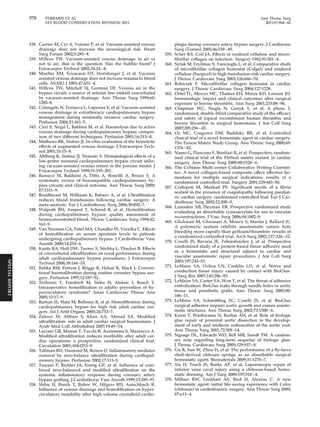
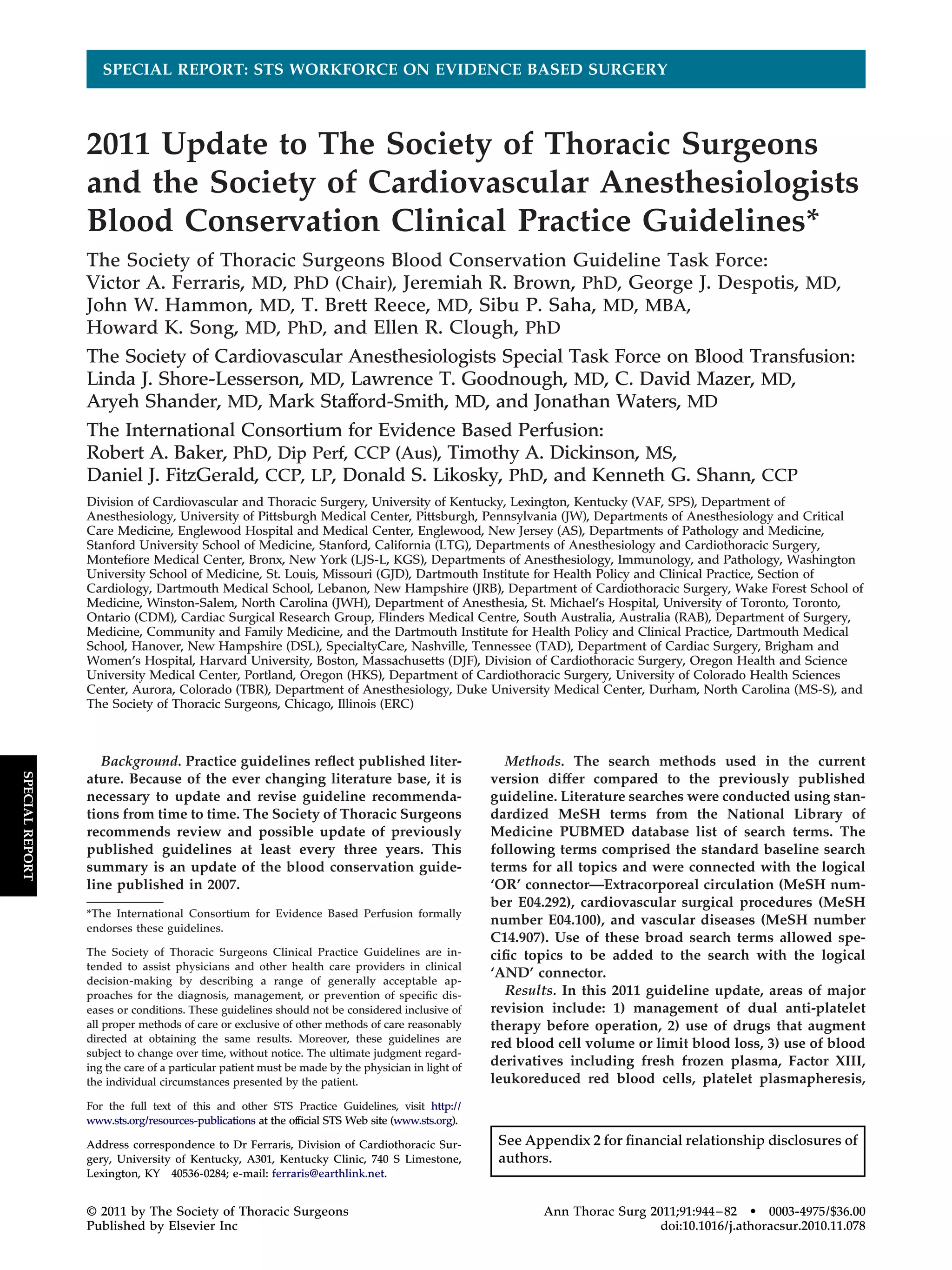
Fig 1. Meta-analysis comparing mortality between aprotinin and tranexamic acid or epsilon aminocarpoic acid. The relative risks (RR) of
mortality by antifibrinolytic agent compared head-to-head are plotted. The RR for each study is plotted (blue box) with 95% confidence inter-val
(horizontal bar). A pooled estimate RR (diamond) and 95% confidence intervals (width of diamonds) summarize the effect using a fixed
effects model. Effects to the left of 1.0 favors aprotinin over tranexamic acid or epsilon aminocaproic acid; effects to the right favors tranexamic
acid or epsilon aminocaproic acid over aprotinin. When the horizontal bars cross 1.0, the effect is not significantly different from the compari-son
group. The I2 test for heterogeneity was not significant, demonstrating homogeneity in mortality effects across the independent randomized
[66]. Figure 1 shows a summary of the head-to-head
comparisons between aprotinin and lysine analogues.
Aprotinin carries a significant increased risk of death up
to 30 days after operation compared with tranexamic acid
(RR 1.49; 95% CI: 1.02 to 2.16) and a similar effect with
epsilon-aminocaproic acid (RR 1.50; 95% CI: 0.97 to 2.32).
By pooling the mortality results, there is a significant
increased mortality (RR 1.49, 95% CI:1.12 to 1.98) with
aprotinin (4.4%) compared with lysine analogues (2.9%).
The randomized trial data and meta-analyses support the
FDA analysis of the i3 Drug Safety Study of 135,611
patients revealing an increased adjusted risk of death
(RR 1.54; 95% CI: 1.38 to 1.73), stroke (RR 1.24; 95% CI:
1.07 to 1.44), renal failure (RR 1.82; 95% CI: 1.61 to 2.06),
and heart failure (RR 1.20; 95% CI: 1.14 to 1.26) [70].
Because of these safety concerns, risks of aprotinin out-weigh
the benefits, and its routine use in low to moderate
risk cardiac operations is not recommended.
Since discontinued use of aprotinin occurred abruptly
in the United States in 2008, comparisons of bleeding risk
before and after aprotinin availability appeared in the
literature [71–73]. Some of these studies suggest that
blood product transfusion increased after aprotinin dis-appeared
from clinical use, but others do not. New
reports suggested benefit from aprotinin in reducing
blood transfusion without significant increased risk.
Aprotinin may be beneficial in infants [74,75], and in
patients at greatest risk for postoperative bleeding [76].
Aprotinin reduces the need for blood transfusion in
cardiac operations by about 40% [73], and this benefit
may be substantially greater in the patients at highest
risk for bleeding [76]. Aprotinin availability is limited in
many countries including the United States, except for
compassionate and special circumstances. It remains for
the clinician and patient to determine the risk benefit
profile for use of this drug in cardiac operations. The
highest risk patients and in those patients with no blood
transfusion alternatives (ie, Jehovah’s Witness) may be
potential candidates for use of aprotinin, but usage in this
setting is likely to be rare. Since the abrupt discontinua-tion
of aprotinin in the United States, further cautionary
reports appeared in the literature [77]. Aprotinin is still
used in fibrin sealant preparations, and anaphylactic
reactions occur in this setting [78].
Case reports and cohort studies identified a possible
risk of seizure after the injection of tranexamic acid
during cardiac procedures [79]. However, a subsequent
randomized trial did not confirm this finding [80]. An
upcoming trial (Aspirin and Tranexamic Acid for Coronary
Artery Surgery trial) enrolling 4,600 patients may provide
trials (a trend toward increased death risk with aprotinin treatment).
SPECIAL REPORT](https://image.slidesharecdn.com/crxclgf4szi52kwjal69-signature-a407141102b4d26dab61dec9d826bc3f976f09084ffbd565106a4e626689d9ba-poli-141208100715-conversion-gate01/85/2011-blood-conservationupdateguidelines-citazione-11-320.jpg)
![Ann Thorac Surg FERRARIS ET AL 955
2011;91:944–82 STS BLOOD CONSERVATION REVISION 2011
answers about the safety profile of this drug [81]. Large-scale
head-to-head randomized trials and postmarketing
surveillance identify potentially harmful side effects and
this information is incomplete for lysine analogs, despite
approval of these agents by regulatory organizations.
c) Blood Derivatives Used for Blood Conservation
PLASMA TRANSFUSION
Class IIa.
1. Plasma transfusion is reasonable in patients with
serious bleeding in context of multiple or single
coagulation factor deficiencies when safer fraction-ated
products are not available. (Level of evidence B)
2. For urgent warfarin reversal, administration of pro-thrombin
complex concentrate (PCC) is preferred,
but plasma transfusion is reasonable when ade-quate
levels of Factor VII are not present in PCC.
(Level of evidence B)
Class IIb.
1. Transfusion of plasma may be considered as part of
a massive transfusion algorithm in bleeding pa-tients
requiring substantial amounts of red-blood
cells. (Level of evidence B)
Class III.
1. Prophylactic use of plasma in cardiac operations in
the absence of coagulopathy is not indicated, does
not reduce blood loss and exposes patients to
unnecessary risks and complications of allogeneic
blood component transfusion. (Level of evidence A)
2. Plasma is not indicated for warfarin reversal or
treatment of elevated international normalized ra-tio
in the absence of bleeding. (Level of evidence A)
Plasma transfusions share many of the risks and com-plications
associated with RBC transfusions. Some risks
of blood product transfusion (eg, transfusion-related
acute lung injury) are specifically linked to plasma trans-fusions.
Moreover, the same cost and logistic challenges
faced by other blood components plague plasma trans-fusions
as well. Therefore, reduction or avoidance of
plasma transfusions should be among objectives of blood
conservation strategies.
Similar to other blood components, substantial varia-tions
exist in the plasma transfusion practices in general
and specifically in cardiac surgery, with many centers not
following any available guidelines [82, 83]. Conversely,
available guidelines are dated and often based on limited
low-quality evidence and do not specifically address
cardiac surgery [84, 85].
Current clinical indications for plasma are mainly
limited to serious bleeding or surgical procedures in the
context of multiple or single coagulation factor deficien-cies
when safer fractionated products are not available
[85– 87]. Factor deficiencies that justify plasma transfu-sion
can be congenital, acquired, dilutional, or due to
consumption (disseminated intravascular coagulation),
but usually occur in the presence of serious bleeding [84,
87]. In patients with microvascular bleeding after CPB,
plasma may limit coagulopathy [88].
Prothrombin complex concentrate (PCC) is preferred
for reversing warfarin. Plasma should not be considered
for warfarin reversal unless PCC is not available and/or
severe bleeding is present [89 –91]. The PCC contains
relatively high levels of factors II, IX, and X, and in some
preparations, factor VII (see below). Compared with
plasma, PCC is administered in much smaller volumes
without consideration of blood groups, is free of many
safety issues of plasma as an allogeneic blood compo-nent,
and acts faster. Recent evidence suggests that PCC
can be used in bleeding patients without coagulopathy,
but more evidence is needed to establish this as an
indication in bleeding patients undergoing cardiac oper-ations
[92]. Prothrombin complex concentrate poses
some thrombotic risks, usually in patients with other
prothrombotic risk factors [93]. Information regarding
thrombotic risks in patients having cardiac operations is
lacking. It should be noted that current PCC preparations
available in the United States contain reduced or negli-gible
amounts of factor VII compared with the PCC units
available in Europe possibly reducing their effectiveness
in warfarin reversal [94, 95].
Approximately one fifth of patients undergoing CPB
have an inadequate response to heparin, a condition
known as heparin resistance. Fresh frozen plasma (FFP)
may be used to treat heparin resistance, but antithrombin
concentrate is preferred since there is reduced risk of
transmitting infections and other blood complications
and antithrombin concentrate does not result in volume
overload (see below) [96].
Plasma units are commonly included in massive transfu-sion
protocols. Although some studies indicate better out-comes
with higher FFP:RBC ratios, conflicting reports exist
on the benefits of FFP in this context. The presence of a
survival bias resulting in apparent beneficial effect of FFP
cannot be ruled out [97–100]. Plasma is not useful for
volume expansion, plasma exchange (with possible excep-tion
of thrombotic thrombocytopenic purpura), or correc-tion
of coagulopathy in the absence of bleeding [85].
Available evidence does not support prophylactic use
of plasma transfusion as part of blood conservation
strategies, in the absence of the above evidence-based
indications. A systemic review of six clinical trials includ-ing
a total of 363 patients undergoing cardiac procedures
showed no improvement in blood conservation with
prophylactic use of plasma [101]. Importantly, significant
heterogeneity exists in the studies included in this anal-ysis.
Differences in controls used for these studies and in
the type of plasma included in the experimental groups
(allogeneic FFP versus autologous plasma) account for
this heterogeneity. Another review identified seven ad-ditional
randomized trials of FFP used in cardiovascular
procedures (including pediatric operations), and found
no association between use of FFP and reduced surgical
blood loss [102]. These reviews contain several method-ological
limitations, but constitute the only available
evidence to address the use of plasma in cardiac proce-dures
[102].
SPECIAL REPORT](https://image.slidesharecdn.com/crxclgf4szi52kwjal69-signature-a407141102b4d26dab61dec9d826bc3f976f09084ffbd565106a4e626689d9ba-poli-141208100715-conversion-gate01/85/2011-blood-conservationupdateguidelines-citazione-12-320.jpg)
![956 FERRARIS ET AL Ann Thorac Surg
STS BLOOD CONSERVATION REVISION 2011 2011;91:944–82
Fig 2. Forest plot displaying (A) the 24-hour chest tube drainage and (B) packed red cell transfusion requirement in patients undergoing car-diopulmonary
bypass with standard filters (control) compared with leukodepletion filters. (Reprinted from Warren O, et al, ASAIO J 2007;53:
Trials that examined use of FFP in cardiac operations
suffer from small sample size and lack of modern-day
perspective. Consten et al conducted a more contempo-rary
study that evaluated 50 elective CPB patients (mean
age 63 years; 70% male) who were randomly assigned to
receive 3 units of FFP after operation or an equal amount
of Gelofusine plasma substitute [103]. They found no
significant differences between the two study groups
with regard to blood loss, transfusion requirement, coag-ulation
variables, or platelet counts. In contrast, Kasper
and associates [104] randomly assigned 60 patients un-dergoing
elective primary CABG to receive 15 mL/kg
autologous FFP (obtained by platelet-poor plasmaphere-sis
several weeks before surgery) or 15 mL/kg of 6%
hydroxyethyl starch 450/0.7 after CPB, and noted that
postoperative and total perioperative RBC transfusion
requirements were not different between the two groups.
In another study, Wilhelmi and colleagues [105] com-pared
60 patients undergoing elective CABG surgery
who received 4 units of FFP intraoperatively with 60
controls who did not receive FFP, and demonstrated that
avoidance of routine intraoperative FFP in cardiac sur-gery
does not lead to increased postoperative blood loss
and does not increase postoperative FFP requirements.
Despite limitations, the results of FFP trials are congru-ent,
and the available evidence suggests that the prophy-lactic
use of plasma in routine cardiac surgeries is not
associated with reduced blood loss or less transfusion
requirement, and this practice is not recommended.
FACTOR XIII
Class IIb.
1. Use of factor XIII may be considered for clot stabi-lization
after cardiac procedures requiring cardio-pulmonary
bypass when other routine blood con-servation
measures prove unsatisfactory in
bleeding patients. (Level of evidence C)
Several derivatives of plasma coagulation factor pro-teases
have hemostatic properties and are currently un-der
clinical investigation. Coagulation factor XIII is one
such drug that may reduce bleeding and transfusion
requirement after cardiovascular operations. Factor XIII
is an enzyme that acts upon fibrin, the final component of
the common coagulation pathway. Factor XIII is neces-sary
for cross-linking of fibrin monomers to form a stable
fibrin clot [106]. The activity of factor XIII in building
fibrin polymers is similar to the activity of antifibrinolytic
agents. The former builds fibrin cross-links while the
latter prevents them from being broken down. It is not
known whether factor XIII can be used in conjunction
with antifibrinolytic agents or if this combination is safe
and effective.
514-21 [139], with permission.)
SPECIAL REPORT](https://image.slidesharecdn.com/crxclgf4szi52kwjal69-signature-a407141102b4d26dab61dec9d826bc3f976f09084ffbd565106a4e626689d9ba-poli-141208100715-conversion-gate01/85/2011-blood-conservationupdateguidelines-citazione-13-320.jpg)
![Ann Thorac Surg FERRARIS ET AL 957
2011;91:944–82 STS BLOOD CONSERVATION REVISION 2011
Factor XIII levels are reduced by 30% to 50% in patients
who are supported with cardiopulmonary bypass [107–
109]. In a randomized study in adult CABG patients given
factor XIII at two different doses, there was no difference
in bleeding rates compared with controls. However, in
this study, increased bleeding occurred in patients with
low postoperative plasma levels of factor XIII, irrespec-tive
of group assignment [108]. Other types of surgical
procedures including neurosurgical, general surgical,
and congenital cardiac operations found similar associa-tion
between decreased factor XIII levels and increased
bleeding [110 –112]. In a randomized study involving 30
children with congenital cardiac disease, the administra-tion
of factor XIII resulted in less myocardial edema, and
less total body fluid weight gain [111]. This, along with
some basic science research, suggests that factor XIII has
antiinflammatory properties. Studies with recombinant
fibrinogens identified molecular mechanisms of clot sta-bilizing
effects of factor XIII. Interestingly, thromboelas-tography
identified differences in strength of fibrin cross-linking
between the various recombinant forms of fibrin
[113, 114].
These findings provided a rationale for the study of
factor XIII in patients undergoing cardiovascular proce-dures.
Two small trials of coronary artery bypass patients
studied plasma-derived factor XIII concentrates and
found reduced postoperative blood loss and transfusion
in patients with low preoperative plasma factor XIII
levels [108, 109]. Recombinant factor XIII is the subject of
a phase II study of patients at moderate risk for hemor-rhage
after cardiovascular surgery with cardiopulmonary
bypass. The phase I trial is complete and provides prom-ising
results, but further studies are justified [115]. While
the existing literature does not support the routine use of
factor XIII at this time, its role as a clot stabilizer is
appealing as part of a multimodality treatment in high-risk
patients because of its hemostatic properties and low
risk of thrombotic complications.
LEUKOREDUCTION
Class IIa.
1. When allogeneic blood transfusion is needed, it is
reasonable to use leukoreduced donor blood, if
available. Benefits of leukoreduction may be more
pronounced in patients undergoing cardiac proce-dures.
(Level of evidence B)
Class III.
1. Currently available leukocyte filters placed on the
CPB circuit for leukocyte depletion are not indi-cated
for perioperative blood conservation and may
prove harmful by activating leukocytes during CPB.
(Level of evidence B)
Adverse effects of transfused blood attributed to the
presence of leukocytes in the packed cells include proin-flammatory
and immunomodulatory effects. In addition,
white blood cells can harbor infectious agents (eg, cyto-megalovirus).
Removing white blood cells from trans-fused
packed red blood cells (leukoreduction or leu-kodepletion)
may reduce the risk of disease transmission
and harmful immunomodulation. Indeed, fears over the
transmission of prions responsible for variant Creutzfeldt-
Jakob disease through transfusion of white blood cell-contaminated
red cells triggered the establishment of leu-koreduction
protocols in the United Kingdom, although
later studies demonstrated that red blood cells, platelets,
and plasma may also contain prions [116]. Despite this and
uncertain literature, leukoreduction filters are used increas-ingly
in various stages of blood processing.
Canada and most European countries perform univer-sal
prestorage leukoreduction of donated packed cells. In
the United States, most transfused allogeneic blood units
are leukoreduced, but local variations exist. Despite
widespread use and associated costs, evidence on the
effectiveness of universal prestorage leukoreduction of
donor blood is equivocal. Reduction of infectious com-plications
and human leukocyte antigen (HLA) immuni-zation
are among the most established benefits of leu-koreduction,
although contradictory results exist in
published studies, possibly attributable to the analysis
approach (intention-to-treat versus as-treated analysis)
[117–124]. Some randomized trials suggest lower mortal-ity
with transfusion of prestorage leukoreduced alloge-neic
blood compared with transfusion of standard buffy-coat-
depleted blood in patients having cardiac
operations [122–124]. A reanalysis of some of these stud-ies
concluded that higher mortality in the nonleukore-duced
group and related higher infections are colinear in
the patient population, and a cause-effect relationship is
uncertain [122]. Regardless of the mechanism, pooled
analysis of the data suggests that mortality reduction
associated with transfusion of leukoreduced packed cells
is greater in patients undergoing cardiac operations com-pared
with other noncardiac operations [124]. Evidence
implies that the incidence of posttransfusion purpura
and transfusion-associated graft-versus-host disease is
lower among patients transfused with leukoreduced
blood. Further, leukoreduction is unlikely to eliminate
the risk of transfusion-associated graft-versus-host dis-ease
in at-risk populations, and other methods (eg, irra-diation)
are required [125]. More debated potential ben-efits
of leukoreduction include reduced incidence of
febrile reactions, minimized multiorgan failure with lung
injury, and decreased length of hospital stay [126 –135].
Some evidence suggests that the presence of white blood
cells in stored blood enhances the deleterious effects of
prolonged storage [123].
Given the overall safety of the procedure and the
possibility of improving outcomes, the current rising
trend in use of leukoreduced donor blood appears to be
justified. Concerns with the cost-effectiveness of univer-sal
leukoreduction persist [136]. Use of leukoreduced
blood likely benefits special groups of patients, especially
those patients who receive four or more transfusions
[137]. No evidence suggests that use of leukoreduced allo-geneic
blood reduces transfusion requirements in bleeding
patients. Moreover, a large trial demonstrated that leu-
SPECIAL REPORT](https://image.slidesharecdn.com/crxclgf4szi52kwjal69-signature-a407141102b4d26dab61dec9d826bc3f976f09084ffbd565106a4e626689d9ba-poli-141208100715-conversion-gate01/85/2011-blood-conservationupdateguidelines-citazione-14-320.jpg)
![958 FERRARIS ET AL Ann Thorac Surg
STS BLOOD CONSERVATION REVISION 2011 2011;91:944–82
kodepletion of preoperatively donated autologous whole
blood does not improve patient outcomes [138].
Another application of leukocyte filters is in extracor-poreal
circuit during CPB. During CPB, white blood cells
are activated and links exist between activation of white
cells and some of the postoperative complications. Leu-kocytes
play an important role in ischemia-reperfusion
injury. Therefore, removal of leukocytes from blood at
various stages of CPB may reduce inflammatory response
and improve organ function. However, the process of
in-line leukoreduction during CPB may activate leuko-cytes
even more, and the efficacy of leukocyte removal by
the filters is uncertain. Based on available evidence, the
previous edition of the STS Blood Conservation Guide-lines
concluded that none of the putative beneficial
effects of leukodepletion during CPB provide tangible
clinical benefits, and recommend against its routine use
for blood management in CPB [9]. Two recent meta-analyses
of rather small and heterogeneous studies pub-lished
from 1993 to 2005 concluded that leukodepletion
during CPB does not reduce 24-hour chest tube drainage
or the number of total packed red cell transfusions.
Leukodepletion does not attenuate lung injury in pa-tients
undergoing CPB (Fig 2) [139, 140].
Limited evidence suggests that in-line leukodepletion
during CPB may be beneficial in specific patient popula-tions
(eg, patients with left ventricular hypertrophy,
prolonged ischemia, chronic obstructive airways disease,
pediatric patients undergoing cardiac operations, and
patients in shock requiring emergency CABG proce-dures)
[141]. A small trial on patients with renal impair-ment
undergoing on-pump CABG showed that leu-kodepletion
is associated with better renal function [142],
and another trial in CABG patients indicated that use of
leukofiltration together with polymer-coated circuits is
associated with a lower incidence of post-CPB atrial
fibrillation in high-risk patients (EuroSCORE [European
System for Cardiac Operative Risk Evaluation] of 6 or
more) but not in the low-risk patients [143]. Conversely,
another trial of CPB leukodepletion in high-risk patients
undergoing CABG demonstrated that use of leukocyte
filters translates to more pronounced activation of neu-trophils,
and this intervention did not provide clinical
benefits [144].
Although improvements in commercially-available
leukocyte filters are likely to occur, the results of more
recent studies remain controversial and high quality and
consistent evidence to recommend leukocyte filters in
routine cardiac operations does not exist [139]. It is likely
that specific patient populations benefit to some degree
from these devices, but more investigation is needed.
Until further studies are available, leukocyte filters at-tached
to the CPB circuit are not indicated because of the
lack of clear benefit and the potential for harm.
PLATELET PLASMAPHERESIS
Class IIa.
1. Use of intraoperative platelet plasmapheresis is
reasonable to assist with blood conservation strat-egies
as part of a multimodality program in high-risk
patients if an adequate platelet yield can be
reliably obtained. (Level of evidence A)
Platelet plasmapheresis is a continuous centrifugation
technique, that when employed using a fast, followed by
a slow centrifugation speed, allows for selective removal
of a concentrated autologous platelet product from whole
blood. During the centrifugation process, the harvested
RBCs and platelet-poor plasma are immediately returned
to the patient. The concentrated platelet-rich-plasma
(PRP) is stored, protected from CPB, and reinfused after
completion of CPB. Since platelet dysfunction contrib-utes
to CPB-induced bleeding, this strategy of removing
platelets from the circulation and “sparing” them from
CPB has a theoretical advantage of providing more
functional platelets for hemostasis at the end of CPB.
Centrifugation at high speeds only produces a platelet-poor
plasma product whereas a fast centrifugation fol-lowed
by a slow centrifugation sequesters more of the
platelet fraction and produces PRP [145].
At least 20 controlled trials studied platelet-rich plas-mapheresis
during cardiac procedures using CPB [145–
164]. Most of the controlled trials are prospective and
randomized, but with a complex technical procedure
such as platelet pheresis, blinding is difficult. Only one
study to date “blinded” the platelet pheresis by subject-ing
control patients to a sham pheresis procedure [160].
The results of that study demonstrated no differences
between patients who had PRP harvested and reinfused,
and those that did not. Across all studies with regard to
hemostasis, the results vary from “no effect” [150, 151,
156, 157, 161] to a significant effect in reducing bleeding
and transfusion requirements [149, 153–155, 158, 162–
165]. Other studies compared PRP to control groups
and demonstrated improved platelet aggregation stud-ies
and improved thromboelastographic parameters in
the patients who received PRP [145, 159]. Other bene-ficial
effects of PRP harvest and transfusion include an
improvement in intrapulmonary shunt fraction and,
when PRP harvest is supplemented with platelet gel
use, a reduced risk of infection [147, 162, 164, 166]. It
seems that an adequate platelet yield obtained from
the pheresis procedure is a critical determinant of the
efficacy of platelet pheresis in promoting blood conser-vation.
Each study did not specifically report the plate-let
yield in their PRP product, but in those that did
report it, harvest of at least 3 1011 platelets, or 28% of
the patients circulating platelet volume, is a minimum
to achieve a product with optimal hemostasis. Limited
value of this procedure accrues to patients taking
antiplatelet drugs.
The fact that prophylactic transfusion of platelet con-centrates
does not improve hemostasis after cardiac
operations [167, 168] provides concerns that preoperative
harvest of PRP and retransfusion might lack efficacy. The
effect of preoperative harvest of fresh whole blood dem-onstrated
a beneficial platelet-protective effect [169, 170].
However, the volume of fresh blood needed to sequester
SPECIAL REPORT](https://image.slidesharecdn.com/crxclgf4szi52kwjal69-signature-a407141102b4d26dab61dec9d826bc3f976f09084ffbd565106a4e626689d9ba-poli-141208100715-conversion-gate01/85/2011-blood-conservationupdateguidelines-citazione-15-320.jpg)
![Ann Thorac Surg FERRARIS ET AL 959
2011;91:944–82 STS BLOOD CONSERVATION REVISION 2011
a large complement of platelets is prohibitively high in
most cardiac surgical patients.
The PRP harvest procedure is time- and resource-consuming.
Technical errors and possible contamination or
wastage of the platelet product are possible and add to the
cost and risk of the procedure [171]. Reports of hemody-namic
instability during the harvest procedure and during
reinfusion of the citrate-containing product exist, but the
majority of studies using this technique report no errors or
mishaps. It is possible that patients who would benefit most
from PRP harvest are the poorest candidates for the proce-dure,
either because of clinical instability or because of low
volumes of PRP harvest. Given that PRP harvest is operator
dependent, its use as an adjunct to blood conservation
strategies for cardiac procedures may be considered if a
large yield of platelets is sequestered.
RECOMBINANT ACTIVATED FACTOR VII
Class IIb.
1. Use of recombinant factor VIIa concentrate may be
considered for the management of intractable non-surgical
bleeding that is unresponsive to routine
hemostatic therapy after cardiac procedures using
CPB. (Level of evidence B)
Recombinant activated factor VII (r-FVIIa) is used to treat
refractory bleeding associated with cardiac operations, al-though
no large randomized trial data exists to support this
use. A recent randomized controlled trial of r-FVIIa of 172
patients with bleeding after cardiac surgery reported a
significant decrease in reoperation and allogeneic blood
products, but a higher incidence of critical serious adverse
events, including stroke, with r-FVIIa treatment [172]. Mul-tiple
case reports, meta-analyses, registry reviews, and
observational studies appear in the published literature in
hopes of providing clarity to the indications for use and
associated side-effects [173–178]. No clear cut consensus
results from these reviews. There is little doubt that r-FVIIa
is associated with reduction in bleeding and transfusion in
some patients. However, which patients are appropriate
candidates for r-FVIIa are uncertain and neither the appro-priate
dose nor the thrombotic risks of this agent are totally
clear. Despite multiple new reports, we did not find con-vincing
evidence to support a change to our original rec-ommendation
in previously published guidelines.
ANTITHROMBIN III
Class I.
1. Antithrombin III (AT) concentrates are indicated to
reduce plasma transfusion in patients with AT-mediated
heparin resistance immediately before
cardiopulmonary bypass. (Level of evidence A)
Class IIb.
1. Administration of AT concentrates is less well es-tablished
as part of a multidisciplinary blood man-agement
protocol in high-risk patients who may
have AT depletion or in some, but not all, patients
who are unwilling to accept blood products for
religious reasons. (Level of evidence C)
Plasma-derived or recombinant antithrombin III (AT)
concentrates are approved for use in patients with he-reditary
AT deficiency to prevent perioperative throm-botic
complications. These agents are used off-label to
either manage AT mediated heparin resistance or to
prevent perioperative thrombotic complications and tar-get
organ injury in patients with acquired AT deficiency.
Heparin is by far the most commonly used anticoagulant
for the conduct of CPB. Heparin binds to the serine pro-tease
inhibitor AT causing a conformational change that
results in dramatic change in affinity of AT for thrombin
and other coagulation factors. After binding to its targets,
the activated AT inactivates thrombin and other proteases
involved in blood clotting, including factor Xa. Heparin’s
anticoagulant effect correlates well with plasma AT activity.
Impaired heparin responsiveness or heparin resistance is
often attributed to AT deficiency [179]. Antithrombin activ-ity
levels as low as 40% to 50% of normal, similar to those
observed in patients with heterozygotic hereditary defi-ciency
[180], are commonly seen during and immediately
after CPB [181, 182]. Acquired perioperative reductions in
plasma AT concentrations are related to a time-dependent
drop associated with preoperative heparin use (ie, approx-imately
5% to 7% decrease in AT levels per preoperative
day of heparin), hemodilution, or consumption during CPB
[183, 184].
Heparin resistance is defined as the failure to achieve
activated clotting time of at least 400 s to 480 s after a
standard heparin dose, although there is a substantial
variability in the definition of the “standard” heparin
dose, ranging from less than 400 to 1,200 U/kg [185].
Plasma proteins and the formed elements of blood mod-ify
the anticoagulant effect of heparin [186, 187]. As a
result, the most common cause of heparin resistance in
patients not receiving preoperative heparin is likely due
to inactivation of heparin by bound proteins or other
blood elements [188 –192].
A minority of patients undergoing CPB are resistant to
heparin. Antithrombin depletion causes heparin resis-tance
in some of these patients receiving preoperative
heparin [193–195]. Empiric treatment for this type of
heparin resistance often involves administration of either
FFP, plasma-derived AT, or recombinant human AT.
Twelve of 13 previous reports indicate that AT supple-mentation
by either FFP [196, 197] recombinant AT [8,
198, 199], or plasma-derived AT concentrates [198, 200–
206] effectively manage heparin resistance. The use of
factor concentrates or recombinant AT offers unique
advantages because of reduced blood-borne disease
transmission and reduced incidence of fluid overload
compared with FFP. Two recent randomized, controlled
studies demonstrated that recombinant AT treats hepa-rin
resistance [8, 199]. These two studies demonstrated
that recombinant human AT effectively restores heparin
responsiveness before CPB in the majority of patients
with heparin resistance [8, 199]. The primary endpoint of
these trials was the proportion of patients requiring
SPECIAL REPORT](https://image.slidesharecdn.com/crxclgf4szi52kwjal69-signature-a407141102b4d26dab61dec9d826bc3f976f09084ffbd565106a4e626689d9ba-poli-141208100715-conversion-gate01/85/2011-blood-conservationupdateguidelines-citazione-16-320.jpg)
![960 FERRARIS ET AL Ann Thorac Surg
STS BLOOD CONSERVATION REVISION 2011 2011;91:944–82
administration of 2 units FFP to achieve therapeutic
anticoagulation. A significantly (p 0.001) lower percent-age
of patients required FFP in the recombinant human
AT-treated group (20%) compared with the placebo
group (86%). Although these studies achieved the pri-mary
endpoint of reduced FFP transfusion, they did not
assess the effect of AT concentrates on suppression of the
hemostatic system activation during and after CPB.
In addition to the management of prebypass heparin
resistance, restoration of AT levels during CPB may limit
inflammatory activation and replace AT depleted by inad-equate
heparinization during CPB. During CPB, blood
interfaces with the artificial surface of the bypass circuit
which generates a powerful stimulus for thrombin genera-tion
that is not completely suppressed by heparin. In
addition, subtherapeutic anticoagulation can result in ex-cessive
hemostatic system activation leading to generation
of thrombin and plasmin, which then can result in deple-tion
of clotting factors and platelets (a disseminated intra-vascular
coagulationlike consumptive state). The AT-mediated
heparin resistance may be an early warning sign
of inadequate anticoagulation during CPB, leading to an
increase in bleeding and/or thrombotic complications.
Previous studies suggest that AT supplementation atten-uates
thrombin production and fibrinolytic activity during
CPB [184, 198]. This suppression of thrombin production
and fibrinolytic activity during CPB may represent better
inhibition of hemostatic activation than that achieved with
heparin but without AT supplementation [198]. Other stud-ies
show an inverse relationship between plasma AT con-centration
and markers of both thrombin activation (eg,
fibrinopeptide A, fibrin monomer) and fibrinolytic activa-tion
(D-dimer) [184, 207]. Preservation of the hemostatic
system by AT supplementation may reduce bleeding and
provide a better hemostatic profile after CPB. In general,
less bleeding and blood transfusion occur in patients whose
hemostatic system is better preserved after the physiologic
stresses of CPB [208–211].
Thromboembolic complications early after cardiac oper-ations
are potentially lethal. Antithrombin is a major in vivo
inhibitor of thrombin, and a deficiency of AT in the pres-ence
of excess thrombin after CPB provides a potent pro-thrombotic
environment that may cause thromboembolic
complications. This concept is supported by case reports
describing catastrophic thrombosis in a few patients with
low AT III levels in the perioperative period [212, 213].
Additionally, observational studies suggest an indirect re-lationship
between AT levels and perioperative complica-tions
[214–216]. Ranucci and coworkers [216] showed that
patients who suffered adverse neurologic outcomes, throm-boembolic
events, surgical reexploration for bleeding, or
prolonged intensive care unit (ICU) stay after cardiac oper-ations
had reduced AT III levels on admission to the ICU
compared with patients without complications [216]. Two
other observational studies confirm an inverse relationship
between AT III activity and adverse thromboembolic pa-tient
outcomes [214, 215]. Thrombotic complications involv-ing
target organ injury may be under reported after cardiac
procedures, especially in high-risk patients [217]. This tar-get
organ injury may be related to microvascular thrombo-sis
from a procoagulant postoperative environment, at least
in part due to AT deficiency. Based on these published
reports, it may be reasonable to add AT, preferably in
concentrated form, in patients at increased risk for end-organ
thrombotic complications after CPB.
FACTOR IX CONCENTRATES, PROTHROMBIN COMPLEX CONCENTRATES,
AND FACTOR VIII INHIBITOR BYPASSING ACTIVITY
Class IIb.
1. Use of factor IX concentrates, or combinations of
clotting factor complexes that include factor IX, may
be considered in patients with hemophilia B or who
refuse primary blood component transfusion for reli-gious
reasons (eg, Jehovah’s Witness) and who re-quire
cardiac operations. (Level of evidence C)
An intermediate step in clot formation involves activation
of factor IX by the tissue factor/factor VIIa complex [218]. In
some patients requiring cardiac procedures, factor IX con-centrates
are used for one of four purposes: (1) control of
perioperative bleeding in patients with hemophilia B [219–
221]; (2) prophylaxis in high-risk patients unable to accept
primary component transfusions for religious reasons (eg,
Jehovah’s Witness) [222]; (3) as part of prothrombin com-plex
concentrate (Beriplex) for reversal of warfarin before
operative intervention; and (4) part of the factor VIII inhib-itor
bypassing activity in patients with factor VIII inhibitors
requiring operative intervention [92, 223, 224]. Of these
options, use of factor IX concentrates is most reasonable for
Jehovah’s Witness patients [222].
Jehovah’s Witness patients refuse transfusion on reli-gious
grounds. Blood products encompassed with this
refusal are defined by each individual Jehovah’s Witness
patient confronted with a need for cardiac procedure.
Typically primary components include packed red blood
cells, platelets, and fresh frozen plasma. Changes in the
Jehovah’s Witness blood refusal policy now give mem-bers
the personal choice to accept certain processed
fractions of blood, such as factor concentrates and cryo-precipitate
[225, 226]. In Jehovah’s Witness patients, a
multimodality approach to blood conservation is reason-able
[227], and addition of factor concentrates augments
multiple other interventions. Fractionated factor concen-trates,
like factor IX concentrates or one of its various
forms (Beriplex or factor VIII inhibitor bypassing activ-ity),
are considered “secondary components” and may be
acceptable to some Jehovah’s Witness patients [222].
Addition of factor IX concentrates may be most useful in
the highest risk Jehovah’s Witness patients.
d) Blood Salvage Interventions
EXPANDED USE OF RED CELL SALVAGE USING CENTRIFUGATION
Class IIb.
1. In high-risk patients with known malignancy who
require CPB, blood salvage using centrifugation of
blood salvaged from the operative field may be
considered since substantial data support benefit in
patients without malignancy, and new evidence
SPECIAL REPORT](https://image.slidesharecdn.com/crxclgf4szi52kwjal69-signature-a407141102b4d26dab61dec9d826bc3f976f09084ffbd565106a4e626689d9ba-poli-141208100715-conversion-gate01/85/2011-blood-conservationupdateguidelines-citazione-17-320.jpg)
![Ann Thorac Surg FERRARIS ET AL 961
2011;91:944–82 STS BLOOD CONSERVATION REVISION 2011
suggests worsened outcome when allogeneic trans-fusion
is required in patients with malignancy.
(Level of evidence B)
In 1986, the American Medical Association Council on
Scientific Affairs issued a statement regarding the safety
of blood salvage during cancer surgery [228]. At that
time, they advised against its use. Since then, 10 obser-vational
studies that included 476 patients who received
blood salvage during resection of multiple different tumor
types involving the liver [229–231], prostate [232–234],
uterus [235, 236], and urologic system [237, 238] support the
use of salvage of red cells using centrifugation in cancer
patients. In seven studies, a control group received no
transfusion, allogeneic transfusion, or preoperative autolo-gous
donation. In all studies, the centrifugation salvage
group received less allogeneic blood and had equivalent
long-term outcomes compared with control patients. None
of the studies found evidence of subsequent wide-spread
metastasis from salvaged red cells harvested using centrif-ugation
in operations for malignancy.
Because operations involving tumor removal may have
less allogeneic blood transfusion with blood salvage, the
theoretical risk of avoiding blood salvage needs to be
examined more closely. First, there are numerous reports
suggesting that the presence of circulating tumor has no
prognostic significance, and one author suggested that
outcome was better in patients with higher circulating
tumor cells [239, 240]. Second, multiple reports indicate
that allogeneic transfusion increases the rates of recur-rence
after tumor operations. Two recent meta-analyses
of these reports suggest that patients suffer nearly a
twofold increase in recurrence when exposed to alloge-neic
transfusion [241, 242].
There are numerous reports of leukocyte depletion
filters or radiation being used to eliminate malignant
cells from salvaged blood [243–245]. These studies sug-gest
that a 3-log to 4-log reduction in tumor burden is
achieved with leukocyte depletion filters whereas a 12-
log reduction is achieved with radiation [246]. While
debate exists among advocates of either removal tech-nique
as to the best strategy, in six of the seven studies
referenced above where control groups existed, neither
technique was used. In the seventh manuscript, a leu-kodepletion
filter was used. The bulk of evidence sug-gests
that the addition of tumor cells into circulation from
salvaged red cells is of little prognostic significance.
No data exist to contradict the use of blood salvage in
malignant surgery and substantial data exist to support
worsened outcome when the alternative therapy (alloge-neic
transfusion) is used. The use of centrifugation of
salvaged blood in patients with malignancy who require
cardiac procedures using CPB is less well established but
may be considered.
PUMP SALVAGE
Class IIa.
1. Consensus suggests that some form of pump sal-vage
and reinfusion of residual pump blood at the
end of CPB is reasonable as part of a blood man-agement
program to minimize blood transfusion.
(Level of evidence C)
2. Centrifugation instead of direct infusion of residual
pump blood is reasonable for minimizing post-CPB
allogeneic RBC transfusion. (Level of evidence A)
Most surgical teams reinfuse blood from the extracor-poreal
circuit (ECC) back into patients at the end of CPB
as part of a blood conservation strategy. Currently, two
blood salvaging techniques exist: (1) direct infusion of
post-CPB circuit blood with no processing; and (2) pro-cessing
of the circuit blood, either by centrifugation of by
ultrafiltration, to remove either plasma components or
water soluble components from blood before reinfusion.
Centrifugation of residual CPB blood produces concen-trated
red cells mostly devoid of plasma proteins,
whereas ultrafiltration produces protein-rich concen-trated
whole blood [247].
Two studies compared the clinical effects of direct
infusion, centrifugation and ultrafiltration. Sutton and
colleagues [248] randomly allocated 60 patients undergo-ing
elective CABG using CPB [248]. They observed sig-nificantly
longer partial thromboplastin times in the
centrifugation group compared with the direct infusion
and ultrafiltration groups 20 minutes after circuit blood
administration. Importantly, there were no differences in
blood product usage, total chest tube drainage, or dis-charge
hematocrit values. Eichert and coworkers [249]
prospectively randomized 60 patients undergoing elec-tive
CABG operations into three blood salvage interven-tions
(described above). Among the three groups, there
were no significant differences in postoperative hemoglo-bin
levels, postoperative chest tube drainage, platelet
counts, or partial thromboplastin times at 12 hours after
operation. Allogeneic blood transfusion rates were not
reported.
Two studies compared the effectiveness of centrifuga-tion
compared with ultrafiltration in concentrating post-
CPB ECC blood to minimize blood loss and transfusion
requirements. Boldt and coworkers [247] studied 40 non-randomized
patients undergoing elective CABG proce-dures
and discovered no significant difference in blood
loss or frequency of donor blood transfusion between the
centrifugation and ultrafiltration groups. Samolyk and
coworkers [250] used a case-matched control study to
compare centrifugation and ultrafiltration in 100 patients;
there was no difference in blood utilization or postoper-ative
bleeding between groups.
A number of studies compared the effects of direct
infusion of unprocessed post-CPB blood versus centrifu-gation.
In a prospective randomized study of 40 elective
patients having cardiac procedures, Daane and cowork-ers
[251] found significantly less allogeneic red cell use in
the centrifugation group (2.4 1.3 units of PRBC) versus
the direct infusion group (3.2 1.1 units PRBC; p
0.046). Fresh frozen plasma and platelet administration
were not significantly different between the two groups.
Walpoth and associates [252] randomly assigned 20 pa-tients
undergoing elective CABG procedures to either
SPECIAL REPORT](https://image.slidesharecdn.com/crxclgf4szi52kwjal69-signature-a407141102b4d26dab61dec9d826bc3f976f09084ffbd565106a4e626689d9ba-poli-141208100715-conversion-gate01/85/2011-blood-conservationupdateguidelines-citazione-18-320.jpg)
![962 FERRARIS ET AL Ann Thorac Surg
STS BLOOD CONSERVATION REVISION 2011 2011;91:944–82
direct infusion or centrifugation. There was no significant
difference in postoperative day 1 hemoglobin values or
allogeneic blood transfusions between the two groups.
Wiefferink and associates [253] randomized 30 primary
elective CABG surgery patients and found elevated D-dimer
levels (suggesting increased intravascular fibrin
degradation) in the early postoperative period in the
direct infusion group compared with the centrifugation
group. Chest tube drainage and transfusion require-ments
were also higher in the direct infusion group. In
this study, direct infusion of mediastinal blood by an
autotransfusion system in only the centrifugation group
undoubtedly impacted transfusion requirements. These
findings support earlier results of Moran and colleagues
[254] who studied the safety and effectiveness of centrif-ugation
of residual CPB blood among patients undergo-ing
cardiopulmonary bypass (including CABG and valve
procedures). For 50 randomized patients, they reported
significantly (p 0.05) less allogeneic RBC exposure in
the centrifugation group (1,642 195 mL) compared with
the direct infusion group (2,175 175 mL).
Two limitations to the current body of literature on the
topic of salvaging post-CPB ECC blood are noteworthy:
(1) most of the current literature focuses on elective
CABG patients; and (2) many studies include small
sample sizes. From the available literature, no firm dis-tinction
among these techniques for salvaging post-CPB
ECC blood arises. Additional studies are required to
clarify the effectiveness and efficacy of these approaches
to pump salvage. However, consensus supports the prac-tice
of pump salvage compared with no salvage of resid-ual
blood.
e) Minimally Invasive Procedures
AORTIC ENDOGRAFTS
Class I.
1. Thoracic endovascular aortic repair (TEVAR) of
descending aortic pathology reduces bleeding and
blood transfusion compared with open procedures
and is indicated in selected patients. (Level of
evidence B)
Reports of thoracic endovascular aortic repair (TEVAR)
for descending thoracic aortic pathology appeared in the
literature more than 15 years ago [255]. Since then,
surgeons embraced TEVAR without evidence from ran-domized
comparisons of open repair versus TEVAR. The
uptake of TEVAR outstripped adequate evaluation
largely because of perceived benefit in morbidity, and
possibly mortality, associated with TEVAR. Recently, two
meta-analyses of published nonrandomized compari-sons
of TEVAR to open repair appeared in the literature
[256, 257]. The most recent of these meta-analyses en-compassed
45 nonrandomized studies involving more
than 5,000 patients [256]. The results suggest that TEVAR
may reduce early death, paraplegia, renal insufficiency,
transfusions, reoperation for bleeding, cardiac complica-tions,
pneumonia, and length of stay compared with
open surgery. Anecdotal evidence suggests that this
benefit extends to Jehovah’s Witness patients [258, 259].
The early adoption of TEVAR seems justified based on
these less than rigorous studies. If these results are
confirmed in randomized trials, TEVAR represents a
paradigm shift in the treatment of thoracic aortic disease,
and this shift may have already occurred.
OFF-PUMP PROCEDURES
Class IIa.
1. Off-pump operative coronary revascularization
(OPCABG) is a reasonable means of blood conser-vation,
provided that emergent conversion to on-pump
CABG is unlikely and the increased risk of
graft closure is considered in weighing risks and
benefits. (Level of evidence A)
In the original publication of the STS Blood Conserva-tion
Guidelines, patients having OPCABG had reduced
blood utilization and postoperative bleeding [9]. Since
publication of this practice guideline, new information
suggests that blood transfusion and significant postoper-ative
bleeding is less common among patients treated
with OPCABG compared with conventional CABG using
CPB [260 –262]. These findings support our initial recom-mendation
for the use of OPCABG as a blood conserva-tion
intervention, especially in skilled hands where con-version
to an open CABG using CPB is unlikely [263].
However, several threads of evidence, including a large
multiinstitution comparison, suggest that graft patency is
reduced in OPCABG patients [11, 264].
f) Perfusion Interventions
MICROPLEGIA
Class IIb.
1. Routine use of a microplegia technique may be
considered to minimize the volume of crystalloid
cardioplegia administered as part of a multimodal-ity
blood conservation program, especially in fluid
overload conditions like congestive heart failure.
However, compared with 4:1 conventional blood
cardioplegia, microplegia does not significantly im-pact
RBC exposure. (Level of evidence B)
Blood cardioplegia is the most common form of myocar-dial
preservation during cardiac surgery with the crystalloid
component facilitating cardiac arrest and providing myo-cardial
protection during ischemia and reperfusion [265].
Microplegia is a term used to describe the mixing of blood
from the CPB circuit with small quantities of concentrated
cardioplegia additives [266]. This technique relies on preci-sion
pumps to deliver small and accurate quantities of
concentrated cardioplegia additives. The concentrated ad-ditives
offer the theoretical advantage of minimizing fluid
overload and hemodilutional anemia.
Four studies suggest that microplegia results in less
hemodilution compared with traditional 4:1 blood to crys-talloid
cardioplegia systems [267–270]. These studies
showed an 11-fold to 27-fold increase in delivered cardio-plegia
volume in the 4:1 blood cardioplegia groups, but
SPECIAL REPORT](https://image.slidesharecdn.com/crxclgf4szi52kwjal69-signature-a407141102b4d26dab61dec9d826bc3f976f09084ffbd565106a4e626689d9ba-poli-141208100715-conversion-gate01/85/2011-blood-conservationupdateguidelines-citazione-19-320.jpg)
![Ann Thorac Surg FERRARIS ET AL 963
2011;91:944–82 STS BLOOD CONSERVATION REVISION 2011
similar allogeneic blood product usage compared with the
microplegia groups. In each of these studies, the clinical
outcomes were not statistically different between groups,
although a potential type II error exists because of the low
event rates for adverse sequelae and the small sample sizes.
Minimizing the crystalloid component of blood cardio-plegia
is intuitively advantageous with respect to mini-mizing
hemodilutional anemia and possible subsequent
allogeneic RBC transfusion. The peer-reviewed medical
literature focuses on cardioplegia volume delivered to
patients and not on blood conservation. More data are
required to assess whether microplegia has an apprecia-ble
effect on transfusion rates and blood conservation,
but microplegia use may be considered as part of a
multimodality blood management program.
BLOOD CONSERVATION IN ECMO AND SHORT-TERM VENTRICULAR
SUPPORT
Class I.
1. Extracorporeal membrane oxygenation patients
with heparin-induced thrombocytopenia should be
anticoagulated using alternate nonheparin antico-agulant
therapies such as danaparoid or direct
thrombin inhibitors (eg, lepirudin, bivalirudin, or
argatroban). (Level of evidence C)
Class IIa.
1. It is reasonable to consider therapy with a lysine
analog antifibrinolytic agents (epsilon aminocap-roic
acid, tranexamic acid) to reduce the incidence
of hemorrhagic complications in ECMO patients.
(Level of evidence B)
Class IIb.
1. It may be helpful to use recombinant activated
factor VII as therapy for life-threatening bleeding in
ECMO patients. The potential benefit of this agent
must be weighed against numerous reports of cat-astrophic
acute thrombotic complications. (Level of
evidence C)
Over the last decade patients who require cardiac
operations are sicker and have more comorbidities and
less functional reserve. The increased acuity of patients
presenting for cardiac operations results in more fre-quent
use of prolonged circulatory support. Extracorpo-real
membrane oxygenation and short-term ventricular
support devices can cause additional serious clinical
sequellae that often translates into excess bleeding and
blood transfusion [271, 272]. Hemolysis of red cells lead-ing
to transfusion presents a special problem in patients
supported with these devices [273].
Extracorporeal membrane oxygenation is used to pro-vide
temporary (days to weeks) respiratory (venovenous)
or cardiorespiratory (venoarterial) support, to critically ill
adult, pediatric, and neonatal patients failing conven-tional
therapy because of cardiopulmonary failure. Veno-venous
ECMO is most frequently used for pulmonary
disorders such as adult respiratory distress syndrome,
congenital diaphragmatic hernia, and meconium aspira-tion,
whereas venoarterial ECMO is indicated for revers-ible
cardiogenic shock, or as a bridge to more permanent
ventricular assist device insertion or heart transplanta-tion.
The Extracorporeal Life Support Organization de-veloped
guidelines for the practice of ECMO [274].
Thrombotic and bleeding complications are common
with ECMO. In a recent review of 297 adult ECMO
patients, 11% had serious neurologic events related to
intracranial infarct or hemorrhage and19% had clots in
the ECMO circuit, and other complications included
cardiac tamponade (10%) and serious bleeding from
cannula insertion sites (21%), surgical incisions (24%),
and the gastrointestinal tract (4%) [275]. Other reports
indicate that intracranial hemorrhage is particularly com-mon
in ECMO patients with renal dysfunction or throm-bocytopenia
[276]. Reports document ECMO-related co-agulopathy
due to platelet dysfunction and hemodilution
with consumptive factor depletion [277, 278]. Although
the challenge of balancing the need for sufficient antico-agulation
to prevent ECMO circuit thrombosis while
minimizing bleeding risk exists, there are few objective
data to guide ECMO anticoagulation therapy.
Consensus ECMO guidelines advocate heparin antico-agulation
to achieve a target activated clotting test time
between 160 s and 240 s [107, 274, 279, 280]. However,
recent studies question the activated clotting time as a
reliable indicator of heparin effect with ECMO [281], and
correlate increased heparin dosing with improved sur-vival
[282]. Nonrandomized observational ECMO studies
employing modified heparin protocols report reduced
thrombohemorrhagic complications [283, 284]. Similarly,
many small nonrandomized studies evaluating heparin-coated
ECMO systems with or without heparin adminis-tration
describe reduced blood loss, but also thrombotic
complications [285–287]. Other proposed alterations to
ECMO guidelines include alternate heparin monitoring
tests (eg, activated partial thromboplastin time, anti-Xa,
thromboelastography, heparin/protamine titration, and
direct heparin level measurements) [282].
Prolonged ECMO or ventricular support using heparin
anticoagulation can cause heparin-induced thrombocy-topenia.
In this setting, anticoagulation regimens includ-ing
danaparoid [288] and direct thrombin inhibitors such
as lepirudin, bivalirudin, or argatroban [289 –291] are
useful alternatives.
Several studies advocate the use of antifibrinolytic
agents in ECMO patients to limit bleeding complications
and blood transfusion. One study reports a potentially
blood sparing effect of aprotinin in ECMO patients but
this is unlikely to be useful given the risk/benefit analysis
described above [292]. Retrospective evaluations of use of
epsilon aminocaproic or tranexamic acid therapy in post-cardiotomy
ECMO patients suggest reduced bleeding
complications [293–295]. A randomized study of hemor-rhagic
complications in 29 neonates could not reproduce
these findings [296]. A randomized study of 60 patients
evaluating leukoreduction using a Pall LG6 leukocyte
filter during extracorporeal circulation also found no
reduction in bleeding complications or transfusion [297].
SPECIAL REPORT](https://image.slidesharecdn.com/crxclgf4szi52kwjal69-signature-a407141102b4d26dab61dec9d826bc3f976f09084ffbd565106a4e626689d9ba-poli-141208100715-conversion-gate01/85/2011-blood-conservationupdateguidelines-citazione-20-320.jpg)
![964 FERRARIS ET AL Ann Thorac Surg
STS BLOOD CONSERVATION REVISION 2011 2011;91:944–82
Consensus guidelines for blood component therapy in
ECMO patients advocates transfusion to assure adequate
oxygen carrying capacity, normal AT III activity (80% to
120% of control) and fibrinogen levels (250 to 300 mg/dL),
while maintaining a platelet count greater than 80,000 to
100,000/L with platelet transfusion [274, 279, 280]. No-tably,
although the generalized effects of ECMO on
coagulation, fibrinolysis, and platelet function are well
documented [278], studies in neonates often focus on
platelet number and function. To maintain target platelet
counts, transfusion requirements tend to be higher in
neonates with meconium aspiration or sepsis, and in
those receiving venoarterial compared with venovenous
ECMO [298]. Although some studies advocate prophy-lactic
transfusion to target platelet counts that are higher
than standard guidelines [299], there is concern that
prophylactic rather than therapeutic platelet transfusion
results in excessive platelet transfusion in ECMO pa-tients
with concomitant detrimental effects [300].
Bleeding is a common complication in ECMO patients.
Likely causes of bleeding include overanticoagulation
and decreased platelet number and function. Significant
bleeding occurs from minimal interventions such as
tracheal suction or nasogastric tube placement, or from
minor surgical interventions [301]. Some reports of re-fractory
bleeding with ECMO describe benefit from
r-FVIIa therapy [302–304]. Unfortunately, other reports
describe catastrophic acute thrombotic complications
with this agent, suggesting this therapy should be con-sidered
as a last resort [305, 306].
MINICIRCUITS AND VACUUM-ASSISTED VENOUS DRAINAGE
Class I.
1. Minicircuits (reduced priming volume in the mini-mized
CPB circuit) reduce hemodilution and are
indicated for blood conservation, especially in pa-tients
at high risk for adverse effects of hemodilu-tion
(eg, pediatric patients and Jehovah’s Witness
patients). (Level of evidence A)
Class IIb.
1. Vacuum-assisted venous drainage in conjunction
with minicircuits may prove useful in limiting
bleeding and blood transfusion as part of a multi-modality
blood conservation program. (Level of
evidence C)
Abundant evidence suggests that preoperative anemia
or small body size is a risk for postoperative blood
transfusion [9], but it seems likely that these patients with
reduced red cell volume (either from anemia or from
small body size) risk severe hemodilution with conven-tional
CPB as a cause of blood transfusion [307]. Reduced
priming volume in the CPB circuit (minicircuits) appeals
to surgeons for many reasons. Paramount among these is
the potential for reduced hemodilution and blood utili-zation.
Minicircuits have particular appeal to pediatric
cardiac surgeons since the effects of hemodilution in
conventional CPB circuits are greatest in small patients.
Acceptance of minicircuits is not universal, partly be-cause
most minicircuit perfusion configurations require a
closed venous reservoir or no venous reservoir [308].
Introduction of air into the closed venous system risks an
“air-lock” in the CPB circuit, and absence of a venous
reservoir risks exsanguination from uncontrolled bleed-ing
in the operative field. Experience with minicircuits
includes use in conjunction with beating heart proce-dures
[309], in usual cardiac procedures [310 –314] and in
Jehovah’s Witness patients [315, 316].
Nine of 14 randomized trials [309, 310, 317–327] and a
well-constructed meta-analysis [328] suggest benefit
from minicircuits with reduced transfusion and postop-erative
bleeding. There is near universal agreement that
minicircuits limit markers of inflammation compared
with conventional CPB circuits [312, 313, 329]. Summa-tion
of available evidence suggests that minicircuits pro-vide
a valuable alternative that limits hemodilution and
blood usage after cardiac procedures.
Many reports of use of minicircuits for extracorporeal
circulation use vacuum-assisted venous drainage
(VAVD). A VAVD in conjunction with minicircuits may
improve hemostasis, limit chest tube drainage, and de-crease
blood transfusion, compared with CPB circuits
with gravity venous drainage [330 –333]. The majority of
air microemboli originate in the venous line of the CPB
circuit [334]. Most [335–337], but not all studies [338]
suggest that VAVD entrains air and leads to increased
systemic microemboli compared with gravity venous
drainage [335–341]. The effect of microemboli can be
minimized by flooding the operative field with carbon
dioxide and adjustment of perfusion parameters [336,
339, 341, 342]. A VAVD may [343] or may not [344, 345]
cause hemolysis within the CPB circuit. Given the poten-tial
limitations of VAVD, use of this technology necessi-tates
caution and adjustment of perfusion techniques
[336, 339, 342], but may provide benefit, especially in
pediatric patients [332].
BIOCOMPATIBLE CPB CIRCUITS
Class IIb.
1. Use of biocompatible CPB circuits may be consid-ered
as part of a multimodality program for blood
conservation. (Level of evidence A)
Biocompatible surfaces for CPB circuits are commer-cially
available. More than 200 publications address
the potential benefit of these circuits. A recent meta-analysis
reviewed bleeding outcomes associated with
these biocompatible surfaces [346]. In this review,
Ranucci and coworkers [346] identified a cohort of 128
publications that explored outcomes in patients treated
with these circuits. From these 128 articles, 36 random-ized
clinical trials contained information on more than
4,000 patients. In the preliminary analysis, the authors
found less bleeding and blood transfusion in patients
treated with biocompatible circuits. However, in a
subgroup of 20 of the highest quality randomized
clinical trials, benefits related to blood conservation
SPECIAL REPORT](https://image.slidesharecdn.com/crxclgf4szi52kwjal69-signature-a407141102b4d26dab61dec9d826bc3f976f09084ffbd565106a4e626689d9ba-poli-141208100715-conversion-gate01/85/2011-blood-conservationupdateguidelines-citazione-21-320.jpg)
![Ann Thorac Surg FERRARIS ET AL 965
2011;91:944–82 STS BLOOD CONSERVATION REVISION 2011
disappeared. The authors of this well-done meta-analysis
suggest that use of biocompatible surfaces
without other measures of blood conservation results
in limited clinical benefit [346]. Use of biocompatible
CPB circuits as part of a multimodality program in
conjunction with closed CPB circuits, separation of
cardiotomy suction, minicircuits to reduce priming
volume may be useful for blood conservation.
ULTRAFILTRATION
Class I.
1. Use of modified ultrafiltration (MUF) is indicated
for blood conservation and reducing postoperative
blood loss in adult cardiac operations using CPB.
(Level of evidence A)
Class IIb.
1. Benefit of the use of conventional or zero balance
ultrafiltration (ZBUF) is not well established for
blood conservation and reducing postoperative
blood loss in adult cardiac operations. (Level of
evidence A)
Ultrafiltration is an option to limit hemodilution sec-ondary
to CPB. Ultrafiltration devices can be integrated
in parallel into existing CPB circuits for this purpose.
Ultrafiltration filters water and low-molecular weight
substances from the CPB circuit, producing protein-rich
concentrated whole blood that can be returned to the
patient. In cardiac surgery, three variants are utilized: (1)
conventional ultrafiltration run during CPB but not after;
(2) modified ultrafiltration (MUF) run after completion of
CPB using existing cannulae; and (3) zero balance ultra-filtration
(ZBUF) similar to conventional ultrafiltration
but with replacement of lost volume with crystalloid
solution. A number of investigations, including a meta-analysis
of 10 randomized trials [347], focused on use of
these methods during and after CPB.
Conventional Ultrafiltration. Conventional ultrafiltration is
a method of ultrafiltration used during CPB. One meta-analysis
[347], four randomized trials [348 –351], and two
cohort studies [352, 353] report the use of conventional
ultrafiltration in patients undergoing cardiac procedures
using CPB. Subgroup analysis of five studies included in
the meta-analysis by Boodhwani [347] demonstrated no
advantage in terms of red cell usage or blood loss with
conventional ultrafiltration alone [349 –353].
Modified Ultrafiltration. Modified ultrafiltration is a
method of ultrafiltration used after the cessation of CPB.
Subgroup analysis in the meta-analysis of Boodhwani
[347], involving more than 1,000 patients, found signifi-cantly
reduced bleeding and blood product usage with
MUF. A recent study by Zahoor and colleagues [354],
who randomized 100 patients undergoing CABG or valve
surgery, found a significant reduction in blood loss at 24
hours (p 0.001), and less requirement for PRBC (p
0.001), FFP (p 0.0012), and platelet (p 0.001) transfu-sions
in the MUF-treated group. Luciani and colleagues
[355] (reported in the meta-analysis by Boodhwani)
found significant reduction transfusion in a randomized
group of 573 patients undergoing all types of cardiac
surgery who received MUF compared with control pa-tients
who did not have ultrafiltration [355]. These results
suggest benefit from MUF in reducing hemodilution and
limiting blood transfusion.
Zero Balance Ultrafiltration. With ZBUF, the ultrafiltrate
fluid is replaced with an equal volume of balanced
electrolyte solution during CPB. Patients may benefit
from ZBUF through the removal of mediators and prod-ucts
of systemic inflammatory response syndrome, rather
than as a result of fluid removal [356]. Randomized
controlled trials investigating ZBUF in adult cardiac pro-cedures
did not demonstrate a reduction in blood loss or
transfusion with the application of ZBUF [357, 358].
g) Topical Hemostatic Agents
HEMOSTATIC AGENTS PROVIDING ANASTOMOTIC SEALING OR
COMPRESSION
Class IIb.
1. Topical hemostatic agents that employ localized
compression or provide wound sealing may be
considered to provide local hemostasis at anasto-motic
sites as part of a multimodality blood man-agement
program. (Level of evidence C)
In recent years, the increasing complexity of cardiac
procedures led to the introduction of a number of topical
hemostatic agents intended to reduce or eliminate bleed-ing
from graft-vessel or from graft-cardiac anastomoses.
Multiple topical agents are commercially available and
some evidence suggests varying amounts of benefit from
these agents (Table 3). Two types of topical hemostatic
agents are used at anastomotic sites: (1) compression
hemostatic agents, and (2) anastomotic sealants.
Compression hemostatic agents are the most com-monly
used adjuncts for anastomotic site hemostasis.
These agents provide a scaffold for clot formation. They
provide wound compression as a result of swelling asso-ciated
with clot formation. The two most commonly used
agents are oxidized regenerated cellulose and microfi-brillar
collagen. Oxidized regenerated cellulose which is
known by the brand names Surgicel or Oxycel is a
bioabsorbable gauze available in the form of woven
sheets of fibers. How oxidized cellulose accelerates clot-ting
is not completely understood, but it appears to be a
physical effect rather than any alteration of the normal
physiologic clotting mechanism. This agent can be left in
the wound and will be completely resorbed by 6 to 8
weeks. Oxidized regenerated cellulose is bacteriostatic
with broad spectrum antimicrobial activity including
activity against methicillin-resistant Staphylococcus aureus
[359]. These agents proved effective for suture line bleed-ing
in nonrandomized trials, and gained wide spread
acceptance without much evidence base to support their
use. They depend on a normal clotting system and are
less effective in patients with coagulopathy.
Microfibrillar collagen (Avitene, Colgel, or Helitene) is
a water-insoluble salt of bovine collagen that adheres to
SPECIAL REPORT](https://image.slidesharecdn.com/crxclgf4szi52kwjal69-signature-a407141102b4d26dab61dec9d826bc3f976f09084ffbd565106a4e626689d9ba-poli-141208100715-conversion-gate01/85/2011-blood-conservationupdateguidelines-citazione-22-320.jpg)
![Ann Thorac Surg FERRARIS ET AL 967
2011;91:944–82 STS BLOOD CONSERVATION REVISION 2011
anastomotic bleeding sites and provides some hemo-static
effect by initiation of platelet activation and aggre-gation.
A single nonrandomized trial suggests that mi-crofibrillar
collagen reduces postoperative chest tube
drainage compared with oxidized regenerated cellulose
[360]. These compounds have no known bacteriostatic
properties. Microfibrillar collagen can cause end-organ
damage if blood containing this material is returned to
the circulation through pump suction or cell salvage
devices [361].
Multiple forms of commercially available anastomotic
sealants exist. Sealants attempt to create an air-tight seal
at anastomotic sites to prevent localized bleeding. Many
anastomotic sealants use some form of thrombin to
cleave fibrinogen and form a clot to seal the anastomotic
site. In the past, bovine plasma served as the most
common source of thrombin. Purified bovine thrombin
(Thrombin JMI) generates antibodies when infused into
humans much as any foreign protein would do [362]. In
2008, the FDA approved recombinant human thrombin
(Recothrom). This compound provides an additional po-tential
safety benefit compared with bovine thrombin
because of the generation of fewer antibodies compared
with the bovine product [362, 363]. A single randomized
trial suggests that Recothrom has equivalent hemostatic
efficacy compared with bovine thrombin but with signif-icantly
less antibody production [363].
One anastomotic sealant is a combination of bovine-derived
gelatin and human-derived thrombin. It is
known as FloSeal and, upon contact with blood proteins,
the gelatin-based matrix swells while high thrombin
levels hasten clot formation with a combination of pres-sure
and enhanced clotting to seal bleeding sites. The
thrombin in this product is manufactured by heating
human thrombin to reduce viral load although it is not
effective in totally eliminating the risk of infection. Two
randomized studies suggest improved anastomotic he-mostasis
and reduced blood transfusion with FloSeal
compared with compression alone when applied to ac-tively
bleeding sites in patients with difficult-to-control
bleeding [364, 365]. In one of these studies, a significant
number of patients developed antibodies to both bovine
thrombin and other bovine clotting factors, raising con-cerns
about delayed coagulopathy and potential safety
risk with repeat exposure [364].
One topical sealant that is commercially available
under the trade name of Costasis, is a composite of
bovine microfibrillar collagen and bovine thrombin
mixed with autologous plasma obtained at the time of
operation. This mixture is delivered as a spray onto the
bleeding site. One randomized controlled study suggests
that Costasis is superior to microfibrillar collagen alone
for multiple surgical indications [366].
Tissue sealants containing fibrinogen are used for
hemostasis at anastomotic sites. These compounds have
a variety of trade names (Tisseel, Beriplast, Hemaseel,
Crosseal) and are collectively known as fibrin glue. They
contain two separate components, freeze-dried clotting
proteins (especially fibrinogen) and freeze-dried throm-bin,
which combine to form a clot. There are numerous
studies of fibrin glues with primarily positive results in
nonrandomized trials but with indirect endpoints that do
not address blood transfusion reduction. A single ran-domized
controlled trial in pediatric patients with coagu-lopathy
demonstrated a marked reduction in blood prod-uct
use and time in the operating room when fibrin glue
was used for local hemostasis [367]. Of interest, Tisseel
and Beriplast contain bovine aprotinin, whereas Crosseal
contains tranexamic acid. These antifibrinolytic agents
slow the breakdown of the artificial clot by limiting
generation of plasmin. Aprotinin is a bovine protein that
can cause anaphylactic reactions in rare instances. Al-though
aprotinin is not available for intravenous use in
the United States, its use in these topical products is
unaffected.
Synthetic polymers are used as topical sealants. One
compound known as Omnex is a polymer synthesized by
combining two monomers of cyanoacrylate. This forms a
film over the bleeding site that is independent of the
patient’s clotting processes. It is fully biodegradable. A
randomized controlled study that measured hemostasis
at vascular anastomoses in arteriovenous grafts and fem-oral
bypass grafts in 151 patients demonstrated less
bleeding with this compound compared with oxidized
cellulose [368].
Synthetic polymers of polyethylene glycol (CoSeal and
DuraSeal) that cross link with local proteins to form a
cohesive matrix sealant that adheres to vascular anasto-moses
and seals potential bleeding sites. Randomized
trials with these commercially available synthetic poly-mers
demonstrated significantly greater immediate seal-ing
in vascular grafts compared with thrombin/gelfoam
preparations [369]. These products have limited efficacy
against actively bleeding surfaces and use is limited to
vascular grafts after anastomotic creation but before the
resumption of blood flow.
A sealant composed of bovine albumin and glutaral-dehyde
is also commercially available. This compound
known as BioGlue is dispensed from a double syringe
system that dispenses the two compounds with mixing
within the applicator tip. The mixture is placed on the
repair site creating a seal independent of the body’s
clotting mechanism. This type of sealant adheres to
synthetic grafts by forming a covalent bond to the inter-stices
of the graft matrix. A randomized controlled trial
comparing BioGlue with standard pressure control of
anastomotic bleeding showed superior hemostasis in the
BioGlue group but without evidence of decreased blood
transfusion or diminished chest tube bleeding [370]. A
significant disadvantage to the use of this compound is
the toxicity of glutaraldehyde, with reports of nerve
injury, vascular growth injury, and embolization of poly-mers
through graft materials, with subsequent down-stream
organ damage [371–373]. BioGlue is specifically
contraindicated in growing tissue, limiting its use in
pediatric procedures. There are anecdotal reports of
tissue damage discovered many years after its use [374,
375].
Two new topical sealants (Arista, HemoStase) were
recently approved for human use by the FDA without
SPECIAL REPORT](https://image.slidesharecdn.com/crxclgf4szi52kwjal69-signature-a407141102b4d26dab61dec9d826bc3f976f09084ffbd565106a4e626689d9ba-poli-141208100715-conversion-gate01/85/2011-blood-conservationupdateguidelines-citazione-24-320.jpg)
![968 FERRARIS ET AL Ann Thorac Surg
STS BLOOD CONSERVATION REVISION 2011 2011;91:944–82
much direct proof of efficacy in cardiac procedures.
Nevertheless, these compounds are approved for most
types of surgery including cardiac surgery. These are
plant-based compounds with a very large surface area.
They are delivered as a powder and are designed to
rapidly dehydrate blood by concentrating serum pro-teins,
platelets, and other blood elements on the surface
of contact. These compounds are applied to a bleeding
surface and are fully absorbed from the wound site
within 24 to 48 hours.
Chitin is a naturally occurring polysaccharide polymer
used by many living organisms to form a hard crystalline
exoskeleton around their soft bodies. Chitin preparations
(Celox, HemCon, Chitoseal) form clots in defibrinated or
heparinized blood by a direct reaction between Chitin
and the cell membranes of erythrocytes. Additionally,
Chitin causes the release of local growth factors that
promote healing [376]. This topical agent is used as
hemostatic dressings for traumatic wounds [377]. A sin-gle
human case report found that Celox is effective as a
topical hemostatic agent in a cardiac procedure [378].
Despite widespread use in cardiac procedures over
many years, no single topical preparation emerges as the
agent of choice for localized bleeding that is difficult to
control. There is a distinct need for randomized control
trials of the newer agents, especially microporous poly-saccharide
hemospheres and Chitin-based compounds.
TOPICAL ANTIFIBRINOLYTIC SOLUTIONS
Class IIa.
1. Antifibrinolytic agents poured into the surgical
wound after CPB are reasonable interventions to
limit chest tube drainage and transfusion require-ments
after cardiac operations using CPB. (Level of
evidence B)
During cardiac procedures using CPB thrombin is
generated in the surgical wound mainly by activation of
tissue factor [379, 380]. Cell-bound TF is elaborated by
cells within the surgical wound and combines with factor
VII to form a complex that activates other clotting factors
(eg, factors IX and X) to ultimately generate thrombin
despite massive doses of heparin and despite multiple
attempts to “passivate” the artificial surfaces of the
extracorporeal circuit [10, 381, 382]. Thrombin cleaves
fibrinogen into fibrin. This process is normally limited by
the presence of antithrombin but is rapidly overwhelmed
by ongoing wound trauma and the insult of CPB. In the
wound, thrombin generation stimulates small vessel en-dothelial
cells at sites of injury to produce tissue-type
plasminogen activator, which binds fibrin with high
affinity and the zymogen, plasminogen with high speci-ficity
[383, 384]. Ongoing thrombin production and fibri-nolysis
during CPB causes varying levels of consumptive
coagulopathy. The wound is also the site of extensive
fibrinolysis [385]. High concentrations of fibrin and fi-brinogen
degradation products are present in shed me-diastinal
blood [386]. All of these mechanisms highlight
the importance of the surgical wound in contributing to
perioperative coagulopathy, and suggest that limitation
of bleeding should start within the surgical wound dur-ing
and shortly after CPB.
Two randomized trials and one meta-analysis investi-gated
the efficacy of topical antifibrinolytic agents in-stilled
into the wound after CPB in low-risk cardiac
operations [387–389]. In 1993, Tatar and coworkers [387]
studied topical aprotinin in 50 elective patients undergo-ing
CABG surgery using CPB. They randomly assigned
patients to receive saline or 1 million KIU aprotinin in 100
mL saline poured into the surgical wound immediately
before closure. They clamped chest drains during wound
closure in the experimental group. Chest drainage within
the first 24-hour period was 650 225 mL in the control
group and 420 150 mL in the aprotinin group (p 0.05).
De Bonis and coauthors [388] found similar benefit of 1 g
tranexamic acid poured into the wound at closure in
patients having primary coronary arterial bypass. Inter-estingly,
no tranexamic acid could be detected in the
circulation 2 hours after CPB ended. Abrishami and
coauthors [389] performed a meta-analysis on the topical
application of antifibrinolytic drugs used in cardiac pro-cedures
using CPB. The meta-analysis of seven trials
included 525 first-time cardiac operations, and it was
concluded that topical tranexamic acid or aprotinin sig-nificantly
reduced 24-hour chest drainage [389]. The
preponderance of evidence favors topical antifibrinolytic
agents, independent of intravenous antifibrinolytics, to
limit bleeding related to generation of tissue factor and
thrombin in the surgical wound after cardiac procedures
using CPB.
h) Management of Blood Resources
SURGICAL TEAMS
Class IIa.
1. Creation of multidisciplinary blood management
teams (including surgeons, perfusionists, nurses,
anesthesiologists, ICU care providers, housestaff,
blood bankers, cardiologists, and so forth) is a
reasonable means of limiting blood transfusion and
decreasing perioperative bleeding while still main-taining
safe outcomes. (Level of evidence B)
There is significant practice variation associated with
blood transfusion and management of bleeding in pa-tients
having operative procedures [83, 390–392]. This
variation persists despite available transfusion and blood
management guidelines. For example, many surgeons
transfuse blood products based on hemoglobin levels
rather than based on patients’ clinical status, despite
evidence to suggest that clinical bleeding should guide
transfusion decisions [393]. There are several reasons
why guidelines are not accepted by surgeons [394, 395].
One important component of failure of guideline accep-tance
is failure to recognize the role of teams in care
delivery. In modern medicine, the doctor-patient rela-tionship
viewed as a one-on-one interaction is more
apparent than real. Teams, not individuals, care for
patients in the ICU, in the operating room, and on the
SPECIAL REPORT](https://image.slidesharecdn.com/crxclgf4szi52kwjal69-signature-a407141102b4d26dab61dec9d826bc3f976f09084ffbd565106a4e626689d9ba-poli-141208100715-conversion-gate01/85/2011-blood-conservationupdateguidelines-citazione-25-320.jpg)
![Ann Thorac Surg FERRARIS ET AL 969
2011;91:944–82 STS BLOOD CONSERVATION REVISION 2011
ward. Practice guidelines dealing with blood manage-ment
during cardiac procedures focus on cardiac sur-geons’
actions without much consideration for the poten-tial
contributions of all members of the health care team.
Sharing of responsibilities for blood management among
all providers leads to a division of labor that brings other
providers into the mix [396]. This is important as key
patient events and interventions are often supervised by
nonsurgeons (eg, perfusionists, nurses, anesthesiologists,
ICU care providers, housestaff, and so forth). Accumulat-ing
evidence suggests that nonsurgeon-driven guidelines
and protocols are usually more successful than those that
rely on surgeons [394, 397]. Multidisciplinary teams make
better decisions about postoperative bleeding and blood
transfusion, resulting in low transfusion rates and safe
outcomes [398–404]. Team building for management of
blood resources including transfusion and perioperative
bleeding is a reasonable intervention that is likely to
provide patient benefit.
7) Summary of Recommendations
A starting point for blood management in patients hav-ing
cardiac operations is risk assessment. An exhaustive
review of the literature suggests three important preop-erative
risk factors are linked to bleeding and blood
transfusion: (1) advanced age (age 70 years); (2) low
RBC volume either from preoperative anemia or from
small body size of from both; and (3) urgent or complex
operations usually associated with prolonged CPB time
and non-CABG procedures.
Unfortunately, the literature does not provide a good
method of assigning relative value to these three risk
factors. Nonetheless, these three risks provide a simple
qualitative means of stratifying patients according to
preoperative risk of bleeding and blood transfusion. Not
all patients undergoing cardiac procedures have equal
risk of bleeding or blood transfusion. An important part
of blood resource management is recognition of patients’
risk of bleeding and subsequent blood transfusion. Al-though
there is almost no evidence in the literature to
stratify blood conservation interventions by patient risk
category, logic suggests that patients at highest risk for
bleeding are most likely to benefit from the most aggres-sive
blood management practices. It is necessary to point
out that the interventions listed in Table 1 only have
evidence to support their use, not necessarily to support
their use in each risk category. Blood conservation rec-ommendations
among the various risk categories are
based on expert consensus of the authors, whereas each
of the recommendations in Table 1 has literature support
but not necessarily support for each of the risk categories.
Table 1 provides a qualitative summary of new or revised
recommendations developed in the current document,
while Table 2 contains previously published guideline
recommendations supported by current evidence in the
literature [9]. High-risk patients will likely benefit from
interventions in Tables 1 and 2 by conserving valuable
blood resources and limiting transfusion.
References
1. Sullivan MT, Cotten R, Read EJ, Wallace EL. Blood collec-tion
and transfusion in the United States in 2001. Transfu-sion
2007;47:385–94.
2. Spiess BD. Blood transfusion: the silent epidemic. Ann
Thorac Surg 2001;72(Suppl):1832–7.
3. Ascione R, Williams S, Lloyd CT, et al. Reduced postoper-ative
blood loss and transfusion requirement after beating-heart
coronary operations: a prospective randomized study.
J Thorac Cardiovasc Surg 2001;121:689 –96.
4. Mehta RH, Sheng S, O’Brien SM, et al. Reoperation for
bleeding in patients undergoing coronary artery bypass
surgery: incidence, risk factors, time trends and outcomes.
Circ Cardiovasc Qual Outcomes 2009;2:583–90.
5. Mora Mangano CT, Neville MJ, Hsu PH, et al. Aprotinin,
blood loss, and renal dysfunction in deep hypothermic
circulatory arrest. Circulation 2001;104:I276–81.
6. Genovese EA, Dew MA, Teuteberg JJ, et al. Incidence and
patterns of adverse event onset during the first 60 days after
ventricular assist device implantation. Ann Thorac Surg
2009;88:1162–70.
7. Engoren MC, Habib RH, Zacharias A, et al. Effect of blood
transfusion on long-term survival after cardiac operation.
Ann Thorac Surg 2002;74:1180–6.
8. Koch CG, Li L, Sessler DI, et al. Duration of red-cell storage
and complications after cardiac surgery. N Engl J Med
2008;358:1229 –39.
9. Ferraris VA, Ferraris SP, Saha SP, et al. Perioperative blood
transfusion and blood conservation in cardiac surgery: The
Society of Thoracic Surgeons and the Society of Cardiovas-cular
Anesthesiologists clinical practice guideline. Ann
Thorac Surg 2007;83(Suppl):27– 86.
10. Edmunds LH, Colman RW. Thrombin during cardiopulmo-nary
bypass. Ann Thorac Surg 2006;82:2315–22.
11. Shroyer AL, Grover FL, Hattler B, et al. On-pump versus
off-pump coronary-artery bypass surgery. N Engl J Med
2009;361:1827–37.
12. Edmunds LH, Colman RW. Bleeding that won’t stop. Ann
Thorac Surg 2008;85:1153– 4.
13. Berger JS, Frye CB, Harshaw Q, et al. Impact of clopidogrel
in patients with acute coronary syndromes requiring coro-nary
artery bypass surgery: a multicenter analysis. J Am
Coll Cardiol 2008;52:1693–701.
14. Mahla E, Metzler H, Tantry US, Gurbel P. Controversies in
oral antiplatelet therapy in patients undergoing aortocoro-nary
bypass surgery. Ann Thorac Surg 2010;90:1040 –51.
15. Haydar AA, Abchee AB, El H, et al. Bleeding post coronary
artery bypass surgery. Clopidogrel—cure or culprit? J Med
Liban 2006;54:11– 6.
16. Vaccarino GN, Thierer J, Albertal M, et al. Impact of
preoperative clopidogrel in off pump coronary artery by-pass
surgery: a propensity score analysis. J Thorac Cardio-vasc
Surg 2009;137:309 –13.
17. Ebrahimi R, Dyke C, Mehran R, et al. Outcomes following
pre-operative clopidogrel administration in patients with
acute coronary syndromes undergoing coronary artery by-pass
surgery: the ACUITY (Acute Catheterization and Ur-gent
Intervention Triage Strategy) trial. J Am Coll Cardiol
2009;53:1965–72.
18. Maltais S, Perrault LP, Do QB. Effect of clopidogrel on
bleeding and transfusions after off-pump coronary artery
bypass graft surgery: impact of discontinuation prior to
surgery. Eur J Cardiothorac Surg 2008;34:127–31.
19. Mehta RH, Roe MT, Mulgund J, et al. Acute clopidogrel use
and outcomes in patients with non-ST-segment elevation
acute coronary syndromes undergoing coronary artery by-pass
surgery. J Am Coll Cardiol 2006;48:281– 6.
20. Wolff T, Miller T, Ko S. Aspirin for the primary prevention
of cardiovascular events: an update of the evidence for the
U.S. Preventive Services Task Force. Ann Intern Med 2009;
150:405–10.
SPECIAL REPORT](https://image.slidesharecdn.com/crxclgf4szi52kwjal69-signature-a407141102b4d26dab61dec9d826bc3f976f09084ffbd565106a4e626689d9ba-poli-141208100715-conversion-gate01/85/2011-blood-conservationupdateguidelines-citazione-26-320.jpg)
![970 FERRARIS ET AL Ann Thorac Surg
STS BLOOD CONSERVATION REVISION 2011 2011;91:944–82
21. U.S. Preventive Services Task Force. Aspirin for the pre-vention
of cardiovascular disease: U.S. Preventive Services
Task Force recommendation statement. Ann Intern Med
2009;150:396–404.
22. Ferraris VA, Ferraris SP, Moliterno DJ, et al. The Society of
Thoracic Surgeons practice guideline series: aspirin and
other antiplatelet agents during operative coronary revas-cularization
(executive summary). Ann Thorac Surg 2005;
79:1454–61.
23. Wallentin L, Becker RC, Budaj A, et al. Ticagrelor versus
clopidogrel in patients with acute coronary syndromes.
N Engl J Med 2009;361:1045–57.
24. Montalescot G, Wiviott SD, Braunwald E, et al. Prasugrel
compared with clopidogrel in patients undergoing percu-taneous
coronary intervention for ST-elevation myocardial
infarction (TRITON-TIMI 38): double-blind, randomised
controlled trial. Lancet 2009;373:723–31.
25. Marcucci R, Gori AM, Paniccia R, et al. Cardiovascular
death and nonfatal myocardial infarction in acute coronary
syndrome patients receiving coronary stenting are pre-dicted
by residual platelet reactivity to ADP detected by a
point-of-care assay: a 12-month follow-up. Circulation 2009;
119:237– 42.
26. Guha S, Sardar P, Guha P, et al. Dual antiplatelet drug
resistance in patients with acute coronary syndrome. In-dian
Heart J 2009;61:68 –73.
27. Angiolillo DJ, Suryadevara S, Capranzano P, et al. Anti-platelet
drug response variability and the role of platelet
function testing: a practical guide for interventional cardi-ologists.
Catheter Cardiovasc Interv 2009;73:1–14.
28. Kereiakes DJ, Gurbel PA. Peri-procedural platelet function
and platelet inhibition in percutaneous coronary interven-tion.
J Am Coll Cardiol Intv 2008;1:111–21.
29. Feher G, Feher A, Pusch G, et al. The genetics of antiplatelet
drug resistance. Clin Genet 2009;75:1–18.
30. Gladding P, Webster M, Zeng I, et al. The pharmacogenet-ics
and pharmacodynamics of clopidogrel response: an
analysis from the PRINC (Plavix Response in Coronary
Intervention) trial. J Am Coll Cardiol Intv 2008;1:620 –7.
31. Shantsila E, Lip GY. “Aspirin resistance” or treatment
non-compliance: which is to blame for cardiovascular com-plications?
J Transl Med 2008;6:47.
32. Gremmel T, Steiner S, Seidinger D, et al. Comparison of
methods to evaluate clopidogrel-mediated platelet inhibi-tion
after percutaneous intervention with stent implanta-tion.
Thromb Haemost 2009;101:333–9.
33. Oestreich JH, Smyth SS, Campbell CL. Platelet function
analysis: at the edge of meaning. Thromb Haemost 2009;
101:217–9.
34. Breet NJ, van Werkum JW, Bouman HJ, et al. Comparison
of platelet function tests in predicting clinical outcome in
patients undergoing coronary stent implantation. JAMA
2010;303:754–62.
35. Bonello L, Camoin-Jau L, Arques S, et al. Adjusted clopi-dogrel
loading doses according to vasodilator-stimulated
phosphoprotein phosphorylation index decrease rate of
major adverse cardiovascular events in patients with clopi-dogrel
resistance: a multicenter randomized prospective
study. J Am Coll Cardiol 2008;51:1404 –11.
36. Patel JH, Stoner JA, Owora A, Mathew ST, Thadani U.
Evidence for using clopidogrel alone or in addition to
aspirin in post coronary artery bypass surgery patients.
Am J Cardiol 2009;103:1687–93.
37. Bevilacqua S, Alkodami AA, Volpi E, et al. Risk stratifica-tion
after coronary artery bypass surgery by a point-of-care
test of platelet function. Ann Thorac Surg 2009;87:496 –502.
38. Manufacturer removes remaining stocks of trasylol: access
limited to investigational use. In: U. S. Food and Drug
Administration; 2008.
39. Helm RE, Gold JP, Rosengart TK, et al. Erythropoietin in
cardiac surgery. J Card Surg 1993;8:579–606.
40. Vitale MG, Stazzone EJ, Gelijns AC, Moskowitz AJ, Roye
DP. The effectiveness of preoperative erythropoietin in
averting allogenic blood transfusion among children
undergoing scoliosis surgery. J Pediatr Orthop B 1998;7:
203–9.
41. Monk TG. Preoperative recombinant human erythropoietin
in anemic surgical patients. Crit Care 2004;8(Suppl 2):45– 8.
42. Goldberg MA. Perioperative epoetin alfa increases red
blood cell mass and reduces exposure to transfusions:
results of randomized clinical trials. Semin Hematol 1997;
34:41–7.
43. Hardy JF. Pharmacological strategies for blood conserva-tion
in cardiac surgery: erythropoietin and antifibrinolytics.
Can J Anaesth 2001;48(Suppl):24 –31.
44. Alghamdi AA, Albanna MJ, Guru V, Brister SJ. Does the use
of erythropoietin reduce the risk of exposure to allogeneic
blood transfusion in cardiac surgery? A systematic review
and meta-analysis. J Card Surg 2006;21:320–6.
45. Madi-Jebara SN, Sleilaty GS, Achouh PE, et al. Postopera-tive
intravenous iron used alone or in combination with
low-dose erythropoietin is not effective for correction of
anemia after cardiac surgery. J Cardiothorac Vasc Anesth
2004;18:59–63.
46. Osaka M, Fukuda I, Ohuchi H. [Aprotinin and recombinant
human erythropoietin reduce the need for homologous
blood transfusion in cardiac surgery]. Jpn J Thorac Cardio-vasc
Surg 1998;46:846 –53.
47. Yoshikawa Y, Niwaya K, Hasegawa J, et al. [Effect of blood
conservation in open-heart surgery: a comparison of 3
different methods]. Kyobu Geka 1994;47:1059–62.
48. Price S, Pepper JR, Jaggar SI. Recombinant human eryth-ropoietin
use in a critically ill Jehovah’s witness after
cardiac surgery. Anesth Analges 2005;101:325–7, table of
contents.
49. Sonzogni V, Crupi G, Poma R, et al. Erythropoietin therapy
and preoperative autologous blood donation in children
undergoing open heart surgery. Br J Anaesth 2001;87:
429–34.
50. Shimpo H, Mizumoto T, Onoda K, Yuasa H, Yada I.
Erythropoietin in pediatric cardiac surgery: clinical efficacy
and effective dose. Chest 1997;111:1565–70.
51. Sowade O, Warnke H, Scigalla P, et al. Avoidance of
allogeneic blood transfusions by treatment with epoetin
beta (recombinant human erythropoietin) in patients un-dergoing
open-heart surgery. Blood 1997;89:411– 8.
52. Rosengart TK, Helm RE, Klemperer J, Krieger KH, Isom
OW. Combined aprotinin and erythropoietin use for blood
conservation: results with Jehovah’s Witnesses. Ann Thorac
Surg 1994;58:1397– 403.
53. Fullerton DA, Campbell DN, Whitman GJ. Use of human
recombinant erythropoietin to correct severe preoperative
anemia. Ann Thorac Surg 1991;51:825– 6.
54. Gaudiani VA, Mason HD. Preoperative erythropoietin in
Jehovah’s Witnesses who require cardiac procedures. Ann
Thorac Surg 1991;51:823– 4.
55. Hoynck van Papendrecht MA, Jeekel H, Busch OR, Mar-quet
RL. Efficacy of recombinant erythropoietin for stimu-lating
erythropoiesis after blood loss and surgery. An
experimental study in rats. Eur J Surg 1992;158:83–7.
56. Beholz S, Liu J, Thoelke R, Spiess C, Konertz W. Use of
desmopressin and erythropoietin in an anaemic Jehovah’s
Witness patient with severely impaired coagulation capac-ity
undergoing stentless aortic valve replacement. Perfu-sion
2001;16:485–9.
57. Yamagishi I, Sakurada T, Abe T. Cardiac surgery using only
autologous blood for a patient with hereditary spherocyto-sis:
a case report. Ann Thorac Cardiovasc Surg 1998;4:
294–7.
58. Spiess BD. Transfusion of blood products affects outcome
in cardiac surgery. Semin Cardiothorac Vasc Anesth 2004;
8:267– 81.
59. Weltert L, D’Alessandro S, Nardella S, et al. Preoperative
very short-term, high-dose erythropoietin administration
diminishes blood transfusion rate in off-pump coronary
SPECIAL REPORT](https://image.slidesharecdn.com/crxclgf4szi52kwjal69-signature-a407141102b4d26dab61dec9d826bc3f976f09084ffbd565106a4e626689d9ba-poli-141208100715-conversion-gate01/85/2011-blood-conservationupdateguidelines-citazione-27-320.jpg)
![Ann Thorac Surg FERRARIS ET AL 973
2011;91:944–82 STS BLOOD CONSERVATION REVISION 2011
diac surgery? A systematic review and meta-analysis.
ASAIO J 2007;53:514 –21.
140. Warren OJ, Alcock EM, Choong AM, et al. Recombinant
activated factor VII: a solution to refractory haemorrhage in
vascular surgery? Eur J Vasc Endovasc Surg 2008;35:145–52.
141. Lim HK, Anderson J, Leong JY, et al. What is the role of
leukocyte depletion in cardiac surgery? Heart Lung Circ
2007;16:243–53.
142. Bolcal C, Akay HT, Bingol H, et al. Leukodepletion improves
renal function in patients with renal dysfunction undergoing
on-pump coronary bypass surgery: a prospective randomized
study. Thorac Cardiovasc Surg 2007;55:89–93.
143. Gunaydin S, Ayrancioglu K, Dikmen E, et al. Clinical effects
of leukofiltration and surface modification on post-cardiopulmonary
bypass atrial fibrillation in different risk
cohorts. Perfusion 2007;22:279–88.
144. Bakhtiary F, Moritz A, Kleine P, et al. Leukocyte depletion
during cardiac surgery with extracorporeal circulation in
high-risk patients. Inflamm Res 2008;57:577– 85.
145. Boldt J, Zickmann B, Ballesteros M, et al. Influence of acute
preoperative plasmapheresis on platelet function in cardiac
surgery. J Cardiothorac Vasc Anesth 1993;7:4 –9.
146. Al-Rashidi F, Bhat M, Pierre L, Koul B. Acute plateletphere-sis
and aprotinin reduces the need for blood transfusion
following Ross operation. Interact Cardiovasc Thorac Surg
2007;6:618 –22.
147. Trowbridge CC, Stammers AH, Woods E, et al. Use of
platelet gel and its effects on infection in cardiac surgery. J
Extracorpor Technol 2005;37:381– 6.
148. Karkouti K, Beattie WS, Wijeysundera DN, et al. Hemodi-lution
during cardiopulmonary bypass is an independent
risk factor for acute renal failure in adult cardiac surgery.
J Thorac Cardiovasc Surg 2005;129:391– 400.
149. Tomar AS, Tempe DK, Banerjee A, et al. Preoperative
autologous plateletpheresis in patients undergoing open
heart surgery. Ann Card Anaesth 2003;6:136–42.
150. Ford SM, Unsworth-White MJ, Aziz T, et al. Platelet phere-sis
is not a useful adjunct to blood-sparing strategies in
cardiac surgery. J Cardiothorac Vasc Anesth 2002;16:321–9.
151. Wajon P, Gibson J, Calcroft R, Hughes C, Thrift B. Intraop-erative
plateletpheresis and autologous platelet gel do not
reduce chest tube drainage or allogeneic blood transfusion
after reoperative coronary artery bypass graft. Anesth An-alges
2001;93:536–42.
152. Yokomuro M, Ebine K, Shiroma K, et al. Safety and efficacy
of autologous platelet transfusion in cardiac surgery: com-parison
of cryopreservation, blood collection on the day
before surgery, and blood collection during surgery. Cryo-biology
1999;38:236–42.
153. Menges T, Welters I, Wagner RM, et al. The influence of
acute preoperative plasmapheresis on coagulation tests,
fibrinolysis, blood loss and transfusion requirements in
cardiac surgery. Eur J Cardiothorac Surg 1997;11:557– 63.
154. Christenson JT, Reuse J, Badel P, Simonet F, Schmuziger M.
Plateletpheresis before redo CABG diminishes excessive
blood transfusion. Ann Thorac Surg 1996;62:1373–79.
155. Christenson JT, Reuse J, Badel P, et al. Autologous platelet
sequestration in patients undergoing coronary artery by-pass
grafting. Eur J Cardiothorac Surg 1996;10:1083–9.
156. Shore-Lesserson L, Reich DL, DePerio M, Silvay G. Autol-ogous
platelet-rich plasmapheresis: risk versus benefit in
repeat cardiac operations. Anesth Analges 1995;81:229 –35.
157. Wong CA, Franklin ML, Wade LD. Coagulation tests, blood
loss, and transfusion requirements in platelet-rich plas-mapheresed
versus nonpheresed cardiac surgery patients.
Anesth Analges 1994;78:29 –36.
158. Stover EP, Siegel LC. Platelet-rich plasmapheresis in car-diac
surgery: efficacy may yet be demonstrated. J Thorac
Cardiovasc Surg 1994;108:1148 –9.
159. Stammers AH, Kratz J, Johnson T, Crumbley J, Merrill J.
Hematological assessment of patients undergoing plas-mapheresis
during cardiac surgery. J Extracorpor Technol
1993;25:6 –14.
160. Ereth MH, Oliver WC, Beynen FM, et al. Autologous
platelet-rich plasma does not reduce transfusion of homol-ogous
blood products in patients undergoing repeat valvu-lar
surgery. Anesthesiology 1993;79:540 –7, discussion 527A.
161. Boey SK, Ong BC, Dhara SS. Preoperative plateletpheresis
does not reduce blood loss during cardiac surgery. Can J
Anaesth 1993;40:844 –50.
162. Davies GG, Wells DG, Sadler R, et al. Plateletpheresis and
transfusion practice in heart operations. Ann Thorac Surg
1992;54:1020.
163. Jones JW, McCoy TA, Rawitscher RE, Lindsley DA. Effects
of intraoperative plasmapheresis on blood loss in cardiac
surgery. Ann Thorac Surg 1990;49:585–90.
164. Boldt J, von Bormann B, Kling D, et al. Preoperative
plasmapheresis in patients undergoing cardiac surgery
procedures. Anesthesiology 1990;72:282– 8.
165. Li S, Ji H, Lin J, et al. Combination of acute preoperative
plateletpheresis, cell salvage, and aprotinin minimizes
blood loss and requirement during cardiac surgery. J Ex-tracorpor
Technol 2005;37:9 –14.
166. Harke H, Tanger D, Furst-Denzer S, Paoachrysanthou C,
Bernhard A. [Effect of a preoperative separation of platelets
on the postoperative blood loss subsequent to extracorpo-real
circulation in open heart surgery (author’s transla-tion)].
Anaesthesist 1977;26:64 –71.
167. Simon TL, Akl BF, Murphy W. Controlled trial of routine
administration of platelet concentrates in cardiopulmonary
bypass surgery. Ann Thorac Surg 1984;37:359–64.
168. Harding SA, Shakoor MA, Grindon AJ. Platelet support for
cardiopulmonary bypass surgery. J Thorac Cardiovasc Surg
1975;70:350 –3.
169. Lavee J, Martinowitz U, Mohr R, et al. The effect of
transfusion of fresh whole blood versus platelet concen-trates
after cardiac operations. A scanning electron micro-scope
study of platelet aggregation on extracellular matrix.
J Thorac Cardiovasc Surg 1989;97:204 –12.
170. Mohr R, Martinowitz U, Lavee J, et al. The hemostatic effect
of transfusing fresh whole blood versus platelet concen-trates
after cardiac operations. J Thorac Cardiovasc Surg
1988;96:530–4.
171. Ferraris VA, Berry WR, Klingman RR. Comparison of blood
reinfusion techniques used during coronary artery bypass
grafting. Ann Thorac Surg 1993;56:433– 40.
172. Gill R, Herbertson M, Vuylsteke A, et al. Safety and efficacy
of recombinant activated factor VII: a randomized placebo-controlled
trial in the setting of bleeding after cardiac
surgery. Circulation 2009;120:21–7.
173. Ranucci M, Isgro G, Soro G, Conti D, De Toffol B. Efficacy
and safety of recombinant activated factor vii in major
surgical procedures: systematic review and meta-analysis
of randomized clinical trials. Arch Surg 2008;143:296 –304.
174. von Heymann C, Jonas S, Spies C, et al. Recombinant
activated factor VIIa for the treatment of bleeding in major
abdominal surgery including vascular and urological sur-gery:
a review and meta-analysis of published data. Crit
Care 2008;12:R14.
175. Levi M, Peters M, Buller HR. Efficacy and safety of recom-binant
factor VIIa for treatment of severe bleeding: a
systematic review. Crit Care Med 2005;33:883–90.
176. Hardy JF, Belisle S, Van der Linden P. Efficacy and safety of
recombinant activated factor VII to control bleeding in
nonhemophiliac patients: a review of 17 randomized con-trolled
trials. Ann Thorac Surg 2008;86:1038–48.
177. Karkouti K, Beattie WS, Arellano R, et al. Comprehensive
Canadian review of the off-label use of recombinant acti-vated
factor VII in cardiac surgery. Circulation 2008;118:
331–8.
178. Dunkley S, Phillips L, McCall P, et al. Recombinant acti-vated
factor VII in cardiac surgery: experience from the
Australian and New Zealand Haemostasis Registry. Ann
Thorac Surg 2008;85:836–44.
179. Despotis GJ, Levine V, Joist JH, Joiner-Maier D, Spitznagel
E. Antithrombin III during cardiac surgery: effect on re-
SPECIAL REPORT](https://image.slidesharecdn.com/crxclgf4szi52kwjal69-signature-a407141102b4d26dab61dec9d826bc3f976f09084ffbd565106a4e626689d9ba-poli-141208100715-conversion-gate01/85/2011-blood-conservationupdateguidelines-citazione-30-320.jpg)