This document presents the 2019 ESC Guidelines for the management of patients with supraventricular tachycardia. It was developed by a Task Force of experts and provides recommendations on the definitions, classifications, mechanisms, epidemiology, clinical presentation, diagnosis, and acute and long-term management of supraventricular tachycardias. Key changes from the 2003 guidelines include revised concepts and new recommendations on the differential diagnosis, evaluation and treatment of narrow and wide complex tachycardias using electrocardiographic, pharmacological and electrophysiological testing approaches.

![.
.
.
.
.
.
.
.
.
.
.
.
.
.
.
.
.
.
.
.
.
.
.
.
.
.
.
.
.
.
.
.
.
.
.
.
.
.
.
.
.
.
.
.
.
.
.
.
.
.
.
.
.
.
.
.
.
.
.
.
.
.
.
.
.
.
.
.
.
.
.
.
.
.
.
.
.
.
.
.
.
.
.
.
.
.
.
.
.
.
Document Reviewers: Tom De Potter [Committee for Practice Guidelines (CPG) Review Coordinator]
(Belgium), Christian Sticherling (CPG Review Coordinator) (Switzerland), Victor Aboyans (France),
Cristina Basso (Italy), Mario Bocchiardo (Italy), Werner Budts (Belgium), Victoria Delgado
(Netherlands), Dobromir Dobrev (Germany), Donna Fitzsimons (United Kingdom), Sofie Gevaert
(Belgium), Hein Heidbuchel (Belgium), Gerhard Hindricks (Germany), Peter Hlivak (Slovakia),
Prapa Kanagaratnam (United Kingdom), Hugo Katus (Germany), Josef Kautzner (Czech Republic),
Thomas Kriebel1
(Germany), Patrizio Lancellotti (Belgium), Ulf Landmesser (Germany),
Christophe Leclercq (France), Basil Lewis (Israel), Yury Lopatin (Russian Federation), Béla Merkely
(Hungary), Thomas Paul (Germany), Nikola Pavlovi
c (Croatia), Steffen Petersen (United Kingdom),
Anna Sonia Petronio (Italy), Tatjana Potpara (Serbia), Marco Roffi (Switzerland), Daniel Scherr (Austria),
Evgeny Shlyakhto (Russian Federation), Iain A. Simpson (United Kingdom), Katja Zeppenfeld
(Netherlands)
The disclosure forms of all experts involved in the development of these Guidelines are available on the
ESC website www.escardio.org/guidelines
For the Supplementary Data which include background information and detailed discussion of the data
that have provided the basis for the Guidelines see https://academic.oup.com/eurheartj/article-lookup/doi/
10.1093/eurheartj/ehz467#supplementary-data
...................................................................................................................................................................................................
Keywords Guidelines • arrhythmia • tachycardia • supraventricular • flutter • atrioventricular • re-entrant • focal
• macrore-entrant • junctional • nodal • pre-excitation • ablation
Table of contents
1 Preamble . . . . . . . . . . . . . . . . . . . . . . . . . . . . . . . . . . . . . . . . . . . . . . . . . . . . . . . . . 5
2 Introduction . . . . . . . . . . . . . . . . . . . . . . . . . . . . . . . . . . . . . . . . . . . . . . . . . . . . . . 7
2.1 Evidence review . . . . . . . . . . . . . . . . . . . . . . . . . . . . . . . . . . . . . . . . . . . . . . 7
2.2 Relationships with industry and other conflicts of interest . . . . . . 7
2.3 What is new in the 2019 Guidelines? . . . . . . . . . . . . . . . . . . . . . . . . . . 7
2.3.1 Change in recommendations from 2003 to 2019 . . . . . . . . . . 7
2.3.2 New recommendations in 2019 . . . . . . . . . . . . . . . . . . . . . . . . . . . 8
2.3.3 New revised concepts . . . . . . . . . . . . . . . . . . . . . . . . . . . . . . . . . . . . 8
3 Definitions and classification . . . . . . . . . . . . . . . . . . . . . . . . . . . . . . . . . . . . . . . 8
4 Electrophysiological mechanisms of supraventricular
tachycardia . . . . . . . . . . . . . . . . . . . . . . . . . . . . . . . . . . . . . . . . . . . . . . . . . . . . . . . . . 9
5 Cardiac anatomy for the electrophysiologist . . . . . . . . . . . . . . . . . . . . . . 10
6 Epidemiology of supraventricular tachycardia . . . . . . . . . . . . . . . . . . . . . 10
7 Clinical presentation . . . . . . . . . . . . . . . . . . . . . . . . . . . . . . . . . . . . . . . . . . . . . 10
8 Initial evaluation of patients with supraventricular tachycardia . . . . . . 11
9 Differential diagnosis of tachycardias . . . . . . . . . . . . . . . . . . . . . . . . . . . . . . 11
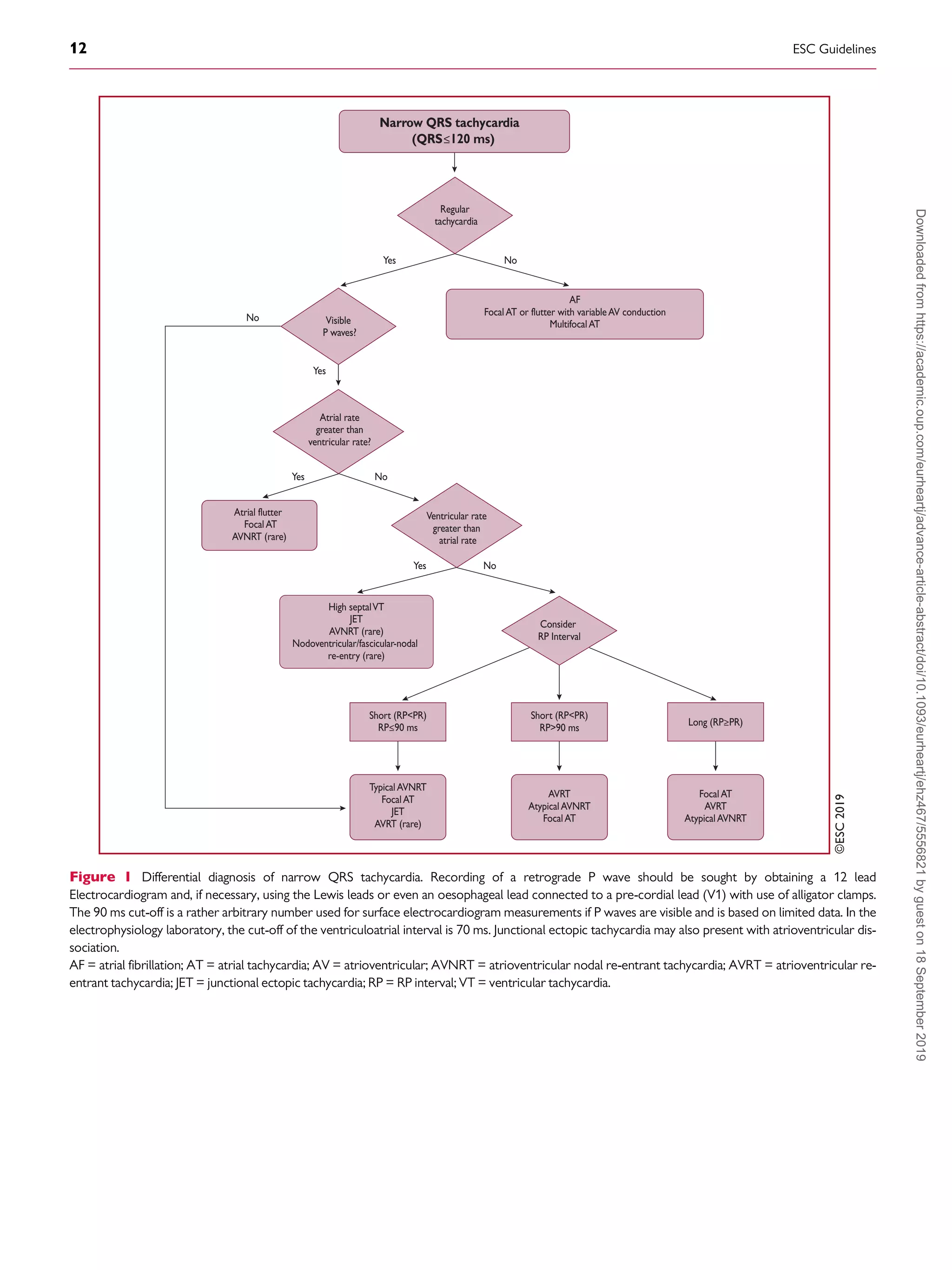
9.1 Narrow QRS (=120 ms) tachycardias . . . . . . . . . . . . . . . . . . . . . . . . 11
9.1.1 Electrocardiographic differential diagnosis . . . . . . . . . . . . . . . . 11
9.1.1.1 Initiation and termination of the tachycardia . . . . . . . . . . . . 11
9.1.1.2 Regularity of tachycardia cycle length . . . . . . . . . . . . . . . . . . 11
9.1.1.3 P/QRS relationship . . . . . . . . . . . . . . . . . . . . . . . . . . . . . . . . . . . 11
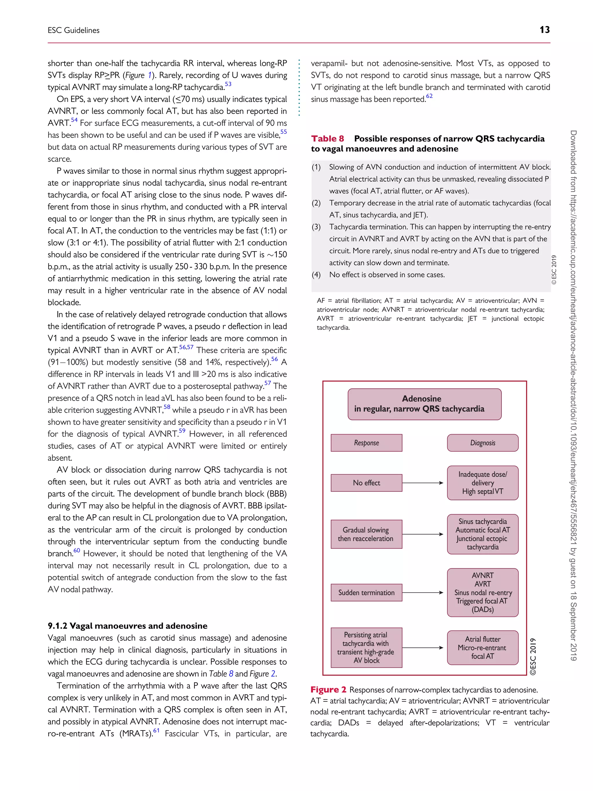
9.1.2 Vagal manoeuvres and adenosine . . . . . . . . . . . . . . . . . . . . . . . . 13
9.1.3 Electrophysiology study . . . . . . . . . . . . . . . . . . . . . . . . . . . . . . . . . 14
9.2 Wide QRS (120 ms) tachycardias . . . . . . . . . . . . . . . . . . . . . . . . . . . 14
9.2.1 Electrocardiographic differential diagnosis . . . . . . . . . . . . . . . . 14
9.2.1.1 Atrioventricular dissociation . . . . . . . . . . . . . . . . . . . . . . . . . . . 14
9.2.1.2 QRS duration . . . . . . . . . . . . . . . . . . . . . . . . . . . . . . . . . . . . . . . . 14
9.2.1.3 QRS axis . . . . . . . . . . . . . . . . . . . . . . . . . . . . . . . . . . . . . . . . . . . . 14
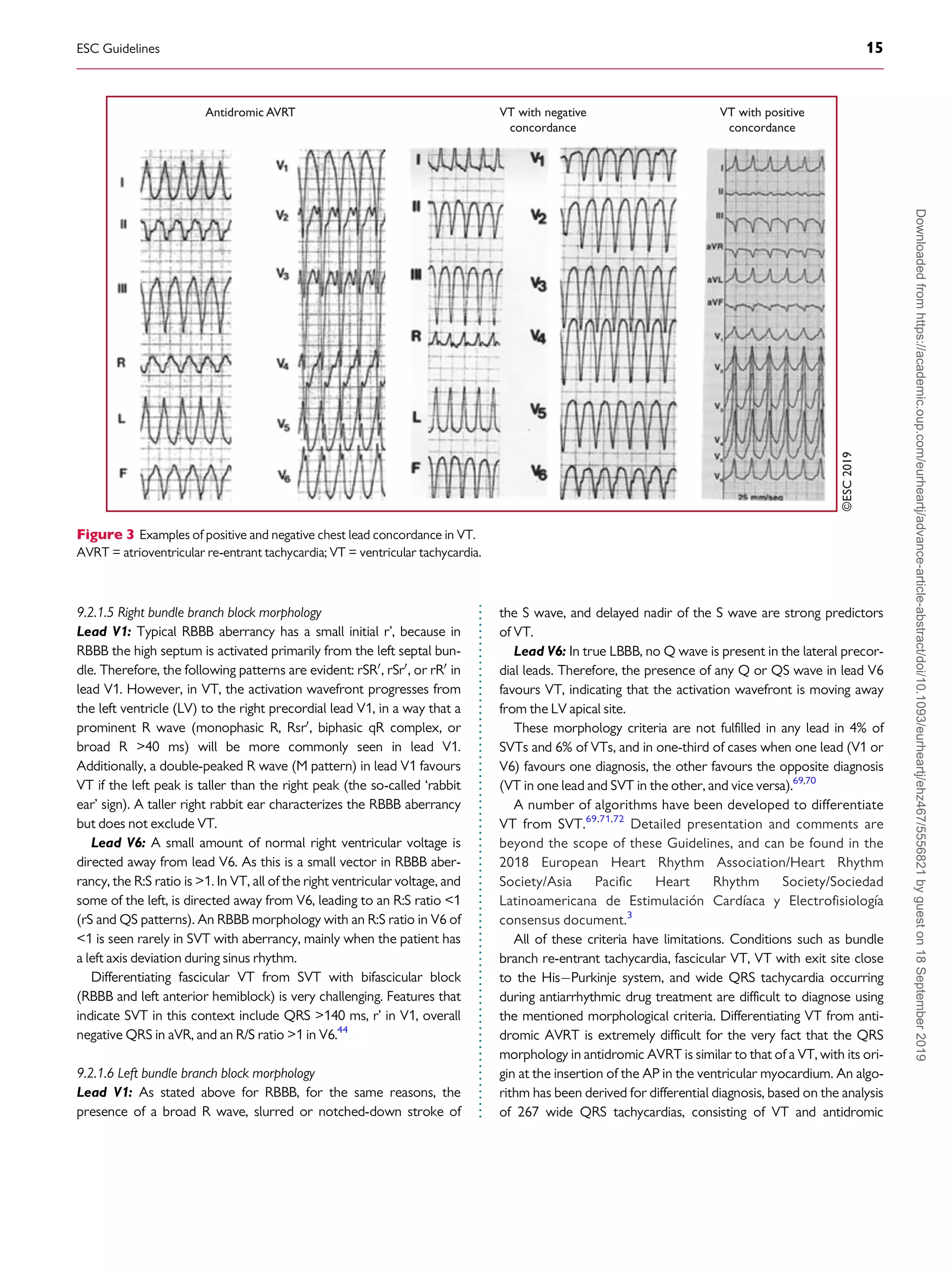
9.2.1.4 Chest lead concordance . . . . . . . . . . . . . . . . . . . . . . . . . . . . . . 14
9.2.1.5 Right bundle branch block morphology . . . . . . . . . . . . . . . . . 15
9.2.1.6 Left bundle branch block morphology . . . . . . . . . . . . . . . . . . 15
9.2.2 Electrophysiology study . . . . . . . . . . . . . . . . . . . . . . . . . . . . . . . . . 16
9.3 Irregular tachycardias . . . . . . . . . . . . . . . . . . . . . . . . . . . . . . . . . . . . . . . . 16
10 Acute management in the absence of an established diagnosis . . . . 16
10.1 Regular tachycardias . . . . . . . . . . . . . . . . . . . . . . . . . . . . . . . . . . . . . . . . 16
10.1.1 Narrow QRS (=120 ms) tachycardias . . . . . . . . . . . . . . . . . . . 16
10.1.1.1 Haemodynamically unstable patients . . . . . . . . . . . . . . . . 16
10.1.1.2 Haemodynamically stable patients . . . . . . . . . . . . . . . . . . . 16
10.1.2 Wide QRS (120 ms) tachycardias . . . . . . . . . . . . . . . . . . . . . 18
10.1.2.1 Haemodynamically unstable patients . . . . . . . . . . . . . . . . 18
10.1.2.2 Haemodynamically stable patients . . . . . . . . . . . . . . . . . . . 18
10.2 Irregular tachycardias . . . . . . . . . . . . . . . . . . . . . . . . . . . . . . . . . . . . . . . 19
11 Specific types of supraventricular tachycardia . . . . . . . . . . . . . . . . . . . . 19
11.1 Atrial arrhythmias . . . . . . . . . . . . . . . . . . . . . . . . . . . . . . . . . . . . . . . . . . 19
11.1.1 Sinus tachycardia . . . . . . . . . . . . . . . . . . . . . . . . . . . . . . . . . . . . . . . 19
11.1.1.1 Physiological sinus tachycardia . . . . . . . . . . . . . . . . . . . . . . . 19
11.1.1.2 Inappropriate sinus tachycardia . . . . . . . . . . . . . . . . . . . . . . 19
11.1.1.2.1 Diagnosis . . . . . . . . . . . . . . . . . . . . . . . . . . . . . . . . . 19
11.1.1.2.2 Therapy . . . . . . . . . . . . . . . . . . . . . . . . . . . . . . . . . . 19
11.1.1.3 Sinus node re-entrant tachycardia . . . . . . . . . . . . . . . . . . . . 20
11.1.1.3.1 Diagnosis . . . . . . . . . . . . . . . . . . . . . . . . . . . . . . . . . 20
11.1.1.3.2 Therapy . . . . . . . . . . . . . . . . . . . . . . . . . . . . . . . . . . 20
11.1.1.4 Postural orthostatic tachycardia syndrome . . . . . . . . . . . . 20
11.1.1.4.1 Diagnosis . . . . . . . . . . . . . . . . . . . . . . . . . . . . . . . . . 20
11.1.1.4.2 Therapy . . . . . . . . . . . . . . . . . . . . . . . . . . . . . . . . . . 21
11.1.2 Focal atrial tachycardia . . . . . . . . . . . . . . . . . . . . . . . . . . . . . . . . . 21
2 ESC Guidelines
Downloaded
from
https://academic.oup.com/eurheartj/advance-article-abstract/doi/10.1093/eurheartj/ehz467/5556821
by
guest
on
18
September
2019](https://image.slidesharecdn.com/brugada-2020-esc-guidelines-for-the-management-supraventriculartachycardia-samirrafla-210905063343/75/Brugada-2020-esc-guidelines-for-the-management-supraventricular-tachycardia-samir-rafla-2-2048.jpg)

![.
.
.
.
.
.
.
.
.
.
.
.
.
.
.
.
.
.
.
.
.
.
.
.
.
.
.
.
.
.
.
.
.
.
.
.
.
.
.
.
.
.
.
.
.
.
.
.
.
.
.
.
.
.
.
.
.
.
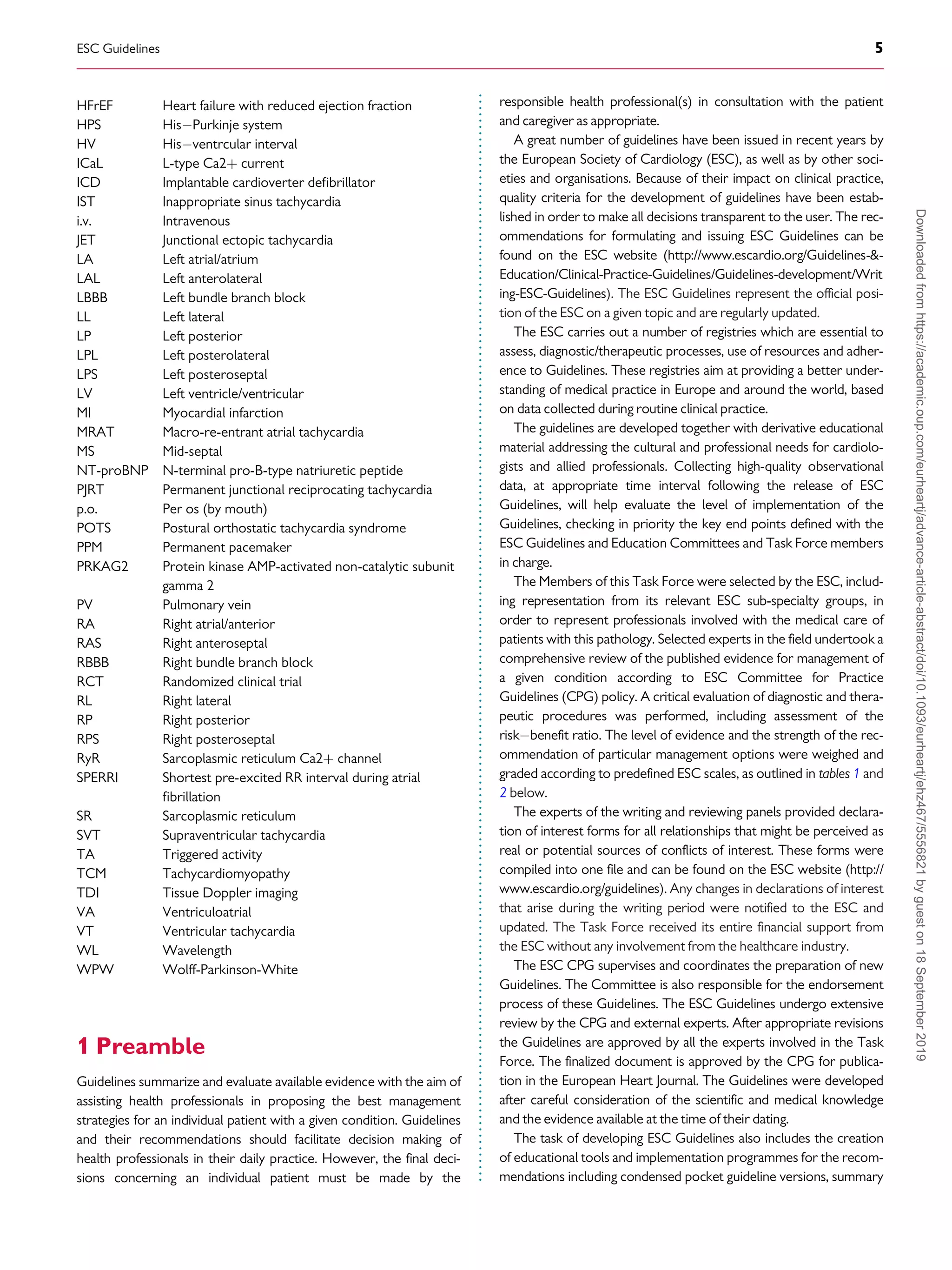
2.3.2 New recommendations in 2019
2.3.3 New revised concepts
• Drug therapy for inappropriate sinus tachycardia and focal atrial
tachycardia.
• Therapeutic options for acute conversion and anticoagulation of
atrial flutter.
• Therapy of atrioventricular nodal re-entrant tachycardia.
• Therapy of antidromic atrioventricular re-entrant tachycardia and
pre-excited atrial fibrillation.
• Management of patients with asymptomatic pre-excitation.
• Diagnosis and therapy of tachycardiomyopathy.
3 Definitions and classification
The term ‘SVT’ literally indicates tachycardia [atrial rates 100 beats
per minute (b.p.m.) at rest], the mechanism of which involves tissue
from the His bundle or above.2,3
Traditionally, SVT has been used to
describe all kinds of tachycardias apart from ventricular tachycardias
(VTs) and AF. It has therefore included tachycardias such as atrioven-
tricular (AV) re-entry due to accessory connections, which is not, in
Table 3 Continued
2003 2019
Verapamil and diltiazem I IIa
Beta-blockers I IIa
Digitalis is not mentioned in the 2019 Guidelines
Chronic
Dofetilide, sotalol, flecainide, propafenone, procai-
namide, quinidine, and disopyramide are not men-
tioned in the 2019 Guidelines
Therapy of AVNRT
Acute
Amiodarone, sotalol, flecainide, and propafenone
are not mentioned in the 2019 Guidelines
Chronic
Verapamil and diltiazem I IIa
Beta-blockers I IIa
Amiodarone, sotalol, flecainide, propafenone, and
the ‘pill-in-the pocket’ approach are not men-
tioned in the 2019 Guidelines
Therapy of AVRT
Flecainide/propafenone IIa IIb
Beta-blockers IIb IIa
Amiodarone, sotalol, and the ‘pill-in-the pocket’
approach are not mentioned in the 2019
Guidelines
SVT in pregnancy
Verapamil IIb IIa
Catheter ablation IIb IIa*
Sotalol, propranolol, quinidine, and procainamide
are not mentioned in the 2019 Guidelines.
*: when fluoroless ablation is available. AT = atrial tachycardia; AVNRT = atrio-
ventricular nodal re-entrant tachycardia; AVRT = atrioventricular re-entrant
tachycardia.
Table 4 New recommendations in 2019
Ivabradine alone or in combination with a beta-
blocker should be considered in symptomatic patients
with inappropriate sinus tachycardia.
IIa
Ibutilide (i.v.) ibutilide may be considered for acute
therapy of focal atrial tachycardia.
IIb
Ivabradine for postural orthostatic tachycardia syn-
drome, and ivabradine with a beta-blocker for chronic
therapy of focal atrial tachycardia, may be considered.
IIb
Patients with atrial flutter without AF should be con-
sidered for anticoagulation, but the threshold for ini-
tiation is not established.
IIa
Ibutilide (i.v.), or i.v. or oral (in-hospital) dofetilide are
recommended for conversion of atrial flutter.
I
High-rate atrial pacing is recommended for termina-
tion of atrial flutter in the presence of an implanted
pacemaker or defibrillator.
I
Continued
i.v. amiodarone is not recommended for pre-excited AF. III
Performance of an EPS to risk-stratify individuals with
asymptomatic pre-excitation should be considered.
IIa
Catheter ablation is recommended in asymptomatic
patients in whom electrophysiology testing with the
use of isoprenaline identifies high-risk properties, such
as SPERRI
_250 ms, AP ERP
_250 ms, multiple APs,
and an inducible AP-mediated tachycardia.
I
Non-invasive evaluation of the conducting properties
of the AP in individuals with asymptomatic pre-excita-
tion may be considered.
IIb
Catheter ablation may be considered in a patient with
asymptomatic pre-excitation and low-risk AP at inva-
sive or non-invasive risk stratification.
IIb
Catheter ablations should be considered in patients
with asymptomatic pre-excitation and LV dysfunction
due to electrical dyssynchrony.
IIa
AV nodal ablation with subsequent pacing (‘ablate and
pace’), either biventricular or His-bundle pacing, is
recommended if a tachycardia responsible for TCM
cannot be ablated or controlled by drugs.
I
During the first trimester of pregnancy, it is recommended
that all antiarrhythmic drugs are avoided, if possible.
I
In pregnant women, beta-1 selective blockers (except
atenolol) or verapamil, in order of preference, should
be considered for prevention of SVT in patients with-
out WPW syndrome.
IIa
In pregnant women, flecainide or propafenone should
be considered for prevention of SVT in patients with
WPW syndrome and without ischaemic or structural
heart disease.
IIa
AF = atrial fibrillation; AP = accessory pathway; AT = atrial tachycardia; AV =
atrioventricular; EPS = electrophysiology study; ERP = effective refractory period;
i.v. = intravenous; LV = left ventricular; POTS: postural orthostatic tachycardia
syndrome; SPERRI = shortest pre-excited RR interval during atrial fibrillation;
SVT = supraventricular tachycardia; TCM = tachycardiomyopathy; WPW =
Wolff-Parkinson-White.
8 ESC Guidelines
Downloaded
from
https://academic.oup.com/eurheartj/advance-article-abstract/doi/10.1093/eurheartj/ehz467/5556821
by
guest
on
18
September
2019](https://image.slidesharecdn.com/brugada-2020-esc-guidelines-for-the-management-supraventriculartachycardia-samirrafla-210905063343/75/Brugada-2020-esc-guidelines-for-the-management-supraventricular-tachycardia-samir-rafla-8-2048.jpg)
![.
.
.
.
.
.
.
.
.
.
.
.
.
.
.
.
.
.
.
.
.
.
.
.
.
.
.
.
.
.
.
.
.
essence, a supraventricular rhythm (Table 5). The term ‘narrow QRS
tachycardia’ indicates those with a QRS duration
_120 ms. A wide
QRS tachycardia refers to one with a QRS duration 120 ms
(Table 6). In clinical practice, SVT may present as narrow or wide QRS
tachycardias, most of which, although not invariably, manifest as regu-
lar rhythms. These Guidelines do not cover AF, which is the subject of
separate clinical Guidelines4
and various consensus documents.57
4 Electrophysiological
mechanisms of supraventricular
tachycardia
Arrhythmia can originate from abnormal impulse initiation in an individ-
ual myocyte or, more realistically, in a close cluster of myocytes. This
can occur in non-pacemaker cells through a mechanism similar to the
physiological automaticity of pacemaker cells [sinus node and AV node
(AVN)], and is thus named ‘abnormal’ or ‘enhanced automaticity’. An
alternative form of abnormal impulse initiation involves oscillations of
membrane potential, named early or delayed ‘after-depolarizations’. In
such cases, the resulting arrhythmias take the name of ‘triggered activ-
ity’.8
Arrhythmias resulting from enhanced automaticity and triggered
activity are defined as ‘non-re-entrant’. Arrhythmias can also arise
when myocardial regions activated later in propagation re-excite
regions that have already recovered excitability. This results from
abnormal propagation of the excitation wavefront and/or of tissue
refractoriness. This mechanism, named ‘re-entry’, is based on the syn-
cytial nature of myocardial tissue and is thus radically different from
focal impulse initiation.8
A detailed discussion and schematic represen-
tation of common SVT circuits is provided in the Supplementary Data.
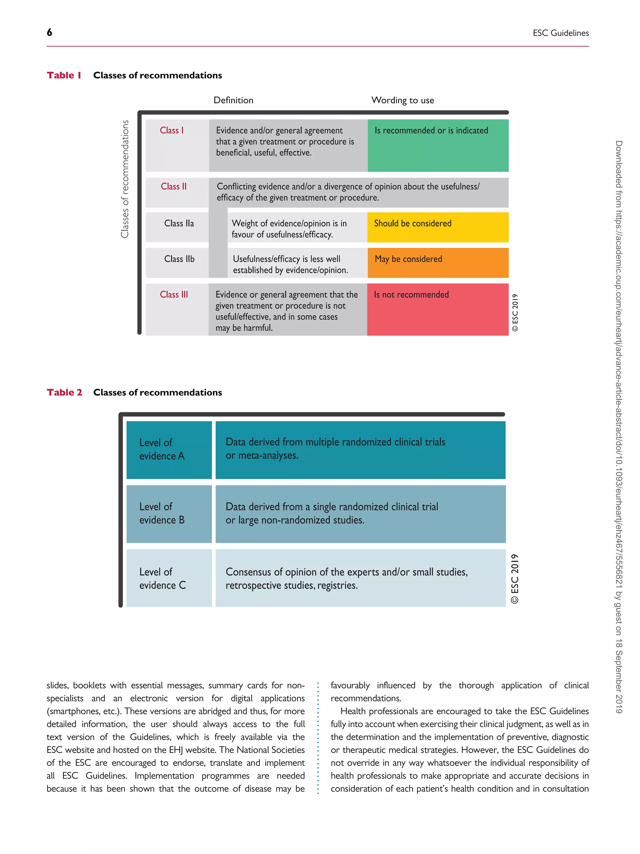
Table 6 Differential diagnosis of narrow and wide QRS
tachycardias
Narrow QRS (120 ms) tachycardias
Regular
• Physiological sinus tachycardia
• Inappropriate sinus tachycardia
• Sinus nodal re-entrant tachycardia
• Focal AT
• Atrial flutter with fixed AV conduction
• AVNRT
• JET (or other non-re-entrant variants)
• Orthodromic AVRT
• Idiopathic VT (especially high septal VT)
Irregular
• AF
• Focal AT or atrial flutter with varying AV block
• Multifocal AT
Wide QRS (120 ms) tachycardias
Regular
• VT/flutter
• Ventricular paced rhythm
• Antidromic AVRT
• SVTs with aberration/BBB (pre-existing or rate-dependent during
tachycardia)
• Atrial or junctional tachycardia with pre-excitation/bystander AP
• SVT with QRS widening due to electrolyte disturbance or antiarrhyth-
mic drugs
Irregular
• AF or atrial flutter or focal AT with varying block conducted with
aberration
• Antidromic AV re-entrant tachycardia due to a nodo-ventricular/fascic-
ular AP with variable VA conduction
• Pre-excited AF
• Polymorphic VT
• Torsade de pointes
• Ventricular fibrillation
Occasionally, AF with very fast ventricular response may apparently
resemble a regular narrow QRS tachycardia.
AF = atrial fibrillation; AP = accessory pathway; AT = atrial tachycardia; AV =
atrioventricular; AVNRT = atrioventricular nodal re-entrant tachycardia; AVRT =
atrioventricular re-entrant tachycardia; BBB = bundle branch block; JET = junc-
tional ectopic tachycardia; SVT = supraventricular tachycardia; VA = ventriculoa-
trial; VT = ventricular tachycardia.
Table 5 Conventional classification of supraventricular
tachycardias
Atrial tachycardias
Sinus tachycardia
• Physiological sinus tachycardia
• Inappropriate sinus tachycardia
• Sinus nodal re-entrant tachycardia
Focal AT
Multifocal AT
MRAT
• Cavotricuspid isthmus-dependent MRAT
Typical atrial flutter, counter-clockwise (common) or clockwise
(reverse)
Other cavotricuspid isthmus-dependent MRAT
• Non-cavotricuspid isthmus-dependent MRAT
RA MRAT
LA MRAT
AF
AV junctional tachycardias
Atrioventricular nodal re-entrant tachycardia (AVNRT)
• Typical
• Atypical
Non-re-entrant junctional tachycardia
• JET (junctional ectopic or focal junctional tachycardia)
• Other non-re-entrant variants
Atrioventricular re-entrant tachycardia (AVRT)
• Orthodromic (including PJRT)
• Antidromic (with retrograde conduction through the AVN or, rarely,
over another pathway)
AF = atrial fibrillation; AT = atrial tachycardia; AV = atrioventricular; AVN =
atrioventricular node; JET = junctional ectopic tachycardia; RA = right atrial; LA
= left atrial; MRAT = macro-re-entrant atrial tachycardia; PJRT = permanent junc-
tional reciprocating tachycardia; RA = right atrial.
ESC Guidelines 9
Downloaded
from
https://academic.oup.com/eurheartj/advance-article-abstract/doi/10.1093/eurheartj/ehz467/5556821
by
guest
on
18
September
2019](https://image.slidesharecdn.com/brugada-2020-esc-guidelines-for-the-management-supraventriculartachycardia-samirrafla-210905063343/75/Brugada-2020-esc-guidelines-for-the-management-supraventricular-tachycardia-samir-rafla-9-2048.jpg)
![.
.
.
.
.
.
.
.
.
.
.
.
.
.
.
.
.
.
.
.
.
.
.
.
.
.
.
.
.
.
.
.
.
.
.
.
.
.
.
.
.
.
.
.
.
.
.
.
.
.
.
.
.
.
.
.
.
.
.
.
.
.
.
.
.
.
.
.
.
.
.
.
.
.
.
.
.
.
.
.
.
.
.
.
.
.
.
.
.
.
.
.
.
.
.
.
.
.
.
.
.
.
.
.
.
.
.
.
5 Cardiac anatomy for the
electrophysiologist
Knowledge of anatomical structures inside and outside the atrial
chambers is of clinical importance, especially when interventional
procedures are being considered. A detailed discussion is provided in
the Supplementary Data.
6 Epidemiology of
supraventricular tachycardia
Epidemiological studies on the SVT population are limited. In the gen-
eral population, the SVT prevalence is 2.25/1000 persons and the
incidence is 35/100000 person-years. Women have a risk of develop-
ing SVT that is two times greater than that of men, and persons aged
_65 years or have more than five times the risk of developing SVT
than younger individuals. Patients with lone paroxysmal SVT vs. those
with cardiovascular disease are younger, have a faster SVT rate, have
an earlier onset of symptoms, and are more likely to have their condi-
tion first documented in the emergency department.9
In a paediatric
cohort study of 1 967 911 live births between 200008, 2021
patients (51.6% male, overall incidence 1.03/1000) had SVT
[Wolff-Parkinson-White (WPW) syndrome accounted for 16.2%].
By the age of 15 years, the annual risk of sudden death was 0.01% per
patient-year.10
In specialized centres, AVNRT is the most frequently treated sub-
strate after AF, followed by atrial flutter and AVRT, in patients
referred for catheter ablation.1113
Women are more likely to be
affected by AVNRT than men (ratio 70:30),1416
while the con-
verse is true for AVRT (ratio 45:55).14
A relationship with the
monthly cycle has been suggested,17
and episodes are more frequent
during pregnancy in women with pre-existing SVT.18
In the general population, the prevalence of a WPW pattern on
surface electrocardiogram (ECG) ranges from 0.150.25%,19
increasing to 0.55% among first-degree relatives of affected
patients.20
However, not all patients develop SVT and
intermittent pre-excitation is not rare. Compared with the remaining
population, the pre-excitation population is generally younger, pre-
dominantly male, and has less comorbidity.21,22
The proportion of
patients with AVRT decreases with age, whereas the proportion of
those with AVNRT and AT increases with age.14
The epidemiology of atrial flutter is not known with certainty, as
atrial flutter and AF can coexist. The prevalence of AF before flutter
ablation ranges from 2462%, and after ablation could be 3070%.
The overall incidence of atrial flutter is 88/100 000 person-years in
the US population annually. Adjusted for age, the incidence of atrial
flutter in men (125/100 000) is 2.5 times that of women (59/
100 000) and increases exponentially with age. Patients with atrial
flutter are more likely to have been smokers, have a longer PR inter-
val, history of myocardial infarction (MI), and history of heart failure
(HF).23
Catheter ablation is now used extensively for most varieties of
SVT, and patient-reported outcome measures have shown that
patients experience significant improvements in their quality of life
following ablation.2427
Patient-reported outcome measures using
various questionnaires are useful in the audit of ablation techniques.
Women are more often prescribed antiarrhythmic drugs before abla-
tion for SVT than men,28
and recurrence rates following AVNRT
ablation are higher in young women.29
However, overall, no signifi-
cant differences in health-related quality of life or access to healthcare
resources between men and women have been reported.28
7 Clinical presentation
The impact of SVT on an individual is based on a range of factors and
may result in palpitations, fatigue, light-headedness, chest discomfort,
dyspnoea, and altered consciousness.30
Rapid rhythms are more
likely to present with a clear history and acutely than less-rapid
rhythms, but SVT usually produces symptoms.31
The duration of
symptoms and the patient’s age at onset are important. An individual
with an onset in the teenage years or younger is less likely to have AT
or AF continuing into adulthood. Such a long history will point
towards a re-entrant mechanism.32
Dyspnoea, or other clinical signs
and symptoms of HF, can occur when the patient has developed
TCM. Light-headedness in association with SVT is not infrequent.30
Presyncope and syncope are less common,33,34
and tend to be asso-
ciated with presentation in older individuals.32
In older patients,
symptoms may be more extreme—with dizziness, presyncope, and
syncope—in view of the less-accommodating characteristics of the
circulation; drops in blood pressure are usually immediate and tend
to recover.35
In some patients, a description of polyuria (possibly due
to atrial stretch-induced atrial natriuretic peptide activity) can be elu-
cidated, although this is infrequent.36
Direct risks due to SVT are
unusual, but in specific situations (e.g. in patients with WPW syn-
drome and AF,22
or after atrial switch operation37
) may lead to sud-
den cardiac death.
A sudden onset more likely points to AVNRT or AVRT, although
an AT may also present in this way.32
Characteristics in terms of the
regularity or irregularity are helpful. The duration of individual epi-
sodes may help in terms of differentiation. Re-entrant tachycardias
Table 7 Initial evaluation of the patient with supraven-
tricular tachycardia
Standard
• History, physical examination, and 12 lead ECG
• Full blood counts, biochemistry profile, and thyroid function
• An ECG during tachycardia should be sought
• Transthoracic echocardiography
Optional
• Exercise tolerance testing
• 24 h ECG monitoring, transtelephonic monitoring, or an implantable
loop recorder
• Myocardial ischaemia testing in patients with risk factors for coronary
artery disease (including men aged 40 years and post-menopausal
women)
• An EPS should be considered for a definitive diagnosis and when cathe-
ter ablation is anticipated
ECG = electrocardiogram; EPS = electrophysiology study.
10 ESC Guidelines
Downloaded
from
https://academic.oup.com/eurheartj/advance-article-abstract/doi/10.1093/eurheartj/ehz467/5556821
by
guest
on
18
September
2019](https://image.slidesharecdn.com/brugada-2020-esc-guidelines-for-the-management-supraventriculartachycardia-samirrafla-210905063343/75/Brugada-2020-esc-guidelines-for-the-management-supraventricular-tachycardia-samir-rafla-10-2048.jpg)
![.
.
.
.
.
.
.
.
.
.
.
.
.
.
.
.
.
.
.
.
.
.
.
.
.
.
.
.
.
.
.
.
.
.
.
.
.
.
.
.
.
.
.
.
.
.
.
.
.
.
.
.
.
.
.
.
.
.
.
.
.
.
.
.
.
.
.
.
.
.
.
.
.
.
.
.
.
.
.
.
.
.
.
.
.
.
.
.
.
.
.
.
.
.
.
.
.
.
.
.
.
.
.
.
.
.
.
.
.
.
.
.
.
.
.
.
.
.
.
.
.
.
.
.
.
.
.
.
.
.
.
.
.
.
.
.
.
.
.
.
.
.
.
.
.
.
.
.
.
.
.
.
.
.
.
.
.
.
.
.
.
.
.
.
.
.
.
.
.
.
.
.
.
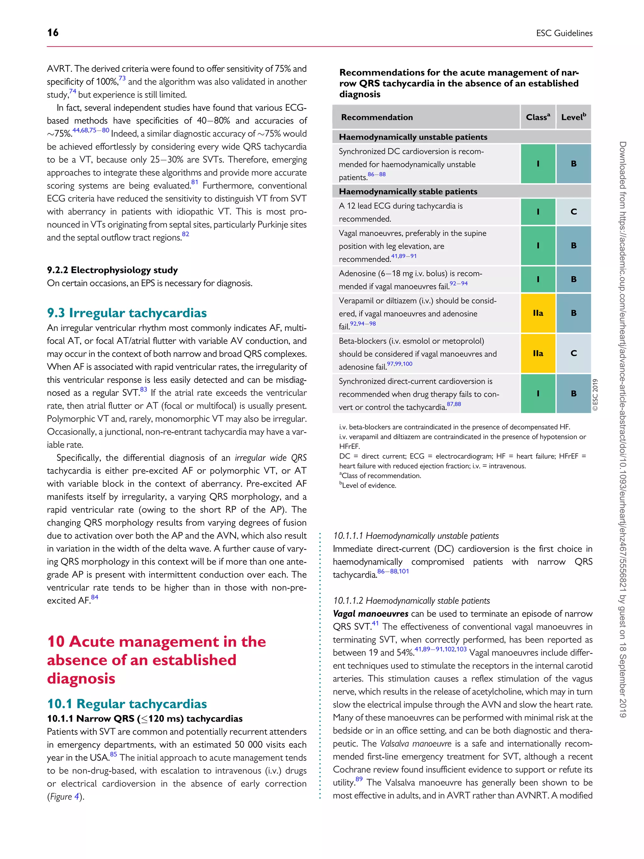
approach to the Valsalva manoeuvre provides a considerable
enhancement of conversion success rates (43 vs. 17% conversion
rate).41
This enhanced method requires the Valsalva to be completed
semi-recumbent, with supine repositioning and passive leg raise after
the Valsalva strain. Blowing into a 10 mL syringe with sufficient force
to move the plunger may standardize the approach.104
Carotid sinus
massage is performed with the patient’s neck in an extended posi-
tion, with the head turned away from the side to which pressure is
applied. It should always be unilateral as there is a potential risk with
bilateral pressure, and it should be limited to 5 s. The patient should
be monitored. This technique should be avoided in patients with pre-
vious transient ischaemic attack or stroke, and in patients with carotid
bruits.3
Other manoeuvres, such as facial immersion in cold water or
forceful coughing, are rarely used now.
Adenosine, an endogenous purine nucleoside (618 mg i.v. bolus)
is the first drug of choice.9294
Pharmacologically relevant electro-
physiological influences are mediated through cardiac adenosine A1
receptors.105,106
Clinical EPSs have documented progressive dose-
related prolongation of AV conduction [due to effects on the
atrialHis (AH) interval, and none on the HV interval], culminating in
transient AV block that is then responsible for tachycardia
termination.107
The mean dose required for termination is 6 mg. To achieve effi-
cient rhythm correction, injection should be as a rapid bolus with
immediate saline flush. Large, centrally located (e.g. antecubital) veins
are likely to deliver more effective drug concentrations to the heart
than smaller distal veins.108
Dosing should then be incremental, start-
ing at 6 mg in adults followed by 12 mg. An 18 mg dose should then
be considered, also taking into account tolerability/side effects in the
individual patient. Adenosine has a very short plasma half-life due to
enzymatic deamination to inactive inosine being achieved in seconds,
with end-organ clinical effects complete within 2030 s.107
Thus,
repeat administration is safe within 1 min of the last dose.2,3
The dose
range between patients may be very wide,107
with 90% success gen-
erally expected.94,109
Some drugs (e.g. dipyridamole and theophyl-
line) may on occasion affect dose requirements, but any influence of
recent intake of caffeinated beverages is disputed.110,111
Transient dyspnoea is common with increased ventilation, and is
more likely to result from the stimulation of pulmonary vagal C
fibres.112
Facial flushing may occur, associated with vasodilatation and
increased skin temperature.107
Chest pain, variable in terms of radia-
tion over the thorax, may suggest ischaemic or oesophageal origins,
and has been associated with increased coronary sinus blood flow so
may well be of cardiac origin.107
Depression of sinoatrial node function is to be expected based on
established pharmacology, but prolonged bradycardia is
unusual.105,107
Nonetheless, adenosine administration should be
approached cautiously in those with known sinus node disease.113
Perceived risks of bradycardia in recipients of denervated orthotropic
heart transplants, in whom SVT is common, have prompted a relative
contraindication.114,115
However, more recent substantive evidence
supports adenosine use in this group with no particular cautions.116
AF may occur following adenosine administration as a result of either
direct pulmonary vein (PV) triggering117
or increasing heterogeneity
of repolarization,118
and appears more commonly associated with
AVRT than AVNRT.93
Adenosine may also occasionally cause or
accelerate pre-excited atrial arrhythmias.119,120
Clinically important bronchoconstriction has been rarely reported
in those receiving i.v. adenosine for SVT,121
and this observation is
further supported by the large experience obtained when adenosine
infusions have been given for cardiac stress testing.105,122,123
Furthermore, despite inhaled adenosine producing bronchoconstric-
tion in people with asthma,124
i.v. administration has had no impact
on the airways in clinical experimental studies.125
There have been
isolated reports of clinically well-documented bronchoconstriction
occurring in patients with or without respiratory disease, thus sug-
gesting that care is required in patients with asthma.121,126,127
However, adenosine can be used cautiously in those with asthma,
although verapamil may be a more appropriate choice in patients
with severe asthma.
Adenosine triphosphate may also be used but clinical experience is
limited.
Calcium channel blockers (verapamil/diltiazem i.v.) and beta-
blockers (e.g. esmolol and metoprolol i.v.) are of value, particularly
in patients with frequent atrial or ventricular premature beats.
Verapamil [0.075 - 0.15 mg/kg i.v. (average 5 - 10 mg) over 2 min] or
i.v. diltiazem [0.25 mg/kg (average 20 mg) over 2 min] has been
shown to terminate SVT in 6498% of patients, but is associated
with a risk of hypotension.92,9498,128
These drugs should be avoided
in patients with haemodynamic instability, HF with reduced LV ejec-
tion fraction (40%), a suspicion of VT, or pre-excited AF. Beta-
blockers (i.v.), such as short-acting esmolol (0.5 mg/kg i.v. bolus or
0.05 - 0.3 mg/kg/min infusion) or metoprolol (2.515 mg given i.v. in
©ESC
2019
Narrow QRS
tachycardia
Vagal manoeuvres
(I B)
i.v.adenosine
(I B)
i.v.verapamil or
diltiazem
(IIa B)
i.v.beta-blocker
(IIa C)
Synchronized
cardioversion
(I B)
Yes
No
If ineffective
If ineffective
If ineffective
Haemodynamic
instability
Figure 4 Acute therapy of narrow QRS tachycardia in the absence of
an established diagnosis.
i.v. = intravenous.
ESC Guidelines 17
Downloaded
from
https://academic.oup.com/eurheartj/advance-article-abstract/doi/10.1093/eurheartj/ehz467/5556821
by
guest
on
18
September
2019](https://image.slidesharecdn.com/brugada-2020-esc-guidelines-for-the-management-supraventriculartachycardia-samirrafla-210905063343/75/Brugada-2020-esc-guidelines-for-the-management-supraventricular-tachycardia-samir-rafla-17-2048.jpg)
![.
.
.
.
.
.
.
.
.
.
.
.
.
.
.
.
.
.
.
.
.
.
.
.
.
.
.
.
.
.
.
.
.
.
.
.
.
.
.
.
.
.
.
.
.
.
.
.
.
.
.
.
.
.
.
.
.
.
.
.
.
.
.
.
.
.
.
.
.
.
.
.
.
.
.
.
.
.
.
.
.
.
.
.
.
.
.
.
.
.
.
.
.
.
.
.
.
.
.
.
.
.
.
.
.
.
.
.
.
.
.
.
.
.
.
.
.
.
.
.
.
.
.
.
.
.
.
.
.
.
.
.
.
.
.
.
.
.
.
.
.
.
.
.
.
.
.
.
.
.
.
.
.
.
.
.
.
.
.
.
.
.
.
.
.
.
.
.
.
.
.
.
.
monitoring. The evaluation of a patient suspected of having POTS
should eliminate other causes of sinus tachycardia such as hypovo-
laemia, anaemia, hyperthyroidism, pulmonary embolus, or pheo-
chromocytoma.178
The clinical history should focus on defining
the chronicity of the condition, possible causes of orthostatic
tachycardia, modifying factors, impact on daily activities, and
potential triggers.
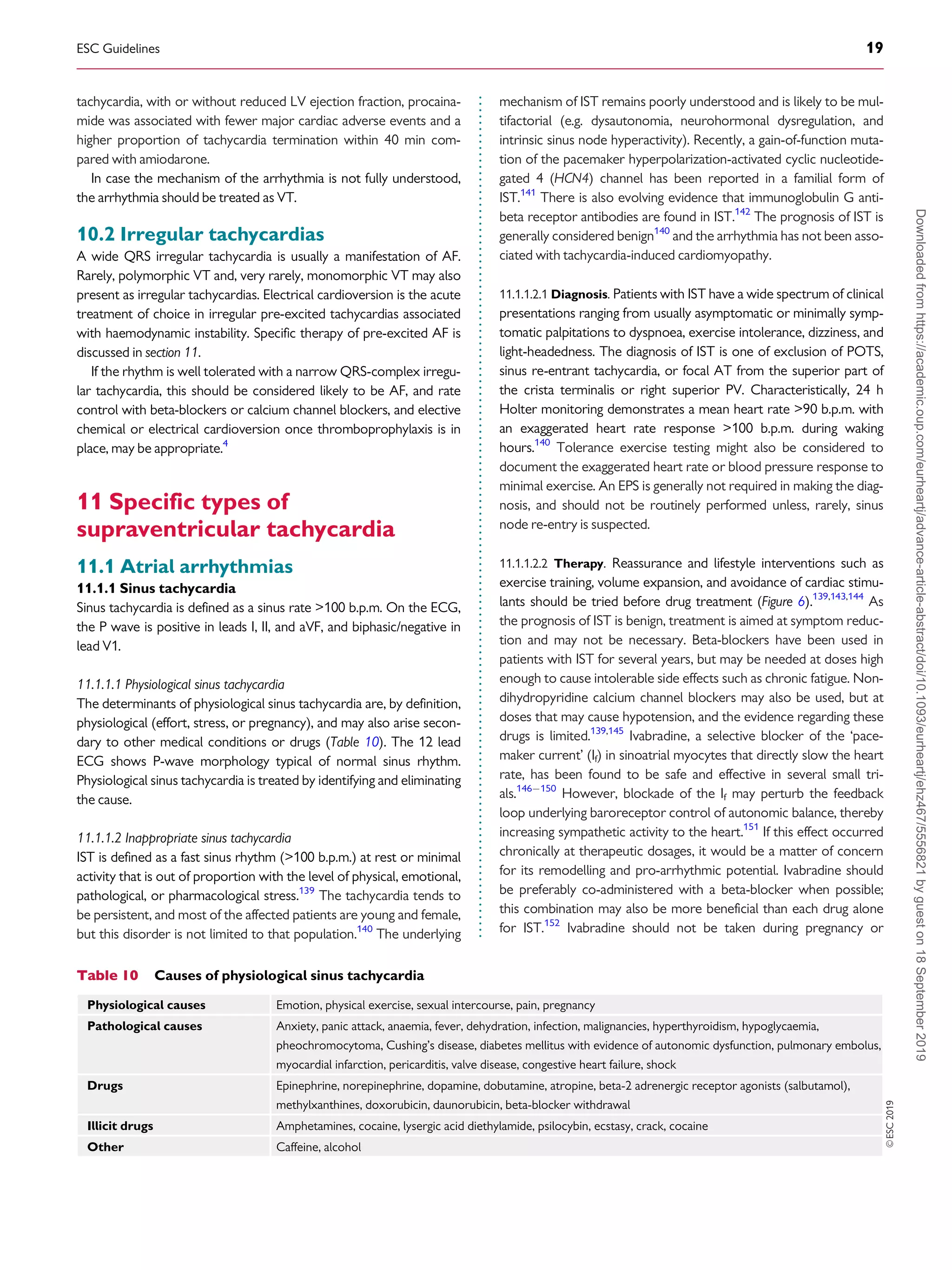
11.1.1.4.2 Therapy. Non-pharmacological treatments should be
attempted first in all patients. These include withdrawing medications
that might worsen POTS, such as norepinephrine transport inhibi-
tors, increasing blood volume with enhanced salt and fluid intake,
reducing venous pooling with compression garments, and limiting
deconditioning. Patients should engage in a regular, graduated, and
supervised exercise programme featuring aerobic reconditioning
with some resistance training for the thighs. Initially, exercise should
be restricted to non-upright exercise, including the use of rowing
machines and swimming, to minimize orthostatic stress on the
heart.180182
If non-pharmacological approaches prove ineffective, pharmaco-
logical therapies may be targeted at specific aspects. Patients strongly
suspected of having hypovolaemia should drink
_23 L of water per
day, and dietary salt intake should be increased to 1012 g/day if
tolerated. Midodrine significantly reduces orthostatic tachycardia but
to a lesser degree than i.v. saline.170
Midodrine has a rapid onset with
only brief effects and is usually administered three times daily. The
drug should only be administered during daytime hours as it can
cause supine hypertension. To reduce unpleasant sinus tachycardia
and palpitations, low-dose propranolol [1020 mg per os (p.o.)]
acutely lowers standing heart rate and improves symptoms in
patients with POTS, while higher doses of propranolol are less well
tolerated.172
Long-acting propranolol does not improve the quality
of life of patients with POTS.167
Non-selective beta-blockers are
preferable because they additionally block epinephrine-mediated
beta-2-vasodilation, but other beta-blockers have not been
adequately studied. Pyridostigmine, a cholinergic agonist that works
by inhibiting acetylcholinesterase, can increase parasympathetic auto-
nomic tone and has a lower risk of hypertension compared with
other medications. Potential side effects include abdominal cramping,
diarrhoea, and muscle cramps.173,174
Ivabradine slows sinus rates
without affecting blood pressure, and in an open-label study 60% of
patients with POTS had symptomatic improvement.175
Ivabradine
should ideally be administered with concomitant beta-blockers for
long-term therapy.151
11.1.2 Focal atrial tachycardia
Focal AT is defined as an organized atrial rhythm
_100 b.p.m. initiated
from a discrete origin and spreading over both atria in a centrifugal
pattern. The ventricular rate varies, depending on AV nodal conduc-
tion. In asymptomatic young people (50 years of age), the preva-
lence of focal AT has been reported to be as low as 0.34% with an
increased prevalence of 0.46% in symptomatic arrhythmia patients.183
Most studies have reported no influence of sex.
Symptoms may include palpitations, shortness of breath, chest
pain, and rarely syncope or presyncope. The arrhythmia may be
sustained or incessant. Dynamic forms with recurrent interruptions
and reinitiations may be frequent.
In patients with PV-related AT, the focus is located at the ostium
of the vein (or within 1 cm of the designated ostium)184
rather than
further distally (24 cm).185
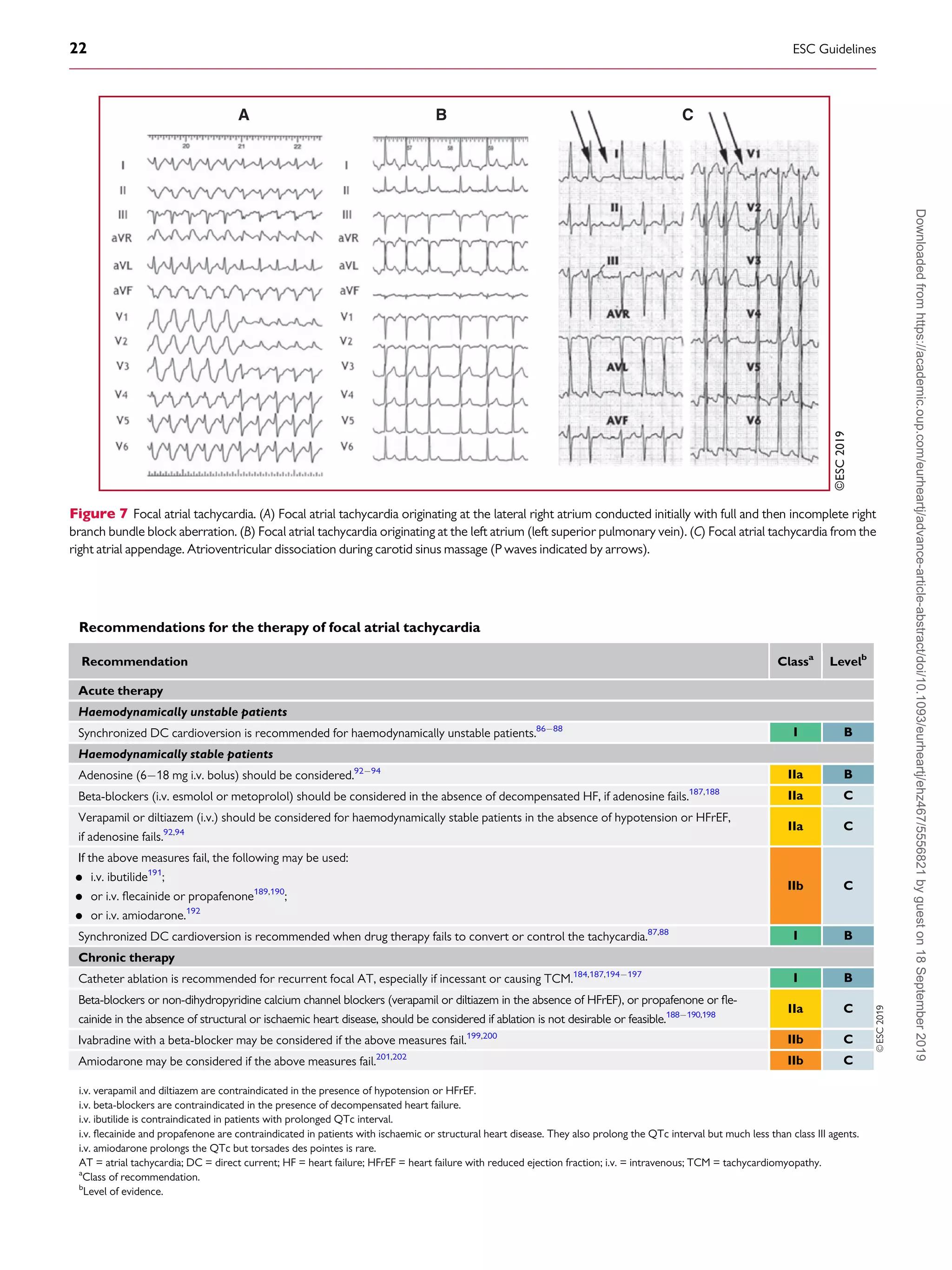
11.1.2.1 Diagnosis
P wave identification from a 12 lead ECG recording during tachycar-
dia is critical (Figure 7). Depending on the AV conduction and AT
rate, the P waves may be hidden in the QRS or T waves. The P waves
are monomorphic with stable CL, which helps to rule out organized
AF. Adenosine injection can help by slowing the ventricular rate or,
less frequently, by terminating focal AT. A discrete P wave with an
intervening isoelectric interval suggests a focal AT. However, distin-
guishing focal from macro-re-entrant arrhythmias by surface ECG is
not always possible. The presence of an isoelectric line does not rule
out a macro-re-entrant mechanism, particularly in the presence of
scar atrial tissue (from structural heart disease or previous extensive
ablation/surgery procedures). In a normal heart and in the absence of
previous ablation, the usual ECG localization rules apply,186
but their
value in localizing the origin of the arrhythmia is also limited in this
context. Focal AT may arise from any site in both atria, but particular
sites of predilection in the normal heart rate are the crista terminalis,
the tricuspid and mitral valve annulus, and within the thoracic veins
joining the atria.46,186
A negative P wave in lead I and aVL suggests an
LA origin. V1 is negative when the arrhythmia source or exit is in the
lateral right atrium, while septal right atrial (RA) and LA origins show
biphasic or positive P waves (Figure 7). Negative P waves in the infe-
rior leads suggest a caudal origin, whereas positive P waves in those
leads favour a superior location.
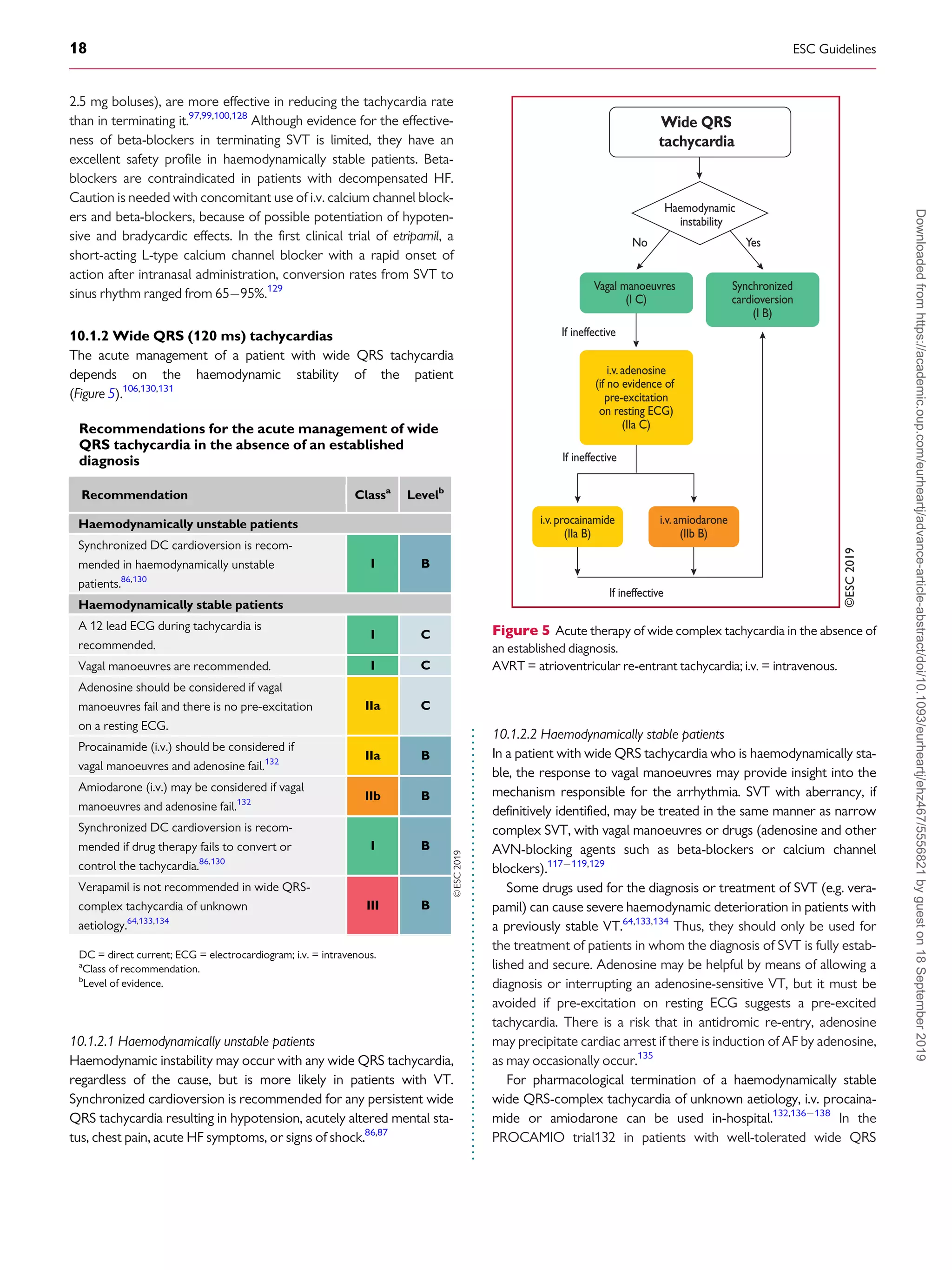
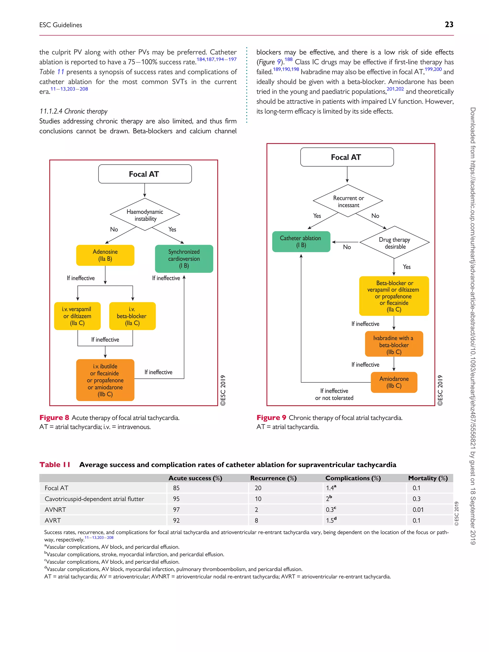
11.1.2.2 Acute therapy
Hard data for an evidence-based choice of drugs for the acute ther-
apy of focal AT are scarce. In general, acute therapy may be initiated
with beta-blockers or calcium channel blockers, which may terminate
focal ATs or slow the ventricular rate (Figure 8).92,94,187,188
Adenosine (i.v.) may terminate AT [delayed after-depolarizations
(DAD)-triggered AT], but the tachycardia may also continue with AV
block. Class IA, IC, and III drugs may also be effective, by prolonging
refractoriness or suppressing automaticity.189191
Amiodarone may
also be used for cardioversion or slowing of the ventricular rate,192
but the efficacy of rate control is unproven in critically ill patients
with atrial arrhythmias.193
DC cardioversion is usually effective in
acutely terminating the tachycardia, irrespective of the mechanism.
However, in incessant forms of focal AT due to enhanced automatic-
ity, the arrhythmia reinitiates, and repeating DC cardioversion is
unlikely to be appropriate.
11.1.2.3 Catheter ablation
Catheter ablation is the treatment of choice for recurrent focal
AT, especially for incessant AT due to which TCM ensues
(Figure 9).196
Distinguishing macro-re-entrant from focal ATs is
critical for the ablation strategy. Focal ATs, as well as localized/
micro-re-entry ATs, display a centrifugal activation pattern that
spreads throughout the atria. Mapping and ablation of focal ATs is
based on determining the earliest activation site. In PV-related AT,
focal ablation may be performed, but electrical isolation of both
ESC Guidelines 21
Downloaded
from
https://academic.oup.com/eurheartj/advance-article-abstract/doi/10.1093/eurheartj/ehz467/5556821
by
guest
on
18
September
2019](https://image.slidesharecdn.com/brugada-2020-esc-guidelines-for-the-management-supraventriculartachycardia-samirrafla-210905063343/75/Brugada-2020-esc-guidelines-for-the-management-supraventricular-tachycardia-samir-rafla-21-2048.jpg)
![.
.
.
.
.
.
.
.
.
.
.
.
.
.
.
.
.
.
.
.
.
.
.
.
.
.
.
.
.
.
.
.
.
.
.
.
.
.
.
.
.
.
.
.
.
.
.
.
.
.
.
.
.
.
.
.
.
.
.
.
.
.
.
.
.
.
.
.
.
.
.
.
.
.
.
.
.
.
.
.
.
.
.
.
.
.
.
.
.
.
.
.
.
.
.
.
.
.
.
.
.
.
.
.
.
.
.
.
.
.
.
.
.
.
.
.
.
.
.
.
.
.
.
.
.
.
.
.
.
.
.
.
.
.
.
.
.
.
.
.
.
.
.
.
.
.
.
.
.
.
.
.
.
.
.
.
.
.
.
.
.
.
.
.
.
.
.
.
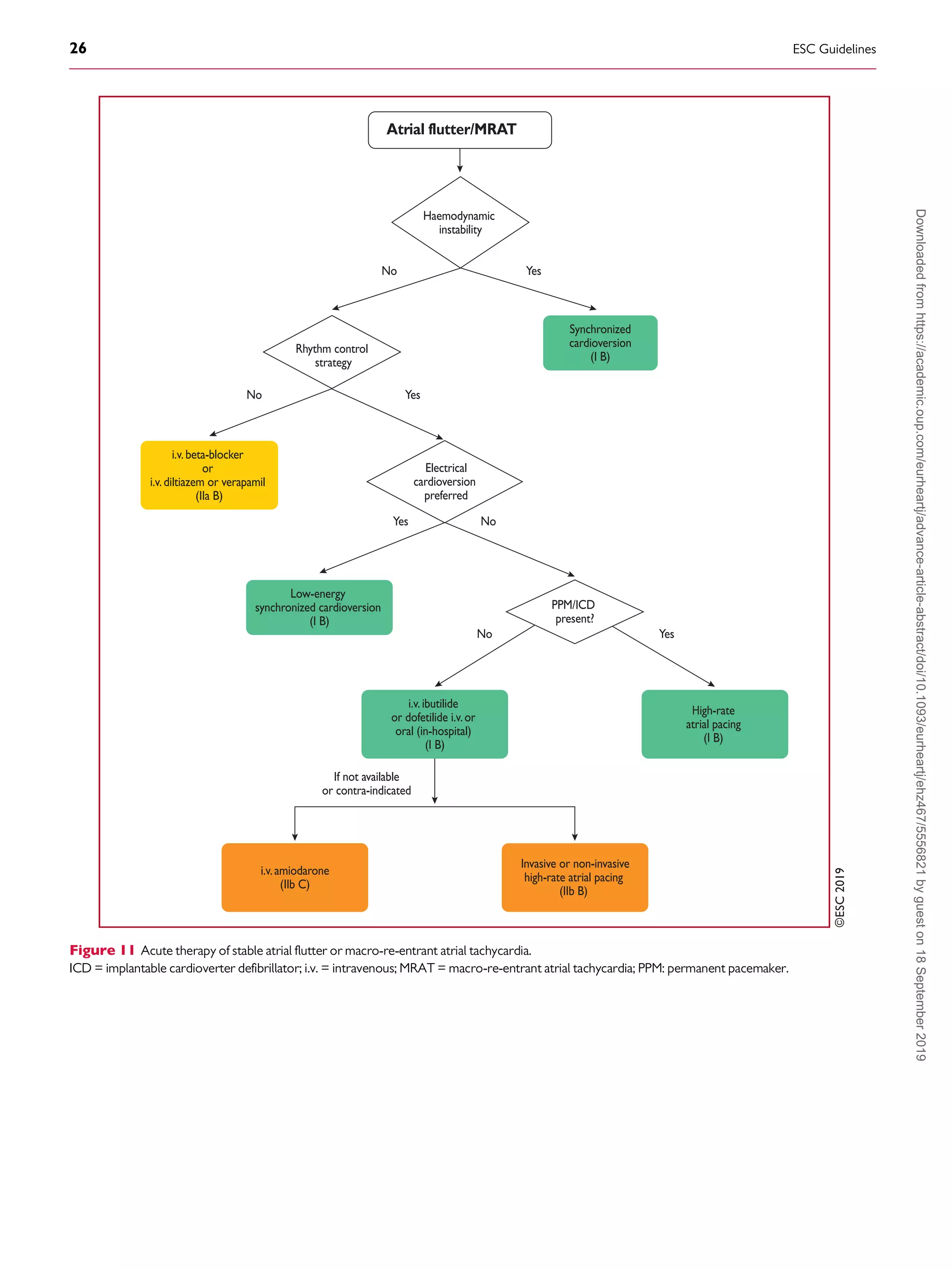
flutter and even the combination of AVN-blocking drugs (digoxin,
beta-blockers, and calcium channel blockers)235238
may fail, mak-
ing cardioversion to sinus rhythm necessary. Dofetilide and ibuti-
lide, pure class III antiarrhythmic drugs, are generally effective in
interrupting atrial flutter in i.v. administration (dofetilide may be
also given orally for this purpose), while class IA and IC drugs have
little or no effect.250257
Class IC antiarrhythmic drugs should not
be used in the absence of AV-blocking agents because of the risk
of slowing the atrial rate, which may result in 1:1 AV conduc-
tion.273,274
Amiodarone may not be very effective acutely to re-
establish sinus rhythm, but it does help to control the ventricular
rate if it is too fast.275,276
Low-energy electrical cardioversion is
commonly used with haemodynamic compromise or after failure
of previous actions, but it could be the first choice due to its high
efficacy. Electrical cardioversion for atrial flutter is more effective
and less energy is required, compared with AF.248,249
When atrial
electrodes are in place, high-rate stimulation can be used to con-
vert flutter, sometimes through AF.258,259
If pacing induces AF,
this may allow better control of the ventricular rate than flutter.
Atrial stimulation can also be done with percutaneous endocardial
electrodes or from the oesophagus; this is mostly done in paediat-
rics.261
Pre-treatment with procainamide may facilitate conver-
sion of atrial flutter by atrial pacing.277
Data on pre-cardioversion
anticoagulation are lacking, but most probably patients should be
treated the same as those with AF.4,278
11.1.4.1.4 Catheter ablation. Catheter ablation is the most effective
therapy to maintain sinus rhythm, and is clearly superior to amio-
darone.262,263
Ablation of CTI with confirmed bidirectional con-
duction block results in a 10% rate of recurrence.279
However,
the incidence of AF is high in the long-term.280
When typical CTI-
dependent atrial flutter ensues during antiarrhythmic drug therapy
(class IC or amiodarone) for AF, CTI ablation is a reasonable
choice to ensure that antiarrhythmic drugs can be continued for
AF control.262,263
Although no procedure-related mortality had been detected in
early studies,203,204
in recent studies, mortality and stroke rates of
0.2 - 0.34 and 0.19 - 0.5%, respectively, have been reported
(Table 11).12,206
In a recent registry, ablation for flutter had a higher
mortality than that for AF (0.3 vs. 0.05%), but this might have been
due to the comorbidities or advanced ages of patients referred for
flutter ablation.207
11.1.4.1.5 Chronic therapy. Rate control is part of the therapeutic
approach, using AV nodal blocking agents such as diltiazem, verapamil,
or beta-blockers (Figure 12). When ablation is not feasible or the
patient’s preference, antiarrhythmic drugs may also be used to maintain
sinus rhythm. Dofetilide257
and sotalol281
are useful, but there are con-
cerns about pro-arrhythmia. Amiodarone may have a role,263
but it
should be restricted to cases of HF or significant structural heart disease.
11.1.4.1.6 Anticoagulation. Data about the embolic risk of atrial
flutter have usually been derived in the presence of concomitant
AF, thus making individualized risk stratification difficult. Left
atrial (LA) appendage ‘stunning’ and thrombi seem to be lower
compared with those in AF.247,282
The thrombo-embolic risk of
atrial flutter, although lower than that of AF,246
is still signifi-
cant.241244
That, together with the association with AF, justifies
thromboprophylaxis, and anticoagulation has been recom-
mended as in AF.2,3
These recommendations extend to the acute
setting for cardioversion when flutter lasts for 48 h.278
However, it should be noted that there is a lack of prospective,
dedicated, randomized studies on the subject. Furthermore, the
value of the CHA2DS2-VASc [Cardiac failure, Hypertension, Age
_75 (Doubled), Diabetes, Stroke (Doubled) Vascular disease,
Age 6574 and Sex category (Female)] score in preventing
ischaemic stroke in patients with atrial flutter has not been estab-
lished,245
and in patients without concomitant AF the threshold
for the initiation of anticoagulation appears to be higher than that
for patients with AF.246
11.1.4.1.7 Other cavotricuspid isthmus-dependent macrore-
entrant atrial tachycardia. An atypical ECG pattern may not
exclude CTI-dependent MRAT.283
Lower-loop re-entry refers to a
circuit rotating around the inferior vena cava instead of around the
tricuspid annulus. It may be clockwise or counter-clockwise.284,285
When rotating counter-clockwise, it might be considered a variant of
typical counter-clockwise flutter with a caudal shift of the cranial
turning point posterior to the entry of the superior vena cava, result-
ing in a similar ECG appearance. ‘Figure-of-eight double-loop re-
entry’ may also occur around the inferior vena cava and tricuspid
annulus, and mimic typical clockwise atrial flutter.285
Other circuits
using part of the CTI or even restricted inside it, are in essence CTI-
dependent with a similar ECG appearance to typical common
flutter.286,287
11.1.4.2 Non-cavotricuspid isthmus-dependent macrore-entrant atrial
tachycardia
The terms non-CTI-dependent MRAT and atypical flutter are
used interchangeably, and describe flutter waves in the ECG not
suggestive of typical circuits. The pitfall with this use comes from
the atypical ECG that may happen when typical circuits develop in
diseased atria, most frequently after surgery or extensive ablation,
or under the effects of antiarrhythmic drugs. Conversely, upper-
loop re-entry may mimic a typical flutter ECG pattern without
being CTI-dependent.283
True atypical flutter is actually a post hoc
diagnosis when the circuit has been outlined and dependence on
CTI has been ruled out.
11.1.4.2.1 Right atrium macro2re-entrant atrial tachycardia. Atrial
sutures and patches used for complex congenital heart disease sur-
gery, together with progressive atrial damage, create multiple
obstacles and protected isthmuses that constitute the substrate for
complex and multiple MRAT.288,289
This usually happens around RA
free wall scars. However, in patients with complex congenital heart
disease, the presence of extensive atrial scars hinders the differential
diagnosis of focal or MRAT.290
Figure-of-eight double-loop tachycardias mimicking the ECG pat-
tern of a common atrial flutter may also occur following surgical
atriotomy.291
ESC Guidelines 27
Downloaded
from
https://academic.oup.com/eurheartj/advance-article-abstract/doi/10.1093/eurheartj/ehz467/5556821
by
guest
on
18
September
2019](https://image.slidesharecdn.com/brugada-2020-esc-guidelines-for-the-management-supraventriculartachycardia-samirrafla-210905063343/75/Brugada-2020-esc-guidelines-for-the-management-supraventricular-tachycardia-samir-rafla-27-2048.jpg)

![.
.
.
.
.
.
.
.
.
.
.
.
.
.
.
.
.
.
.
.
.
.
.
.
.
.
.
.
.
.
.
.
.
.
.
.
.
.
.
.
.
.
.
.
.
.
.
.
.
.
.
.
.
.
.
.
.
.
.
.
.
.
.
.
.
.
.
whereas those that conduct in the retrograde direction only are
more frequent (
_50%). When the AP conducts antegradely, ventric-
ular pre-excitation is usually evident at rest during sinus rhythm and
the AP is referred to as ‘manifest’. Conversely, APs are referred to as
‘concealed’ if they exclusively conduct retrogradely. Concealed APs
may have decremental properties.395
The term ‘latent AP’ denotes
an AP that is not, or is barely, visible due to location or faster conduc-
tion through the AVN.
Multiple APs occur in
_12% of patients with pre-excitation, and in
_50% in patients with Ebstein’s anomaly.396
AVRT is the most common tachycardia associated with APs. Two
mechanisms of re-entry are possible according to the antegrade or
retrograde conduction over the AVNHPS, and are classified as
orthodromic and antidromic AVRT.
11.3.2 WolffParkinsonWhite syndrome
WPW syndrome refers to the presence of an overt (manifest) AP,
thus resulting in the so-called pre-excitation, in combination with
usually recurrent tachyarrhythmias.397
During sinus rhythm, a typ-
ical pattern in the resting ECG with the following characteristics is
present: (i) a short PR interval (
_120 ms); (ii) slurred upstroke (or
downstroke) of the QRS complex (‘delta wave’); and (iii) a wide
QRS complex (120 ms). In most cases, APs giving rise to the
WPW pattern are seen in structurally normal hearts. Rare, familial
forms of pre-excitation associated with LV hypertrophy and multi-
system disease [mutations in the protein kinase adenosine
monophosphate-activated non-catalytic subunit gamma 2
(PRKAG2) gene, Danon and Fabry disease, and others] have also
been described.398
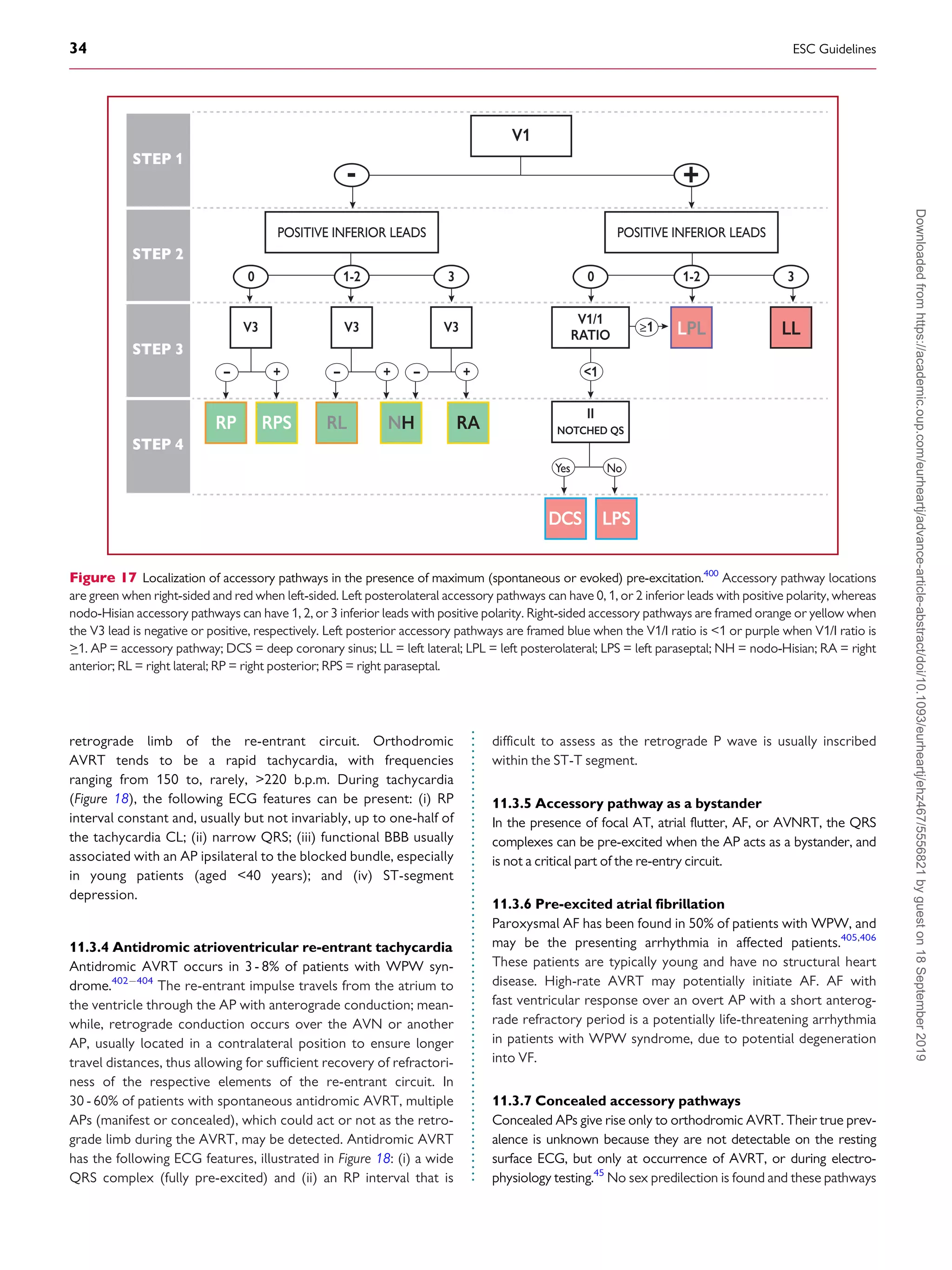
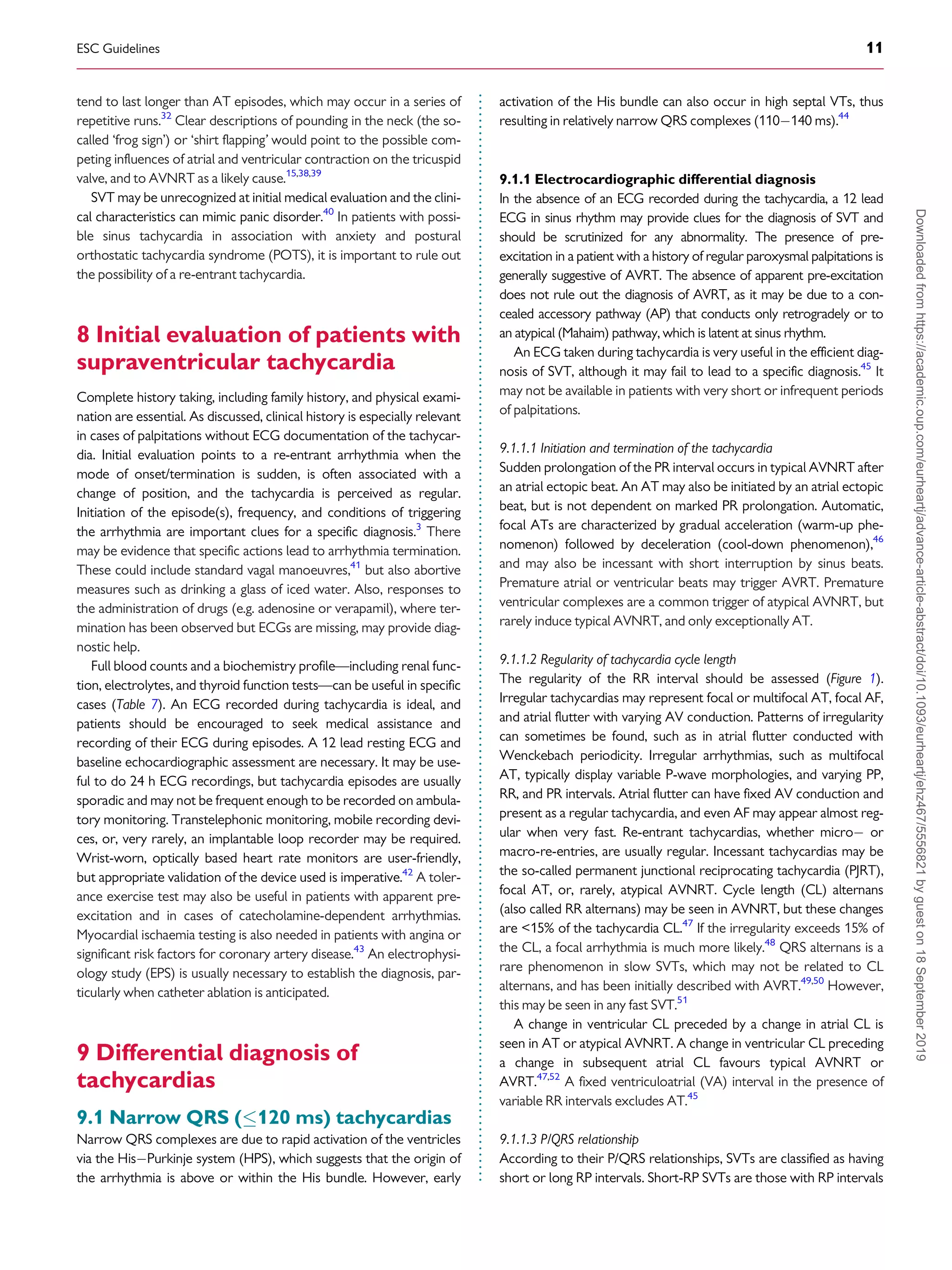
Several surface ECG algorithms have been developed that can be
applied for the localization of APs in the presence of overt pre-
excitation (Figures 16 and 17).399401
Pre-excitation on the surface
ECG can be intermittent and can even disappear permanently (in
_35% of cases) over time. Furthermore, various degrees of pre-
excitation are possible depending on the location of the AP as well as
on AVN conduction properties.
11.3.3 Orthodromic atrioventricular re-entrant
tachycardia
Orthodromic AVRT accounts for 90% of AVRTs and for
20 - 30% of all sustained SVTs. The re-entrant impulse conducts
from the atrium to the ventricle through the AVNHPS, which is
the anterograde limb of the re-entrant circuit, whereas the AP
conducts from the ventricle to the atrium, and serves as the
Figure 16 The St George’s algorithm for the localization of accessory pathways.399
þve = QRS complex-positive; ve = QRS complex-negative; þ/- =
QRS complex equiphasic; AP = accessory pathway; LAL = left anterolateral; LP = left posterior; LPL = left posterolateral; LPS = left posteroseptal; MS =
mid-septal; RAS = right anteroseptal; RL = right lateral; RP = right posterior; RPS = right posteroseptal.
ESC Guidelines 33
Downloaded
from
https://academic.oup.com/eurheartj/advance-article-abstract/doi/10.1093/eurheartj/ehz467/5556821
by
guest
on
18
September
2019](https://image.slidesharecdn.com/brugada-2020-esc-guidelines-for-the-management-supraventriculartachycardia-samirrafla-210905063343/75/Brugada-2020-esc-guidelines-for-the-management-supraventricular-tachycardia-samir-rafla-33-2048.jpg)