Downloaded 11 times


![LIST OF ORIGINAL COMMUNICATIONS
This thesis is based on the following publications, referred to in the text by their
Roman numeral:
I. Stent Retriever Thrombectomy in Different Thrombus Locations of
Anterior Cerebral Circulation. Protto S, Sillanpää N, Pienimäki JP,
Matkaselkä I, Seppänen J, Numminen H. Cardiovasc Intervent Radiol.
2016 Jul;39(7):988-93. doi: 10.1007/s00270-016-1315-4.
II. TREVO and Capture LP have equal technical success rates in
mechanical thrombectomy of proximal and distal anterior circulation
occlusions. Protto S, Pienimäki JP, Seppänen J, Matkaselkä I, Ollikainen
J, Numminen H, Sillanpää N. J Neurointerv Surg. 2016 Jun 17. pii:
neurintsurg-2016-012354. doi: 10.1136/neurintsurg-2016-012354. [Epub
ahead of print]
III. Low Cerebral Blood Volume Identifies Poor Outcome in Stent
Retriever Thrombectomy. Protto, S., Pienimäki, JP., Seppänen, J. et al.
Cardiovasc Intervent Radiol (2017) 40: 502. doi:10.1007/s00270-016-1532-x
IV. Internal Carotid Artery and the Proximal M1 Segment are Optimal
Targets for Mechanical Thrombectomy. Sillanpää N, Protto S,
Saarinen J, T, Pienimäki J, -P, Seppänen J, Numminen H, Rusanen H.
Intervent Neurol 2017;6:207-218](https://image.slidesharecdn.com/strokethrombectomy-180911063514/85/Stroke-thrombectomy-17-320.jpg)
![16
1 INTRODUCTION
Stroke is one of the largest contributors to death and disability worldwide and
furthermore has a major impact to the economy. Stroke is the third-leading cause
of disease burden as measured in disability-adjusted life years (DALYs) in
developed countries and the second leading cause of DALYs in developing
countries [1]. According to the Global Burden of Disease: 2004 Update, stroke
continues to be the second leading cause of death among people aged 15 years and
over.
The Global Burden of Stroke study estimated 6.5 million deaths from stroke and
10.3 million new strokes in 2013. Although the number of people who have
suffered a stroke has increased every year, the global incidence, mortality and the
number of DALYs lost are decreasing. However, this trend is more evident in
developed countries, while the people in developing countries carry a higher
burden of stroke. [1]
In Finland, the number of first-ever stroke episodes in 2007 was 10338. The yearly
medical expenses attributable to the treatment of stroke patients were close to 1.6
billion €, which corresponds to 7 % of all healthcare expenditure. [2]
Most strokes (87%) have an ischemic etiology meaning that they are caused by
insufficient blood flow to the brain tissue. The remainder of strokes is hemorrhagic
in origin, with 10% caused by intracerebral bleedings and 3% by subarachnoidal
hemorrhages [3, 4]. Ischemic stroke (IS) is generally due to a thrombotic or
embolic event in an intracerebral artery, which significantly decreases blood flow to
the tissue distal to the occlusion inducing cell death. Typically, patients present a
sudden onset of symptoms, the most common symptoms being motor disruption,
hemiparesis with or without hemisensory deficits, facial droop, ataxia, aphasia,
dysarthria, visual impairment, and variable decrease of consciousness.
Ischemic stroke is operationally defined as a neurological deficit lasting more than
24 h or an imaging finding in a patient with transient symptoms. Correspondingly,
a transient ischemic attack (TIA) is defined as a self-limiting episode of](https://image.slidesharecdn.com/strokethrombectomy-180911063514/85/Stroke-thrombectomy-18-320.jpg)
![17
neurological dysfunction caused by focal brain, spinal cord, or retinal ischemia,
without acute infarction, lasting less than 24 h. [5] Thus, imaging has a central role
in the evaluation of patients with acute stroke. Generally, multimodal computed
tomography (CT) is performed because it is widely available in most centers and a
fast imaging technique, but stroke magnetic resonance imaging (MRI) has also
been used extensively [6]. Both imaging techniques enable the differential diagnosis
between intracranial or subarachnoidal hemorrhage and ischemic stroke and allow
evaluation of the anatomy of the cerebral and cervical vasculature, the location of
the occlusive lesion and the extent of irreversible and/or reversible changes [7-9].
Prompt and effective revascularization of the affected area is crucial for improving
the patient´s prognosis. The time window between symptom onset and treatment
is a pivotal factor in determining the final outcome [10]. Therefore, in recent
decades, several therapies have been developed, namely, intravenous thrombolysis
(IVT) and intra-arterial interventions that aim to rapidly restore perfusion to the
affected areas. While IVT has been endorsed by different stroke treatment
guidelines for almost two decades, the efficacy of intra-arterial therapy was
demonstrated only recently [11-17]. Based on diverse technical approaches, stent
retriever-based mechanical thrombectomy (MT) provided a breakthrough in the
efficacy and safety of intra-arterial treatment of IS [18].
To better understand the role of the innovative stent retriever technique, this thesis
reports a prospectively collected, observational cohort of patients with acute
anterior circulation stroke who were treated via mechanical thrombectomy (MT)
with a focus on the clinical and imaging parameters that influence the technical and
clinical outcomes. These factors included the location of the clot on admission
computed tomography angiography (CTA) and type of the stent retriever device
used during the intervention. We also investigated the prognostic performance of
data derived from perfusion CT studies (CTP), especially cerebral blood volume
maps (CBV). Moreover, we compared the results in patients treated in our
institution with only IVT to those treated with MT to assess the differences in
clinical outcomes and to clarify the patient subgroups for which MT outperforms
IVT. The 3-month modified Rankin Scale (mRS) was used as the functional
outcome measure and the Thrombolysis in Cerebral Infarction scale (TICI) was
used to define the technical outcome.](https://image.slidesharecdn.com/strokethrombectomy-180911063514/85/Stroke-thrombectomy-19-320.jpg)
![18
2 REVIEW OF THE LITERATURE
2.1 ACUTE ISCHEMIC STROKE
2.1.1 Pathophysiology and etiology
Acute ischemic stroke results from a sudden reduction of cerebral blood flow
(CBF) to the brain parenchyma. Typically, this decrease in blood flow follows
thrombosis or embolism in the artery supplying a part of the brain, inducing
insufficient delivery of oxygen and glucose which leads to cell death in the absence
of sufficient collateral circulation or revascularization (Figure 1). The decrease in
perfusion triggers a cascade of events in cells, which eventually results in
irreversible changes: inhibition of protein synthesis, anaerobic glycolysis,
inflammation, release of excitatory neurotransmitters, disturbance of energy
metabolism and oxidative/nitrative stress, and finally anoxic depolarization and
loss of membrane integrity [19]. In the core of the ischemic area, where the CBF is
drastically impaired (<15 ml/100 g/min), neuronal cell death occurs within a few
minutes, whereas in the periphery, the events leading to irreversible changes take
more time. The so-called ischemic penumbra is an area of the brain parenchyma
where blood flow is moderately reduced (15-20 ml/100 g/min) but the tissue
remains viable if restoration of normal flow is achieved in sufficiently short time.
Benign oligemia (20-55 ml/100 g/min) defines the volume of mild hypoperfusion,
which normally does not lead to infarction even without revascularization. [20, 21]
Hypoperfusion and irreversible ischemic damages can also be caused or aggravated
by systemic hypotension. Hence it is important to control the blood pressure in
patients suffering from IS in order to maintain or improve both antegrade and
collateral circulation. [22]
Ischemic stroke etiology is classified based on the mechanism of the injury. The
Trial of Org 10172 in Acute Stroke Treatment (TOAST) classification defines five
stroke subtypes: large-artery atherosclerotic infarction (19%), embolism from a
cardiac source (9%), small-vessel disease (44%), stroke of other determined](https://image.slidesharecdn.com/strokethrombectomy-180911063514/85/Stroke-thrombectomy-20-320.jpg)
![19
etiology (5%), and infarcts of undetermined cause (23%) [23]. However, because of
the evolution and the precision of the diagnostic tools, more than one cause of
stroke can often be recognized in a single patient. For instance, a potential source
of cardiac embolism can be detected using echocardiography in 50 to 70% of
ischemic stroke patients. Further, 12% of patients with a cardiac source of
embolism and 22% of patients with a lacunar infarction harbor ipsilateral large
artery atherosclerosis causing a narrowing of greater than 50% of the vessel
diameter [24]. Application of the TOAST classification criteria often results in
most strokes being classified to the undetermined category, which led to the
development of new, more refined classification systems. The Stop Stroke Study
TOAST (SSS-TOAST) system uses the same five etiological categories, but based
on the weight of evidence, further classifies each category into “evident”,
“probable” or “possible” [24]. The Causative Classification of Ischemic Stroke
(CCS) is an automated version of SSS-TOAST that maximizes inter-examiner
reliability and limits inter-examiner variability in the classification. [25] The
distribution of ischemic stroke subtypes varies largely between different
populations and reports and is dependent on the availability of modern diagnostic
tools and the age distribution of the population. Recently, an increase in the
prevalence of some stroke subtype over others has been observed, with the
cardioembolic category now being the dominant etiology in western countries (e.g.
48% in Sweden). [3]](https://image.slidesharecdn.com/strokethrombectomy-180911063514/85/Stroke-thrombectomy-21-320.jpg)
![20
Figure 1. Occlusion of the right M1 segment of the MCA. Adapted from
http://www.strokecenter.org/patients/about-stroke/ischemic-stroke/
2.1.2 Risk factors
Numerous risk factors are associated with ischemic stroke. Modifiable risk factors
play an important role in the prevention of stroke. The INTERSTROKE study
recently conducted in 32 countries demonstrated that ten preventable risk factors
(hypertension, current smoking, abdominal obesity, unhealthy diet, lack of regular
physical activity, high alcohol intake, psychosocial stress and depression, cardiac
disease, diabetes mellitus and unfavorable lipid profile) contribute to 91.5% of the
total risk of ischemic stroke. [26] These findings corroborated the results of the
Global Burden of Disease 2013 (GBD) study, in which 90.5% of the stroke burden
(measured in DALYs) was attributable to modifiable risk factors. Both studies
identified hypertension as the most significant factor influencing the Population
Attributable Risk (PAR) with estimates for the parameter ranging from 47.9 to
64.1%.
There is strong evidence that cigarette smoke is intimately related to stroke with a
greater risk for women or younger smokers. Ex-smokers have double the risk of](https://image.slidesharecdn.com/strokethrombectomy-180911063514/85/Stroke-thrombectomy-22-320.jpg)
![21
experiencing a stroke before the age of 75 compared to lifetime non-smokers. [27-
29] The INTERSTROKE study estimated the PAR of cigarette smoking at 12.4%.
[26]
Obesity is also correlated with stroke risk. In the upper body mass index (BMI)
bracket (25 to 50 kg/m2), each 5 kg/m2 increment is associated with 40% higher
stroke mortality. Similar to cigarette smoking, the association of BMI with stroke is
stronger among the younger population. [30-32] Physical activity and healthy
lifestyle have been shown to reduce the risk of stroke. Compared with sedentary
lifestyle, the risk reduction for active individuals has been estimated at 26%. [33,
34] Using the American Heart Association (AHA) recommendation (2.5 hours or
more of exercise per week), increased physical activity was associated with a
reduction in all stroke risk (OR 0.41 [0.35–0.48], PAR 53.0% [47.0–59.0]). [26]
High alcohol consumption increases the risk of all stroke subtypes. However, the
association of alcohol intake and stroke risk appears to be J-shaped. [35] In a recent
study on monozygotic twins, heavy alcohol consumption shortened the time to
first stroke by 5 years. [36]
Psychosocial stress and depression, defined in the INTERHEART study as a
combination of stress (life and work), life events and depression [37], is also
associated with higher risk of stroke. [26] A recent study found that in diabetic
patients there was an association between elevated stress or depressive symptoms
and increased incidence of stroke (Hazard Ratio 1.57 [95% CI 1.05, 2.33]). [38]
Atrial fibrillation (AF), coronary artery disease, and cardiac failure are all well-
known risk factors for stroke [39]. In the INSTROKE study AF was significantly
associated with ischemic stroke (PAR ranging from 3.1% in southern Asia to
17.1% in western Europe, North America and Australia). According to recent
studies, coronary artery calcification (CAC) is a predictor of IS in both genders,
especially among younger patients, independently of AF. CAC was a good
discriminator of stroke risk particularly in subjects presenting a low or intermediate
(< 20%) Framingham risk score. [40, 41]
The relation between diabetes mellitus (DM) and stroke has been widely
demonstrated [42, 43] Many risk factors for stroke such as hypertension,
hypercholesterolemia, ischemic heart disease, and vascular claudication are more
prevalent among diabetic individuals. Subcortical and lacunar infarctions are more
frequent in diabetic patients than in their non-diabetic counterparts. [44]](https://image.slidesharecdn.com/strokethrombectomy-180911063514/85/Stroke-thrombectomy-23-320.jpg)
![22
Several studies have shown that there is also a correlation between atherogenic
lipid profiles and stroke risk. In particular, low levels of high-density lipoprotein
(HDL), high levels of total cholesterol (TC) and a high TC-HDL ratio increase the
risk of IS in both men and women. [45, 46]
Interestingly, at younger age, all the risk factors seem to exert a stronger negative
effect. [26, 47] Pregnancy has been demonstrated to increase risk especially during
the third trimester and the post-partum period, probably because of hormonal
changes and hypercoagulability due to activated protein C resistance, lower levels
of protein S, increased fibrinogen, pregnancy-related hypertension and venous
stasis. [48]
An important non-modifiable risk marker is age, with increased risks each year
after the age of 19 of 9% for men and 10% for women. [47] Previous history of
stroke or TIA, family history of stroke and male gender are associated with
increased risk of IS. [49-51].
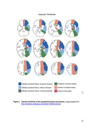
2.1.3 Vascular anatomy and the brain vascular territories
Blood supply to the brain is generally divided into anterior and posterior
circulation. The anterior circulation consists of the middle cerebral artery (MCA)
and the anterior cerebral artery (ACA). The posterior circulation comprises
infratentorially the basilar artery (BA), which branches in the supratentorial space
into the posterior cerebral arteries (PCA). The anterior circulation is supplied by
the common carotid arteries (CCA), which typically originates on the right side
from the brachiocephalic trunk and on the left side directly from the aortic arch.
The common carotid artery is divided into the external carotid artery (ECA) and
the internal carotid artery (ICA), which eventually supplies the intracranial anterior
circulation. The posterior circulation is supplied by the vertebral arteries in both
sides (VA), which usually originate from the subclavian arteries (SA). Normally,
there are many connecting vessels that enable collateral flow and a communication
between the carotid and the vertebrobasilar systems. The ACAs are connected by
the anterior communicating artery (ACommA) and the ICAs and the posterior
cerebral arteries are connected by the posterior communicating arteries
(PCommA). Overall the communicating arteries form the circle of Willis (CW)](https://image.slidesharecdn.com/strokethrombectomy-180911063514/85/Stroke-thrombectomy-24-320.jpg)
![23
together with parts of the ACA, ICA and PCA (Figure 2). The ACA supplies the
medial part of the frontal and parietal lobes. The MCA is divided in named
segments based on distal subdivisions: M1, M2, M3, and M4. The MCA mainly
supplies a large part of the lateral cerebral cortex, the temporal lobes and the
insular cortex, the lateral parts of the frontal cortex and the anterior parts of the
parietal cortex. The lenticulostriate perforating arteries, which perfuse the basal
ganglia, originate from the M1, or sphenoidal, segment. This segment can be
further divided into proximal M1 (M1P) and distal M1 (M1D) subsegments [52,
53]. The M2 segments are variable: The M1 segment normally bifurcates into
superior and inferior divisions but sometimes trifurcates into temporal, parietal and
frontal branches. The M3 and M4 segments are more distal and supply the cortex.
The posterior cerebral arteries (PCAs) supply the occipital lobes and the
posteromedial temporal lobes. The branches of the BA and VAs supply the
cerebellum, the medulla oblongata and the pons. Anatomical variations in the
cerebral vasculature are commonplace. [54, 55] Each artery supplies, in a near end-
artery fashion, a different area of the brain, i.e. their vascular territories [56, 57]
(Figure 3). There is a clear correlation between the vascular territory affected by
stroke and the symptoms presented by the patient. [58-60]](https://image.slidesharecdn.com/strokethrombectomy-180911063514/85/Stroke-thrombectomy-25-320.jpg)
![24
Figure 2. Classical circle of Willis. (1) ACommA, anterior communicating artery, (2)
A1, precommunicating segment of the anterior cerebral artery (ACA), (3)
PCommA, posterior communicating artery, (4) P1, precommunicating
segment of the posterior cerebral artery (PCA), (5) BA, basilar artery, (6)
P2, postcommunicating segment of the PCA, (7) ICA, internal carotid
artery, (8) MCA, middle cerebral artery, (9) A2, postcommunicating
segment of the ACA, and (10) SCA, superior cerebellar artery. From
Gunnal et al. [55]](https://image.slidesharecdn.com/strokethrombectomy-180911063514/85/Stroke-thrombectomy-26-320.jpg)
![26
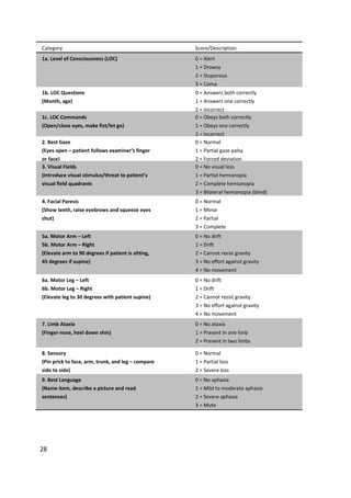
2.1.4 Functional outcome measures
The modified Rankin Scale (mRs) is the most commonly used measure of global
disability when evaluating recovery from stroke. Thus, it is also used as a primary
functional outcome measure in the majority of randomized clinical trials (64%)
[61,62]. The evaluation is usually performed at different time points (1-, 3-, and 6-
months) after the onset of the episode. The scale consists of 6 categories as
depicted in Table 1. The score is often dichotomized using mRS=2 as a cut-off
value because it differentiates between functional independence and dependence.
Thus, mRS≤2 in considered to indicate good clinical outcome. Excellent clinical
outcome is indicated by a mRS ≤1. The use of structured templates for interview
improves the inter-rater reliability [61]. Another rather common score system
(41%) is the Barthel activities of daily living (ADL) index [62]. The
dichotomization cut-off is generally ≥ 90 points.
Table 1. The modified Rankin scale (mRS). mRS is a 6-point scale, with higher scores indicating
a worse functional outcome](https://image.slidesharecdn.com/strokethrombectomy-180911063514/85/Stroke-thrombectomy-28-320.jpg)
![27
2.2 DIAGNOSIS OF ISCHEMIC STROKE
Ischemic stroke is a serious condition that requires fast diagnosis and treatment.
Acute ischemic stroke is generally diagnosed based on signs and symptoms
revealed during neurological and physical examinations, the anamnesis of the
patients and imaging findings. Patients suffering from stroke should be prioritized
in the triage in the emergency department in a similar manner to patients with
hemodynamically significant bleeding or myocardial infarction.
2.2.1 Symptoms and signs of acute ischemic stroke
Typical signs and symptoms of stroke are motor disruption, such as hemiparesis
with or without hemisensory deficits, monocular vision loss, visual field deficits,
diplopia, dysarthria and/or aphasia, facial droop, ataxia, a decrease in the level of
consciousness and vertigo. These symptoms can be better depicted and evaluated
during a fast neurological and physical examination with a standardized, systematic
and uniform stroke scale [63, 64]. The National Institutes of Health Stroke Scale
(NIHSS) is the most widely used assessment tool for evaluating stroke patients
(Table 2). This scale is a 42-point scoring system that quantifies neurological
deficits in 11 categories. A higher score indicates worse deficits; severe stroke is
defined as NIHSS >16, moderate as NIHSS between 8 and 16 and mild as NIHSS
<8. When a patient is suspected of having IS, other possible conditions that can
mimic neurovascular syndrome (i.e., “stroke mimics”) must be excluded. [65-67]
The most common conditions that mimic stroke are postictal states, sepsis,
tumors, toxic and metabolic disturbances, syncope, intracranial abscesses, migraine,
hypoglycemia, hypertensive encephalopathy, neuroimmunologic disease,
polyradiculitis and myasthenia gravis [65-67]](https://image.slidesharecdn.com/strokethrombectomy-180911063514/85/Stroke-thrombectomy-29-320.jpg)
![29
10. Dysarthria
(Evaluate speech clarity by patient repeating
listed words)
0 = Normal articulation
1 = Mild to moderate slurring of words
2 = Near unintelligible or worse
X = Intubated or other physical barrier
11. Extinction and Inattention
(Use information from prior testing to identify
neglect or double simultaneous stimuli testing)
0 = No neglect
1 = Partial neglect
2 = Complete neglect
Table 2. The National Institute of Health Stroke Scale (NIHSS). The NIHSS quantifies
neurological deficits into 11 categories. Higher scores indicate greater deficits. Adapted
from Richardson et al. [68]
2.2.2 Imaging of acute ischemic stroke
Primarily, imaging of a patient suffering symptoms of stroke is performed to
distinguish between hemorrhagic and ischemic stroke, as these categories have
completely different general treatment approaches. Other important information
that can be obtained with imaging includes the extent of the ischemic brain
parenchyma, the site of the occlusion, and an estimate of the volume of the
irreversibly damaged parenchyma.
Diagnostic imaging of acute IS is performed with multimodal computed
tomography (MCT) or MRI [69]. Due to the widespread availability and shorter
scanning durations MCT is used more frequently [70, 71]. MCT is based on a
multidetector technology, which allows continuous scanning of thin section-widths
in a short time. MCT consists of three modalities: 1) non-enhanced CT (NECT),
which permits the exclusion of hemorrhage and highlights early ischemic changes
(EICs); 2) CT angiography (CTA), which gives information about the intra- and
extracerebral vasculature, the location of the clot and possible stenosis of large
vessels; and 3) CT perfusion (CTP), which provides a functional perspective of the
brain circulation by providing information on the perfusion of the parenchyma and
on the regional hemodynamic status.](https://image.slidesharecdn.com/strokethrombectomy-180911063514/85/Stroke-thrombectomy-31-320.jpg)
![30
2.2.2.1 Non-enhanced CT and Alberta Stroke Program Early CT Score (ASPECTS)
NECT is the first imaging modality obtained using an MCT protocol. The main
purpose of the study is to discriminate between hemorrhagic and ischemic stroke.
Secondarily, NECT can be used to detect EICs and to exclude pathologies, which
could mimic a stroke, such as central nervous system (CNS) tumors. NECT can to
some degree predict the possibility of post-treatment hemorrhagic complications
following IVT or intra-arterial interventions. [72-74]
EICs observed in an NECT scan within 8 h of symptom onset include 1) loss of
visualization of the gray-white matter interface, which operationally constitutes
subtle parenchymal hypoattenuation with or without swelling, 2) isolated
parenchymal swelling without hypoattenuation, and 3) focal hyperattenuation of an
arterial trunk, which is an additional sign that can be considered a surrogate for
ongoing parenchymal ischemia. These categories include some well-known imaging
signs such as the dense media sign or hyperdense middle cerebral artery sign
(HMCAS), which indicates M1 and proximal M2 segment thrombosis, the MCA
dot sign, which may indicate thrombosis in the insular branches of the MCA (the
distal M2 and M3 segments), and the insular ribbon sign, which indicates loss of
definition of the gray-white interface of the insula and the obscuration or partial
disappearance of the lentiform nucleus. Unfortunately, the interobserver agreement
and reproducibility of EICs is poor and they are insensitive for detecting acute
ischemic stroke especially in the hyperacute phase (onset to imaging time < 3h).
[73] The presence of EICs depends on the duration of the hypoperfusion. [75]
Nevertheless, these findings cannot reliably differentiate between irreversibly
damaged brain tissue and penumbra. However, isolated focal swelling is associated
with penumbra and frank parenchymal hypoattenuation with the infarct core and
thus poor functional outcome [75-80]. The extent EICs is predictive of the risk of
hemorrhagic transformation. [72-74, 81, 82] The issues arising from the difficulties
to reliably detect EICs have been in part overcome with the use of standardized
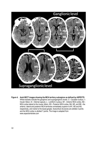
semiquantitative methods such as the Alberta Stroke Program Early CT Score
(ASPECTS). This method is an algorithmic, topographically structured scoring
system that allows semiquantitative assessment of the extent of acute ischemic
changes in the anterior circulation. [83-85] Only parenchymal hypoattenuation is
considered a finding in the scoring system. Each hemisphere is divided into 10
regions (Figure 4) and each of these regions can be scored 1 point. This point is
deducted if the region shows EICs. Thus, a full negative finding yields a score of
10, and extensive ischemia covering the entire MCA region yields a score of 0. If](https://image.slidesharecdn.com/strokethrombectomy-180911063514/85/Stroke-thrombectomy-32-320.jpg)
![31
the ASPECTS score is ≤6, based on volumetric correlates more than one-third of
the MCA territory is affected. When the ASPECTS score is ≤7 (3 or more regions
affected) the patient is unlikely to achieve an independent functional outcome. [86]
Overall, ASPECTS applied to NECT images is predictive of the clinical outcome,
the effectiveness of IVT and IAT and the rate of hemorrhagic complications but
remains a suboptimal technique for intra-arterial treatment decision making
because of the inherent insensitivity of NECT. [84, 87-90]](https://image.slidesharecdn.com/strokethrombectomy-180911063514/85/Stroke-thrombectomy-33-320.jpg)
![33
2.2.2.2 Computed tomography angiography
CTA is generally the second modality acquired in a multimodal CT protocol.
Typically, a volume from the aortic arch up to the vertex of the skull is obtained to
cover both the intra- and extracerebral vasculature. Thin-section slices of isotropic
spatial resolution are calculated to enable the reconstruction of two-dimensional
(2D) reformatted images in arbitrary planes, maximum intensity projection (MIP)
images and three-dimensional (3D) images. They provide detailed information on
the cerebral vasculature that is comparable with that obtained using digital-
subtraction angiography (DSA) [91-96]. Hence, large- and small vessel occlusions
and stenosis can be detected by CTA highly accurately (95-99%) both intracranially
and extracranially [23, 23, 97, 98].
The importance of this imaging tool in the triage of acute stroke patients to
different revascularization therapies was recently demonstrated in five randomized
and controlled trials, which assessed the possible superiority of MT over IVT
treatment. All these trials used vascular imaging with CTA as a diagnostic tool in
their protocol [13-17]. CTA not only allows the detection of a thrombus in the
intracerebral vasculature but also enables the evaluation of the length and density
of the clot, which plays a role in the treatment decision-making process. [95] These
are all independent prognostic factors of acute ischemic stroke for which proximal,
high-volume, organized clots predict poor clinical outcomes compared with distal,
low-volume, fresh clots. [99-101] This is related to the rate of recanalization with
IVT, which is lower in proximal vessel positions, with high clot burden and clots
with lower average Hounsfield Unit (HU) values. These intrinsic factors also
influence the recanalization rate with IAT and should guide therapeutic decision
making and the choice between IVT, intra-arterial interventions or refraining from
revascularization therapy [52, 101-111]
To simplify the evaluation of the location and extent of the thrombus a
semiquantitative clot burden score (CBS) has been developed. [110, 112] The CBS
is a 10-point scoring system. Points are assigned based on the number and location
of arterial segments affected in the anterior circulation (Figure 5). Similar to
ASPECTS, a higher score indicates a lower clot burden. CBS correlates with
technical and clinical outcomes. Patients with higher scores are more likely to
experience better functional outcomes, smaller infarct volumes, and lower
hemorrhagic complication rates. [109, 110, 112]](https://image.slidesharecdn.com/strokethrombectomy-180911063514/85/Stroke-thrombectomy-35-320.jpg)
![34
Other important roles of CTA are facilitating proper planning of the endovascular
procedure for device selection and enabling shorter intervention durations [113,
114] Limitation of CTA include the use of ionized radiation and the use of
iodinated contrast medium, which in rare cases, can lead to contrast induced
nephropathy (CIN).
Figure 5. Schematic representation of the clot burden score (CBS). One or two points each are
subtracted from a total score of 10 when no contrast opacity is detected using CTA in the
infraclinoid ICA (1 point), supraclinoid ICA (2 points), proximal M1 segment (2 points),
distal M1 segment (2 points), M2 segment branches (1 point each) and A1 segment (1
point), as indicated by the numbers next to the corresponding vessel segments. CBS
applies only to the symptomatic hemisphere. From Puetz et al. [110]
2.2.2.3 Computed tomography angiography source images
Unprocessed source images of CTA (CTA-SI) can also be used to estimate EICs
and have increased sensitivity compared to NECT. [115] When capturing the
steady state of the contrast agent, CTA-SI is a surrogate of CBV and, thus,
approximates the extent of irreversible ischemic damage [116, 117]. The](https://image.slidesharecdn.com/strokethrombectomy-180911063514/85/Stroke-thrombectomy-36-320.jpg)
. Therefore, CTA-SI is more sensitive to acute ischemia than NECT
and subsequently more precise in the prediction of infarct extension, hemorrhagic
complications and clinical outcome. [115, 116, 118-121] Rapid CTA image
acquisition at an early time point precludes the contrast agent from fully traversing
collateral vessels and reaching the distal vascular bed, thereby increasing the
volume and severity of hypoattenuation in the ischemic parenchyma and leading to
overestimation of the infarct core [122-124]. In contrast, frank hypoattenuation on
NECT is highly specific for irreversible tissue damage suggesting that CTA-SI
must be interpreted carefully in relation to NECT findings in addition to adjusting
CTA protocols to minimize the overestimation problem [75-80].
Figure 6. Baseline NECT (A) showing early ischemic changes in the right MCA territory (arrows).
CTA-SI image (B) reveals hypoattenuation in the right MCA territory (arrows), which
corresponds to the final infarct (C) on follow-up CT scan (arrows). NECT indicates
noncontrast CT; MCA, middle cerebral artery; CTA-SI, CT angiography source image.
From Bhatia et al. [116]
2.2.2.4 Evaluation of the collateral circulation
The cerebral collateral circulation is a dynamic, interconnected vascular system that
preserves the brain parenchyma from ischemic damage. There is large variation in
this vascular network among individuals. Collateral vasculature can be divided into
primary and secondary pathways. The former pathways make up a system that](https://image.slidesharecdn.com/strokethrombectomy-180911063514/85/Stroke-thrombectomy-37-320.jpg)
![36
allows communication between the two hemispheres and consists of artery-to-
artery communications, i.e., the circle of Willis. The latter pathways become
evident during the occlusion of large intracranial vessels and include the vascular
network between the internal and external carotid arteries and the leptomeningeal
pial arteriolar anastomoses. Sufficient flow through the collateral pathways allows
the brain tissue to remain viable even if the normal antegrade flow supplying the
tissue volume is diminished or interrupted [125-128]. The most important factor
influencing the integrity of collaterals is age, each 10-year increment in patient age
increases the odds of inadequate collateral circulation by 1.8-fold [129-131].
Smoking, hypertension, elevated uric acid and glucose levels at the time of
presentation are other factors that negatively influence the development of
collateral circulation [130, 132]. Even though digital subtraction angiography
(DSA) is the gold standard for anatomical and functional evaluation of collateral
vessels [133], collateral circulation can also be assessed non-invasively. Typically,
CTA is used to evaluate the collateral network before stroke therapy. Many
collateral scoring (CS) systems have been proposed. One of the more widely
adopted systems is that described by Souza et al., which is based on the evaluation
of maximum intensity projection (MIP) reconstructions (Figure 7)[134].
Figure 7. CS system: 0 = absent collaterals >50% of an M2 territory; 1 = diminished collaterals
>50% M2 territory; 2 = diminished collaterals <50% M2 territory; 3 = collaterals equal to
contralateral side; 4 = increased collaterals. From Souza et al. [134]
Adequate collateral flow reduces the baseline infarct core and the expansion of the
core, decreases the risk of hemorrhagic transformation and increases the odds of a](https://image.slidesharecdn.com/strokethrombectomy-180911063514/85/Stroke-thrombectomy-38-320.jpg)
![37
good clinical outcome in-hospital, at discharge and six months after the stroke.
[112, 132, 135-140] The richness of the collateral network predicts the final infarct
volume in both patients with persistent arterial occlusion and those experiencing
recanalization [132, 141, 142]. Two recent randomized trials (ESCAPE and
EXTEND-IA)[14, 17] validate the concept of CS as a central feature in the
evaluation of stroke patients who were candidates for MT by employing CTP
instead of single-phase CTA. Single-phase CTA does not provide temporal
resolution. Therefore, collateral status may be mischaracterized in many patients
and is usually underestimated. Dynamic CTA is a technique that derives time
resolved images of pial arterial filling from CTP images; however, dynamic CTA
requires postprocessing and whole-brain CTP. As an alternative, multiphase
(arterial, arteriovenous, and venous phases) dynamic CTA rapidly provides easily
interpretable information regarding the degree and extent of pial arterial filling in
the whole of brain parenchyma in a time-resolved manner. Supporting this
concept, the inter-rater reliability for multiphase CTA is excellent. In multiphase
collateral scoring (ranging 0 to 5) collateral status on the affected side is considered
to be poor compared to same arterial trunks in the asymptomatic contralateral
hemisphere when there are no vessels visible in any phase within the occluded
vascular territory, only a few vessels visible, or minimal to no collaterals visible in a
region greater than 50% of the MCA territory [143-146].
2.2.2.5 Computed tomography perfusion
The third and final modality acquired in a multimodal protocol is CTP, a dynamic
imaging modality that provides information on cerebral hemodynamics. CTP
consists of serial CT imaging of a volume of brain parenchyma in rapid succession
after an injection of an iodinated contrast agent. This procedure enables
quantification of capillary level phenomena of cerebral blood flow. The
concentration of the iodinated contrast agent in the brain parenchyma and, hence,
the CT density changes time-dependently as depicted by time-density curves. These
curves are used to derive CTP parametric maps, which reflect different aspects
hemodynamics [147-151]:
1) Cerebral Blood Flow (CBF): the volume of blood moving through a brain
volume (mass) of interest per unit time ([CBF] = ml/100 g/min).](https://image.slidesharecdn.com/strokethrombectomy-180911063514/85/Stroke-thrombectomy-39-320.jpg)
![38
2) Cerebral Blood Volume (CBV): the total volume of blood in a given brain
volume (mass) of interest ([CBV] = ml/100 g). This volume includes the
intracellular, intravascular and extravascular interstitial spaces.
3) Mean Transit Time (MTT): the average difference in time between the
arterial inflow and the venous outflow of a brain region-of interest ([MTT]
= s). This time is dependent on the average distance travelled. MTT can be
calculated from the CBF and CBV based on the central volume principle
[152]: MTT = CBV/CBF.
4) Time to Peak (TTP): the time from the beginning of the arterial
enhancement to the peak of the enhancement curve ([TTP] = s).
There is a large variety of algorithms available to calculate the different perfusion
parametric maps from the raw CT data. These algorithms can be classified as 1)
non-deconvolution or 2) deconvolution techniques [150]. Non-deconvolution
techniques use first-pass contrast agent measurements and apply several simplifying
assumptions, which lower the accuracy of the results. Deconvolution techniques
discard some of these assumptions and also take into consideration recirculation of
the contrast agent, collateral flow, delays in the delivery of the contrast agent and
venous output. Thus, the deconvolution algorithms are more complex and
sensitive [153-155]. There are also differences in image acquisition protocols.
Standardization and validation of the quantitation of perfusion parameters across
different vendor platforms or even across different platforms from the same
vendor remain ongoing processes [153, 156-158]. Further, different manual, semi-
automated and fully automated image reconstruction workflows add to the
variation caused by differences in algorithms. In particular, the selection of arterial
input and venous output vessels may vary. Generally, the A2 segment of the
anterior cerebral artery and the sagittal sinus are chosen as arterial input and
venous output because this seems to minimize problems caused by volume
averaging [159-161].
The main goal of an acute stroke perfusion study is to assess the viability of the
ischemic tissue, i.e., to identify the irreversibly damaged tissue (the infarct core), the
tissue that is at risk for progression to infarction if reperfusion is not achieved (the
penumbra) and the normally perfused, benignly hypoperfused or hyperemic tissue
[151, 156, 162, 163]. Experimental studies have demonstrated that these
hemodynamic states are characterized by different functionally defined CBF
thresholds, as follows: 1) The threshold below which cortical function ceases
without an increase in extracellular potassium or reduction in pH (the penumbra)](https://image.slidesharecdn.com/strokethrombectomy-180911063514/85/Stroke-thrombectomy-40-320.jpg)
![39
and 2) the threshold below which there is disruption of cellular integrity (the core)
[20]. These functional definitions have been correlated with advanced
neuroimaging findings— perfusion parametric maps—to define a more clinically
relevant operational penumbra identified as hypoperfused but potentially
salvageable tissue. [162, 164-167] The operational penumbra is the mismatch
(subtraction) volume between the CBF or MTT (or TTP) and the CBV, in which
the CBV lesion reflects the infarct core and the CBF or MTT (or TTP) lesion
reflects the boundaries of the hypoperfused penumbral tissue [153]. This concept
was initially validated for MRI, and MRI and CT results were later correlated [121,
168]. The perfusion parameters that best define the core and the penumbra remain
a topic of discussion. This task is challenging, as both regions are dynamic in
character because of the nature of the disease process. MTT maps potentially
overestimate the size of perfusion defects, while CBV maps may overestimate or
underestimate the volume of the irreversibly damaged brain parenchyma [157,
169]. Some studies suggest that threshold CBF values may assess the core more
accurately than CBV cut-offs [154, 158, 170, 171] and that TTP could be more
closely related to penumbra than MTT [172].
Accurate evaluation of the infarct core at presentation seems to be one of the most
valuable imaging–based prognostic factors. An infarct core volume of 70-100 ml
strongly indicates poor outcome regardless of recanalization and penumbra volume
[173-176]. Moreover, even successful large vessel recanalization reperfusion at the
capillary level can be insufficient. This could be due to downstream embolization
and microvascular obstruction or cytotoxic edema within the penumbral region
precluding tissue perfusion. Hence, the predictive role of penumbra volume at
presentation remains partly unclear and it only becomes a relevant predictive factor
if evaluated in the context of recanalization data [176-180].
In two of the recent pivotal randomized trials comparing MT to IVT (EXTEND-
IA and SWIFT PRIME [15, 16]) CTP evaluation of the size of the infarct core and
the volume of salvageable tissue was successfully used as inclusion criteria.
The ASPECTS scheme has been validated for CTP parametric maps, which
provides another method for quantifying CTP findings in the anterior circulation,
including calculating the perfusion mismatch [21, 180-184]. CTP-ASPECTS,
especially CBV-ASPECTS, better predicts clinical outcome compared to admission
NECT and CTA-SI [17, 86, 86, 180, 185]. Kloska et al. suggested that the optimal
threshold value for CBV-ASPECTS that best differentiated between good and](https://image.slidesharecdn.com/strokethrombectomy-180911063514/85/Stroke-thrombectomy-41-320.jpg)
![40
poor clinical outcomes was ≤7. Furthermore, Aviv et al. found that no patients
with CBV-ASPECTS ≤7 achieved good clinical outcome [21, 89]. In our previous
study patients having CBV-ASPECTS ≥7 performed best in an ROC analysis
[184].
2.2.2.6 Evaluation of recanalization
The Thrombolysis in Cerebral Infarction (TICI) score grading system was
originally described in 2003 by Higashida et al. [133] to evaluate the grade of
reperfusion following IVT in patients suffering from acute stroke. In
neurointerventional radiology, TICI scores are commonly applied to grade the
DSA control runs of endovascularly-performed recanalization.
The original definition was based on the angiographic post-intervention
appearance of the site of the occlusion and the distal branches:
• TICI 0: no perfusion
• TICI 1: penetration of the contrast agent with minimal perfusion
• TICI 2: partial perfusion
o 2a: only partial filling (less than two-thirds) of the entire vascular
territory is visualized
o 2b: complete filling of all of the expected vascular territory is
visualized but the filling is slower than normal
• TICI 3: complete perfusion
Because of marked variability in the application of this score, in 2013 a consensus
paper from three collaborative groups [53] proposed a modified scale, the m-TICI
score:
• m-TICI 0: no perfusion
• m-TICI 1: antegrade reperfusion past the initial occlusion, but limited
distal branch filling with little or slow distal reperfusion
• m-TICI 2:](https://image.slidesharecdn.com/strokethrombectomy-180911063514/85/Stroke-thrombectomy-42-320.jpg)
![41
o 2a: antegrade reperfusion of less than half of the previously
occluded target artery ischemic territory
o 2b: antegrade reperfusion of more than half of the previously
occluded target artery ischemic territory
• m-TICI 3: complete antegrade reperfusion of the previously occluded
target artery ischemic territory, with absence of visualized occlusion in all distal
branches
2.3 ACUTE ISCHEMIC STROKE MANAGEMENT
Acute stroke therapy aims to 1) restore perfusion of the ischemic brain tissue as
rapidly as possible [186-188], 2) limit the amount of damage to the ischemic tissue
whether caused by primary (hypoperfusion) or secondary (for example,
hyperglycemia or hyperthermia) mechanisms, and 3) decrease the probability of
complications (such as hemorrhagic transformation) [189]. Based on data from
both experimental models and clinical trials, the duration and severity of ischemia
determines the extent of irreversible damage [10, 186, 190]. However, potentially
viable ischemic tissue (i.e., the penumbra) has been demonstrated to exist for at
least 24 h after symptom onset [191, 192]. Overall, the time elapsed from the onset
of the symptoms to treatment is a critical determinant of the outcome, which
guides decision–making processes and pre- and in-hospital management. [10, 193-
195]
The following interventions have been explicitly proven to improve the prognosis
after acute IS: 1) management of the patient in a stroke unit [196, 197], 2) use of
aspirin within the first 48 h from onset [198-200], 3) decompressive surgery
(hemicraniectomy) for supratentorial malignant hemispheric cerebral infarction
[201, 202], 4) administration of IVT within 4.5 h from symptom onset [201-203],
and 5) MT with a stent retriever in the case of large vessel occlusion (up to the M1
segment) [204-207]. The goal of the last two therapies is to achieve a prompt
revascularization and reperfusion of the ischemic area by pharmacologically or
mechanically disrupting the occluding thrombus.
Currently available revascularization therapies include IVT, intra-arterial
thrombolysis (IAT) possibly assisted with balloon angioplasty, IVT followed by an
intra-arterial intervention, MT using aspiration, stent retrievers, other specific](https://image.slidesharecdn.com/strokethrombectomy-180911063514/85/Stroke-thrombectomy-43-320.jpg)
![42
retrieval devices or a combination of these therapies, and bypass stenting.
Recently, the completion of five randomized trials on MT performed with stent
retrievers led to a shift in treatment paradigm towards intra-arterial therapies as a
first-line approach [13-17].
2.3.1 Intravenous thrombolysis
IVT is a well-established treatment of acute IS that was approved by the Food and
Drug Administration (FDA) in 1996. It essentially entails the intravenous
administration of a tissue plasminogen activator (TPA), a serine protease that elicits
dissolution of the clot by activating plasmin by conversion of plasminogen [208].
Streptokinase and urokinase were originally used for this purpose, but they were
subsequently replaced by a new generation of recombinant tissue plasminogen
activators (r-tPAs) that selectively activate fibrin-bound plasminogen and thus have
better efficacy and specificity [209].
The most relevant trials on IVT are the Neurological Disorders and Stroke Trial
(NINDS), which proved the feasibility and efficacy of IVT in a time window of 3 h
from symptom onset [203], The European Cooperative Acute Stroke Study
(ECASS), ECASS II, and the Alteplase Thrombolysis for Acute Noninterventional
Therapy in Ischemic Stroke (ATLANTIS) study, which evaluated the efficacy and
safety of IVT within a time window up to 6 h, but did not demonstrate treatment
benefit [210-212]. Nonetheless, two later studies, ECASS III and the Safe
Implementation of Thrombolysis in Stroke–International Stroke Treatment
Registry (SITS-ISTR), succeeded in demonstrating efficacy and safety of IVT
within a prolonged time window of 4.5 h [213, 214]. Further studies have examined
the feasibility of IVT beyond the 4.5 h window using MRI to evaluate the ischemic
penumbra. The Desmoteplase in Acute Ischemic Stroke (DIAS) study used an
extended the time window of 9 h and failed to demonstrate efficacy [215-217]. The
Diffusion-weighted Imaging Evaluation For Understanding Stroke Evolution
(DEFUSE) study demonstrated the utility of different MRI mismatch profiles for
evaluating patients who are likely to have good outcome after IVT when treated
between 3 h and 6 h from symptom onset [218]. The Echoplanar Imaging
Thrombolytic Evaluation Trial (EPITHET) and the Third International Stroke
Trial (IST-3) also evaluated a treatment time window beyond 4.5 h with no positive
results. Recently, the introduction of new MT devices has reduced the interest in
an IVT-only approach [219-224].](https://image.slidesharecdn.com/strokethrombectomy-180911063514/85/Stroke-thrombectomy-44-320.jpg)
![43
A feared complication of IVT is intracerebral hemorrhage (ICH), which can be
fatal [203, 213]. Other potential adverse effects include systemic bleeding,
myocardial rupture if IVT is administered within a few days of AMI, and
immunological reactions such as anaphylaxis or orolingual angioedema, although
these events are rare [65]. Exclusion criteria have been defined to minimize the
possibility of complications (Table 3).
The relative exclusion criteria for IVT include minor or spontaneously rapidly
improving symptoms, pregnancy, seizure at onset with postictal residual
neurological impairments, major surgery or serious trauma within the previous 14
days, recent gastrointestinal or urinary tract hemorrhage (within the previous 21
days) and recent AMI (within the previous 3 months) [65]. Further, relative
exclusion criteria within 3 to 4.5 h after symptom onset include >80 years of age,
severe stroke (NIHSS>25), taking an oral anticoagulant regardless of INR and
histories of both diabetes and prior to IS [65]
Exclusion criteria
Significant head trauma or prior stroke in previous 3 months
Symptoms suggest subarachnoid hemorrhage
Arterial puncture at noncompressible site in previous 7 days
History of previous intracranial hemorrhage
Intracranial neoplasm, arteriovenous malformation, or aneurysm
Recent intracranial or intraspinal surgery
Elevated blood pressure (systolic >185 mm Hg or diastolic >110 mm Hg)
Active internal bleeding, acute bleeding diathesis, including but not limited to
Platelet count <100000/mm
Heparin received within 48 h, resulting in abnormally elevated aPTT greater
than the upper limit of normal](https://image.slidesharecdn.com/strokethrombectomy-180911063514/85/Stroke-thrombectomy-45-320.jpg)
![44
Current use of anticoagulant with INR >1.7 or PT >15 s
Current use of direct thrombin inhibitors or direct factor Xa inhibitors with
elevated sensitive laboratory tests
Blood glucose concentration <50 mg/dL (2.7 mmol/L)
CT demonstrates multilobar infarction (hypodensity >1/3 cerebral
hemisphere)
Table 3. IVT exclusion criteria. Absolute exclusion criteria for patients with ischemic stroke who
could be treated with IVT within 4.5 h of symptom onset
2.3.2 Intra-arterial therapy
The options for endovascular treatment to elicit recanalization of an intracranial or
extracranial artery supplying the brain tissue include balloon angioplasty, (bypass)
stenting, intra-arterial thrombolysis (IAT) and mechanical thrombectomy (MT)
with either aspiration-based or stent-based devices.
Angioplasty and stenting of extracranial ICA are predominantly performed for
secondary or primary prevention rather than to treat acute IS but may be combined
with MT in some cases: when the primary cause of stroke is acute attenuation or
cessation of flow in the extracranial ICA, such as from total or near-total occlusion
caused by an active, severe atherosclerotic lesion or dissection or when catheter
access to an intracranial clot is impeded by severe stenosis of the extracranial ICA
and angioplasty/stenting of the ICA is required prior to treatment of a more distal
intracranial occlusion [189, 225].
Two of the most important studies on IAT were the phase two Prolyse in Acute
Cerebral Thromboembolism (PROACT) and PROACT II trials [11, 12]. In both
studies, the pharmacological intervention consisted of intra-arterial injection of
recombinant prourokinase (r-proUK) with or without intravenous heparin, which
were both administered within 6 h of symptom onset. The recanalization rates and
clinical outcome were better than with IVT only; however, the probability of
intracranial hemorrhagic complication was higher. The Middle Cerebral Artery
Embolism Local Fibrinolytic Intervention Trial (MELT) also suggested the trend
that excellent clinical outcomes were achieved more often with IAT [226]. Further](https://image.slidesharecdn.com/strokethrombectomy-180911063514/85/Stroke-thrombectomy-46-320.jpg)
![45
studies on IAT included the Interventional Management of Stroke (IMS) trials I
and II [227, 228]. They combined intra-arterial and intravenous thrombolysis with
r-tPA administration within the 3-h time window and compared the results with
the NINDS study population. The investigators reported that the 3-month
outcome was significantly better compared to the NINDS placebo arm, whereas
there was only a trend toward improved outcomes compared with the NINDS r-
tPA arm. The Interventional Management of Stroke III (IMS III) trial patients
were administered IVT within 3-h of symptom onset and then randomly assigned
to receive IVT alone or IVT followed by endovascular therapy. Despite the higher
rate of partial or complete recanalization at 24 h in the endovascular group, clinical
outcomes of the two groups were similar, and the trial was stopped early due to
futility [229]. Vagal et al., using the data from the IMS III, demonstrated in a post
hoc model that endovascular therapy after IVT is preferable to IVT alone if the
reperfusion time was less than 347 min [230]. These findings highlight the
importance of developing new techniques to achieve faster and more efficient
recanalization with intra-arterial devices.
The introduction of stent retrievers launched a period of a great progress regarding
the intra-arterial treatment of acute IS. Stent retrievers are soft nitinol stents that
are deployed within the thrombus to push the thrombus against the vessel wall,
immediately reperfusing the distal brain tissue. After a short incubation period,
which is suggested to be up to 5 minutes [231], the thrombus usually adheres to the
struts of the stent, and the stent is then retrieved along with the clot by
withdrawing the stent under aspiration from a more proximal catheter. Removal of
the stent also eliminates the need for acute double-antiplatelet therapy, which is
needed for permanent stent placement in the cerebral vasculature (Figure 8).](https://image.slidesharecdn.com/strokethrombectomy-180911063514/85/Stroke-thrombectomy-47-320.jpg)
![46
A
Figure 8. A timeline depicting the strategies and techniques employed over the time to achieve
recanalization of a large vessel occlusionan ELVO in the setting of acute ischemic stroke.
Adapted, From Spiotta A.M. et al. [232, 233]
Stent retriever technology was preceded by the MERCI retriever system introduced
in the early 2000s (Figure 8). This device included most of the mechanisms of
action of stent retrievers, but the design was not stent-based. The Mechanical
Embolus Removal in Cerebral Ischemia (MERCI) trial was a single-arm study that
included patients between 3-8 h from symptom onset or within 3 h if there was a
contraindication to IVT or if the treatment had failed [234]. This trial was extended
in the Multi MERCI study, an international, multicenter study in which a newer
device (the L5 retriever) was used if available [235]. In the late 2000s, a second
device was approved, the Penumbra endovascular aspiration device. In the
Penumbra Pivotal Stroke Trial 81% of patients achieved recanalization of the
occluded vessel but only 25% had mRS≤2 at three months [236]. Penumbra caused
fewer hemorrhagic complications than MERCI but corresponded to worse
neurological outcome, possibly due to distal embolization [237]. The Mechanical
Retrieval and Recanalization of Stroke Clots Using Embolectomy (MR RESCUE)
trial compared thrombectomy (performed using MERCI or a Penumbra device) to](https://image.slidesharecdn.com/strokethrombectomy-180911063514/85/Stroke-thrombectomy-48-320.jpg)
![47
IVT, and no statistical significant difference was found between the two groups,
but the rate of TICI grade 2b/3 recanalization was low (25%) and the onset to
groin puncture time was 381 +/- 74 (mean+/-SD) [238]. Overall, sufficient proof
of efficacy of these devices was not obtained in these studies or meta-analyses
thereof.
During the past eight years, a number of studies and trials have investigated the
feasibility, safety and efficacy of MT with newer generation stent-based
thrombectomy devices [239-243]. This effort culminated in six multi-center,
prospective, randomized and controlled trials (MPRCTs), which were set up to
compare MT in association with IVT to IVT alone. As a result of these trials,
endovascular therapy with stent retriever thrombectomy is now the standard of
care for patients with acute large-vessel occlusion in the anterior circulation [189].
The first of those trials, the Multicenter Randomized Clinical Trial of Endovascular
Treatment for Acute Ischemic Stroke in the Netherlands (MR CLEAN) enrolled
500 patients. Retrievable stents were used in 190 of the 233 patients (82%) in the
intervention group, and simultaneous acute cervical carotid stenting was performed
in 30 patients (13%). Good reperfusion rate was achieved in 59% of patients in the
intervention group, and there was a significant shift in the distribution of mRS
scores of 0-5 at 90 days in favor of the intervention, regardless of age. There was
no difference in the complication rate. However, in the presence of extracranial
ICA occlusion, admission NECT ASPECTS of <8 or NIHSS <20, the adjusted
odds ratios (ORs) were accompanied by wide confidence intervals (CIs) [13].
In the Endovascular Treatment for Small Core and Anterior Circulation Proximal
Occlusion with Emphasis on Minimizing CT to Recanalization Times (ESCAPE)
trial imaging was used to exclude participants with a large infarct core and/or poor
collateral circulation. Stent retrievers were used in 130 of the 151 participants
(86%) who underwent an endovascular procedure, and the rate of the good
reperfusion was 72%. In contrast, in the control group, successful recanalization
was observed in 43 of 110 patients (31%). The proportion of patients with mRS≤2
at 90 days was 53% in the intervention group and 29% in the control group, and
mortality at 90 days was 10% in the intervention group and 19% in the control
group. However, for the presence of a carotid T- or L- occlusion, admission
NECT ASPECTS of <8 or patient age >80 years, the adjusted ORs were
accompanied by wide CIs. The study was suspended prematurely because of high
efficacy in the intervention group [14].](https://image.slidesharecdn.com/strokethrombectomy-180911063514/85/Stroke-thrombectomy-49-320.jpg)
![48
The Extending the Time for Thrombolysis in Emergency Neurological Deficits —
Intra-Arterial (EXTEND-IA) trial was also stopped early because of high efficacy
in the intervention group. The trial had recruited just 70 patients, 35 in each group.
This study used CT perfusion to identify salvageable brain tissue. The rate of
successful revascularization immediately after the procedure was 86% (with a
restoration of flow of >50% of the affected vascular territory), and the reperfusion
rate at the 24 h follow-up was 100%. In the IVT-only group, the recanalization rate
was 15 of 35 (43%). In addition, early neurologic improvement was observed
significantly more often in the intervention group (80% vs. 37%). Endovascular
therapy improved the functional outcome at 90 days with a significantly larger
proportion of patients achieving functional independence (71% vs. 40%).
Subgroup analyses could not be performed given the small number of patients [17].
The Solitaire with the Intention for Thrombectomy as Primary Endovascular
Treatment (SWIFT PRIME) trial compared IVT followed by MT with the Solitaire FR
or Solitaire 2 stent retriever to IVT alone. The study was also stopped early because of
efficacy and it enrolled 196 patients from 39 different centers, (98 patients in each
group) Similar to EXTEND-IA, viable tissue demonstrated in CTP was an inclusion
criterion. Eighty-eight percent of patients treated with MT exhibited substantial or
complete reperfusion defined as perfusion of 50% or more of the vascular distribution
of the occluded artery. Functional independence at 90 days was significantly more
common in the intervention group compared to the control group (60% vs. 35%).
There was no evidence of heterogeneity in the treatment effect in the subgroups. In
the subgroup corresponding to patients admitted with NECT ASPECTS of 6-7, 10 of
24 patients were independent in the intervention group compared to 5 of 19 in the
IVT-only group [16].
The Randomized Trial of Revascularization with Solitaire FR Device versus Best
Medical Therapy in the Treatment of Acute Stroke Due to an Anterior Circulation
Large Vessel Occlusion Presenting within Eight Hours of Symptom Onset
(REVASCAT) trial compared MT with medical therapy alone in eligible patients
who received IVT within 4.5 h after the onset of symptoms without
revascularization after 30 min of t-PA infusion or who exhibited a contraindication
to IVT. Two-hundred-six patients were enrolled, 103 in each group. A NECT
ASPECTS less than 7 or an MRI ASPECTS less than 6 were exclusion criteria.
Substantial or complete reperfusion in the MT group was achieved in 67 of 102
(66%) patients. The absolute between-group difference in the proportion of
patients who were functionally independent was 15.5%, favoring MT (43.7% vs.](https://image.slidesharecdn.com/strokethrombectomy-180911063514/85/Stroke-thrombectomy-50-320.jpg)
![49
28.2%). The benefits appeared to be least consistent in subgroups with advanced
age (≥70 years) or a long time window after symptoms unset (>4.5 h from onset to
randomization) [15].
The Randomized, Concurrent Controlled Trial to Assess the Penumbra System’s
Safety and Effectiveness in the Treatment of Acute Stroke (THERAPHY) trial is
another notable study and is the only randomized study that utilized an aspiration-
based thrombectomy technique. This trial compared MT and IVT to IVT only.
The trial was terminated when the results of other studies were published and MT
became the standard of care for acute IS. Seventy percent of patients treated with
aspiration only achieved successful reperfusion; the results suggest a potential for
benefit for aspiration thrombectomy compared to IVT only but had a low
statistical power because of early termination. [244]
A recent randomized controlled multicenter trial published in 2016, the
THRombectomie des Artères CErebrales (THRACE) trial compared standard
treatment to IVT plus MT within 4 h of symptom onset. In total 412 patients were
enrolled, 208 in the IVT only group and 204 in the IVT+MT group. This study
had a broad patient selection with randomization within 20 min after the
administration of IVT. The reperfusion rate was 69%, and good clinical outcome
was obtained for 53% of patients in the MT group. This study also demonstrated
the superiority of MT combined with IVT over IVT alone and was terminated
early, because a second unplanned interim analysis showed significantly better
results in the intervention group [245].
In all the trials utilizing stent retrievers mentioned above, there was no sign of
increased risk of bleeding compared to IVT alone. There was an overall trend
toward a reduction in stroke-related mortality in all of the trials [14-17, 244-246].
While the efficacy and safety of MT has now been robustly demonstrated [204-
207], it is still of great interest to study whether there are differences in technical
and clinical outcomes with respect to the site of occlusion, the stent retriever type
used and the evaluation of the admission imaging along with other subgroup
analyses based on different clinical and imaging markers. These data can facilitate
patient selection, avoid unnecessary treatment and improve results by making best
use of devices in relation to the patient imaging and clinical details. The type of
anesthesia (general anesthesia or conscious sedation) used during the
thrombectomy procedure may also influence the patient outcome. This is still an](https://image.slidesharecdn.com/strokethrombectomy-180911063514/85/Stroke-thrombectomy-51-320.jpg)
![50
open issue with some reports supporting the use of conscious sedation. The only
randomized trial so far did not find any significant difference in clinical outcome
between these two types of anesthesia [232, 247-249].
For a comprehensive guideline regarding the selection of patients with acute
ischemic stroke, different revascularization therapies, performing endovascular
procedures, and setting up systems of care to facilitate endovascular treatment,
please refer to the 2015 American Heart Association/American Stroke Association
Focused Update of the 2013 Guidelines for the Early Management of Patients
With Acute Ischemic Stroke Regarding Endovascular Treatment [189].](https://image.slidesharecdn.com/strokethrombectomy-180911063514/85/Stroke-thrombectomy-52-320.jpg)
![52
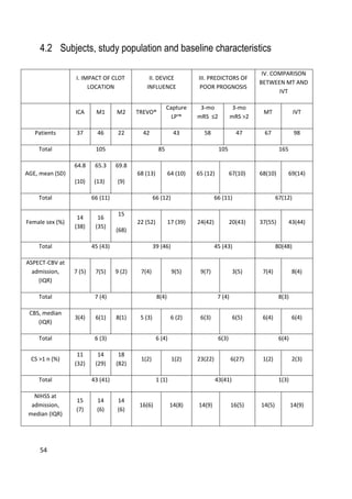
4 SUBJECTS, MATERIALS AND METHODS
4.1 Overview
All studies (I-IV) had an observational prospective cohort design. We prospectively
collected and analyzed the clinical and imaging data of consecutive patients
admitted to Tampere University Hospital from January 2013 to December 2014
due to acute ischemic stroke symptoms and who underwent clinical and imaging
evaluation and proceeded to digital subtraction angiography (DSA) with an
intention to perform MT. In addition, study IV had a retrospective observational
IVT-only cohort as a control group. This cohort had been collected between
January 2004 and December 2007 at our institution [52].
The general inclusion criteria for the studies were occlusion of the internal carotid
artery (ICA) and/or middle cerebral artery (MCA) up to the M2 segment and MT
performed with a stent retriever. The initial imaging evaluation consisted of
NECT, CTA and CTP in the majority of patients. The selection of patients as
candidates for MT was based on absence of extensive irreversible ischemic changes
(frank hypodensity more than 1/3 of the MCA territory) and hemorrhage in
NECT, evaluation of the amount of salvageable tissue in CTP imaging (when
performed) and proximal clot position in CTA (evaluated with raw data and MIP
reconstructions). The decision to proceed to MT was multidisciplinary (stroke
neurologist and neurointerventional radiologist). Patients referred to our institution
from other hospitals were re-evaluated with at least NECT and CTA upon arrival
before proceeding to the angiographic suite to rule out bleeding and extensive
irreversible ischemic lesions. In the case of wake-up strokes, CTP was performed if
no large infarct was seen in NECT. IVT was administered as a bridging therapy to
patients with no contraindications. The r-tPA bolus was given on the CT table. In
one case the bolus was withdrawn, because the time interval between symptom
onset and groin puncture was expected to be minimal (i.e., an inpatient during
office hours). Patients coming from an outside hospital received IVT according to
drip-and-ship protocol. IVT was continued until groin puncture. Majority of
patients were treated under conscious sedation: Dexmedetomidine was chosen in](https://image.slidesharecdn.com/strokethrombectomy-180911063514/85/Stroke-thrombectomy-54-320.jpg)
![53
51 cases (47%) and other combinations of drugs in 33 patients (31%). General
anaesthesia was preferred if the patient was restless rendering the procedure
difficult to perform (21 patients, 20%).
The technical outcome was measured with TICI (Thrombolysis in Cerebral
Ischemia), evaluated with DSA at the end of the procedure. The clinical outcome
measure was the modified Rankin Scale (mRS), evaluated three months after the
stroke based on a follow-up visit with a neurologist or a phone interview with a
neurologist. One patient could not be reached for this control. A follow-up NECT
was performed for all patients 24 h after treatment to assess the infarct volume and
possible hemorrhagic complications.
IVT was administered according to guidelines of the American Heart Association
(AHA) [250]: Actilyse® (Boehringer-Ingelheim, Ingelheim, Germany), total dose
0.9 mg/kg, was administered in a 10% bolus and continued, if necessary, until
groin puncture. Mechanical thrombectomy procedures were performed with
different stent retrievers and sometimes with multiple devices based on the
judgment of the operator. The used devices were TREVO® (Stryker
Neurovascular/Concentric Medical, Mountain View, CA, USA), CAPTURE LP™
(eV3/ COVIDIEN/Medtronic, Santa Rosa, CA, USA), ERIC® (MicroVention,
Tustin, CA,USA), Aperio® (Acandis, Pforzheim, Germany) and REVIVE®
(Codman &Shurtleff, Raynham, MA, USA).](https://image.slidesharecdn.com/strokethrombectomy-180911063514/85/Stroke-thrombectomy-55-320.jpg)
![64
Figure 10. Three-month mRs by the thrombus location. ICA:internal carotid artery, M1:middle
cerebral artery segment 1, M2: middle cerebral artery segment 2. mRS: modified Rankin
Scale.
5.1.2 The clinical benefit of MT is highest for proximal large vessel
occlusions (IV)
Continuing along the lines of location based analysis of study I, in study IV we
further divided the M1 segment in to subsegments of equal length as proposed by
Zaidat et al. [53]. This cut-off location was applied to classify large vessel](https://image.slidesharecdn.com/strokethrombectomy-180911063514/85/Stroke-thrombectomy-66-320.jpg)
![70
6 DISCUSSION
Acute ischemic stroke is a common disease and its natural history, if no treatment
is delivered, often leads to high-grade morbidity and mortality [1]. Even though the
evolution of the disease condition is highly dynamic and somewhat unpredictable
because of its dependence on multiple factors, such as pathophysiological aspects
and promptness of treatment, evaluating the parameters that influence the clinical
and technical outcomes is crucial. Studying the factors related to outcome facilitate
improving our ability to administer more efficient therapies to those patients who
will have the largest benefits from the treatment. The introduction of
interventional radiology techniques with efficacies superior to intravenous
thrombolysis (IVT) has amplified the need to refine our diagnostic methods to
permit more precise risk stratification and treatment decision-making. Technical,
clinical and imaging variables can predict the outcome of the patient and can
provide important information on who would not be a good candidate for diverse
revascularization therapies. While the overall superiority of MT with new
generation stent retrievers over IVT has been largely demonstrated by numerous
randomized trials [13-17, 245], it remains partly unclear if all patients with LVO
and acute ischemic stroke symptoms benefit from MT or if there are subgroups to
whom IVT could be the best first choice of treatment. Furthermore, it has not yet
been unequivocally demonstrated whether different clot locations or device
selection influence the result.
6.1 The effect of the occlusion site and the revascularization
treatment type on the outcome of stroke
In study I, we evaluated the effect of the site of occlusion on the technical and
clinical outcomes of patients presenting anterior circulation occlusion and who
were treated with newer generation stent retrievers. In our study, a statistically
significant difference in the technical and clinical outcomes in different clot
location could not be demonstrated.](https://image.slidesharecdn.com/strokethrombectomy-180911063514/85/Stroke-thrombectomy-72-320.jpg)
![71
In recent randomized trials, the majority of the patients treated with MT had
occlusion of the M1 segment, whereas far fewer patients had a clot in the M2
segment [13-17, 245]. Only some of these trials reported results that were stratified
according to the location of the clot. In the ESCAPE trial, the clinical outcomes
for different clot locations were comparable to our results. However, we had
slightly better outcomes with ICA occlusions: the proportion of patients with
mRS=0 at three months was 15% in our study vs. 13% in the ESCAPE study,
mRS=1: 31% vs. 16%, and mRS=2: 17% vs. 22%, respectively. The finding was
also similar when comparing to the results of the REVASCAT study. In particular,
we had fewer patients who experienced a dismal outcome (mRS 5-6) in the ICA
group (23% in our study vs. 46% in REVASCAT). In the recent THRACE study,
outcomes with respect to the location of the clot were evaluated only for ICA and
M1, where the M2 occlusions were included in the M1 group. Compared to this
trial, our results were slightly better regarding ICA occlusions: mRS=0-2 47% in
our study vs. 23% in the THRACE study. However, these differences may be
partly due to longer average onset-to-recanalization times in these randomized
trials and slightly better recanalization rates in our study. The overall recanalization
rate in our study was 88% vs. 72% in ESCAPE, 66% in REVASCAT, and 69% in
THRACE [13-17, 245].
Coutinho et al. collected the results from SWIFT, STAR and SWIFT PRIME trials
and evaluated the technical and clinical outcomes in M1 and M2 occlusions [251].
They did not find any significant differences between the two locations in terms of
technical or clinical outcomes, and even the complication rate was similar.
Modified TICI >2a was seen in 85% of patients with M2 occlusion and excellent
clinical outcome was achieved in 50% of patients, while in our study we observed
TICI >2a in 77% of patients and mRS 0–1 in just 23% of patients. Further, Dorn
et al. recently showed better technical and clinical outcome in patients presenting
with distal clot, with a recanalization rate in of 76% for M1 occlusions and 93% for
M2 occlusions. The proportions of patients with good clinical outcome at 3
months were 43% and 60%, respectively [252]. Unexpectedly, in our study, there
were no significant differences in the 3-month clinical outcome among the
different clot locations, despite significantly larger thrombus burden and worse
collateral circulation in the more proximal occlusions. In contrary, there was a
trend towards worse results in the M2 group when excellent clinical outcome
(mRS≤1) was considered. Correspondingly, the recanalization outcomes were also
slightly worse in the M2 population (77%). In contrast to the populations of Dorn](https://image.slidesharecdn.com/strokethrombectomy-180911063514/85/Stroke-thrombectomy-73-320.jpg)
![72
et al. and Coutinho et al., in our study the M2 occlusion group had more severe
strokes (median NIHSS 14) than expected based on the literature [52]. In addition,
compared with the other populations, there was a higher average age and a slight
female predominance in the M2 group. Therefore, more symptomatic patients,
patients with occlusions of multiple M2 branches and non-responders to IVT may
have been selected for MT treatment in our population. Finally, the worse technical
outcome may be due to increased tortuosity of the vessels, involvement of multiple
vessel branches, a smaller proportion of patients who had received IVT, and a
thrombus composition less amenable to MT and/or IVT [113, 253]. Nevertheless,
the duration of the intervention was similar in all clot locations and total infarct
volume after 24 h was smaller in the M2 group, as expected. However, this was not
statistically significantly reflected in the 3-month clinical outcome.
These findings emphasize the importance and efficacy of MT for the proximal clot
locations where IVT has been demonstrated to be less effective. However, the role
of MT in more distal occlusions remains somewhat unclear.
We readdressed this problem in study IV, in which we compared our MT results
with the findings from a historical population treated in our hospital with IVT only
and limited the analysis to only those patients with the treatment decision made
within 3 h from symptom onset. In the analysis, we divided the M1 segment in two
subsegments – the proximal M1 and the distal M1, as suggested by a current
consensus statement [53] – and classified occlusions proximal to this point of
division as proximal LVOs and those distal to this point as distal LVOs.
We found that MT was close to equally effective in the treatment of both proximal
and distal LVOs with more than 50% of patients experiencing good clinical
outcome. While MT proved to be superior to IVT in the treatment of proximal
occlusions (ICA and M1P), superiority of MT over IVT in more distal occlusions
was not demonstrated.
Numerous studies have demonstrated the feasibility and efficacy of MT with newer
generation stent retrievers in the M2 segment of the MCA, with reperfusion rates
up to 93% and the proportion of good clinical outcome up to 60% [251, 252, 254,
255]. Four of the recent pivotal randomized trials on MT reported their results
regarding occlusion of the M2 segment. In the MR CLEAN trial 91% of patients in
the control group received IVT [13]. M2 occlusion was seen in 18 patients in the
MT group and 21 patients in the control group. A subgroup analysis with M2](https://image.slidesharecdn.com/strokethrombectomy-180911063514/85/Stroke-thrombectomy-74-320.jpg)
![73
occlusion included in the stratification criteria was not reported. In the ESCAPE
study 79% of patients in the control group were administered IVT [14]. Only six
patients in the intervention group had an isolated M2 occlusion whereas patients
with occlusion of all M2 segment arteries were pooled with M1 occlusions. The
authors stated that the treatment effect (showing benefit from MT) was similar in
the location-based subgroup analyses, but details were not provided. In the SWIFT
PRIME trial, 18 patients had an M2 occlusion, 13 of whom were included in the
intra-arterial intervention group and 5 in the control group corresponding to 53%
and 40% of patients experiencing good clinical outcome, respectively [16]. In the
REVASCAT study 78% of patients in the control group received IVT [15]. Ten
patients in the MT group and 8 in the control group had an M2 occlusion. The
clinical outcome for this clot location was not detailed. Neither of the
abovementioned MT trials had a sufficient number of patients with an M2
occlusion to elucidate the best technique for distal occlusions. Recently, Goyal et
al. published a meta-analysis of 5 randomized trials. When pooled together, there
were 51 patients with an M2 occlusion in the MT population and 44 in the control
population [207]. In this study, the direction of the effect favored MT but the
adjusted OR was not significant (OR=1.28, 95% CI 0.51-3.21). Similar results were
found in a meta-analysis of older studies (OR=1.5, 95% CI 0.8-3.0) [256].
In line with previous studies, our study also could not demonstrate superiority of
MT over IVT (or vice versa) for the treatment of distal MCA occlusions.
Interestingly, in our study, after adjusting for confounders, we observed a non-
significant trend toward worse clinical outcome in the MT group, which was also
observed when the M2 and the distal M1 segments were pooled together (Table 6).
This could be explained by the lower NIHSS, indicating milder stroke in the IVT-
only group (median NIHSS 10 vs. 14); however, the trend remained unchanged
when patients with mild strokes (NIHSS<8) were excluded. Moreover, successful
recanalization of an M2 segment occlusion with MT was achieved in 79% of
subjects, which is comparable to the literature but not optimal. In our data, 77% of
M2 occlusion patients treated with only IVT experienced good clinical outcome, a
considerably higher proportion than in the IMS III trial (44.5%) [100]. This along
with the somewhat suboptimal reperfusion rate in our MT group and the inclusion
of patients with moderate and severe strokes to in the MT group likely led to a
pessimistic estimation of the efficacy of MT. Notwithstanding, there is no
indication in our data that MT would outperform IVT for the treatment of M2
occlusions in a hyperacute setting.](https://image.slidesharecdn.com/strokethrombectomy-180911063514/85/Stroke-thrombectomy-75-320.jpg)
![74
None of the randomized trials reported the distinction of a proximal and distal part
of the M1 segment in their analysis. However, the IMS III trial used this
characterization and 64% of subjects in the IVT-only arm compared to 55% in the
endovascular arm had good clinical outcome of a distal M1 occlusion [100]. The
difference was not statistically significant. According to a recent retrospective study
61% of subjects treated for a distal M1 occlusion with MT experienced good
clinical outcome, and 44% had excellent outcome [257]. These findings are
compatible with our results in that the efficacy of MT and IVT were roughly
equivalent with nearly 60% of patients achieving good clinical outcome in both
groups. When the M1 segment was analyzed as a whole, the result was borderline
significant in favor of MT (OR=2.6, CI 95% 0.90-7.8, p=0.08). This is in line with
the results of a recent meta-analysis considering that, in our study, two-thirds of
the M1 occlusions were in the distal subsegment [207, 256].
Almost half of the patients in the MT group who had a proximal occlusion
experienced excellent clinical outcome compared to only one patient (3%) in the
IVT-only group. Thus, it appears that surviving an ICA or proximal M1 occlusion
without any disability-causing neurological deficits essentially requires MT. This
finding has potential implications, especially in the management of patients with
low pre-stroke mRS (0-1) who are often younger patients in their working years.
We restricted our analysis to hyperacute strokes. The time window of IVT was
extended to 4.5 h in 2008. The efficacy of IVT deteriorates rapidly in the first few
hours resulting in a large number-needed-to-treat figure of 14.9 in the 3- to 4.5 h
time window [258]. Considering the superior reperfusion rates of MT, one would
expect the performance of MT to be at least at the level of IVT in the extended
time window. Extrapolating from the results of a recent meta-analysis, this appears
to be the case [207].](https://image.slidesharecdn.com/strokethrombectomy-180911063514/85/Stroke-thrombectomy-76-320.jpg)
![75
6.2 Influence of the device choice on technical and clinical
outcomes
The superiority of newer generation stent retrievers over older devices in efficacy
and safety has been established in previous studies [240, 242, 243]. Newer
generation devices have different design features that potentially make them more
suitable to certain anatomies and occlusion sites. In the literature, only few studies
have evaluated the differences between newer generation devices. Roth et al.
compared Solitaire AB/FR™ and Aperio™ in a porcine model [259]. They did not
find any significant difference in technical success or in complications between the
two devices. Another study, in a retrospective, non-randomized setup, compared
TREVO® and Solitaire AB/FR™ [260]. Technical and clinical outcomes were
similar in the two groups. Interestingly, there was a trend toward a larger number
of symptomatic ICHs in the Solitaire group (4 vs. 0), but due to the small
population (n=22) the difference was not statistically significant. Zaidat et al.
compared the results of two post-marketing registries and found that the clinical
performance of TREVO® is comparable to that of Solitaire AB/FR™ [261].
In accordance with the literature, there were also no significant differences between
the two stent retrievers in technical and clinical outcomes in our study. Although,
there was a higher clot burden and, thus, potentially more difficult thrombus
removal in the TREVO® group, the average duration of the procedure and the
technical results were similar. This may be because, in a distal position, the
tortuous anatomy and the more difficult catheterization can influence the
deployment and withdrawal of the stent, making the procedure more challenging
and time consuming [113]. Moreover, the technical outcomes were also similar
when proximal and distal occlusions were considered separately. This suggests that
despite different target vessel profiles intended by the manufacturer, the devices
perform similarly in large- and medium- sized vessels.
The 3-month clinical outcome was equivalent in both groups when functional
independence (mRS≤2) was considered. However, in the univariate analyses there
was a trend towards more patients having excellent outcome (mRs≤1) in the
Capture LP™ group and higher mortality in the TREVO® group both overall and
for different clot locations. These findings are likely partially due to larger
perfusion defects in the admission CBV maps in the proximal occlusions,
differences in stroke risk factors between the groups in the distal occlusions, and](https://image.slidesharecdn.com/strokethrombectomy-180911063514/85/Stroke-thrombectomy-77-320.jpg)
![76
consequently larger infarct volumes at 24 h, more hemorrhagic complications and
significantly more severe post-infarct edema. However, in the multivariate analyses
the device selection remained a significant or borderline significant predictor of
excellent outcome. For distal clot locations this may reflect the actual superiority of
Capture LP™, which is specifically designed in terms of size and flexibility of the
system components to enable MT of more distal clots. However, only 6 patients in
the TREVO® group had an M2 occlusion, which inherently has a higher potential
for an excellent 3-month clinical outcome, thus adding uncertainty. However, the
same trend could also be observed for the proximal occlusions in both the
univariate and multivariate analyses.
These results have to be interpreted in the temporal and device specific context, i.e.
only two stent retrievers were compared out of the many in market at the time of
the study and currently. Further, the results may be influenced by the details of the
MT procedure, for example aspiration from a balloon tipped guiding catheter as
opposed to distal access catheter and the type of anesthesia.
6.3 Predicting the outcome of acute ischemic stroke independent
of the type of the revascularization therapy
In study III, we evaluated which factors could influence the clinical outcome of
patients presenting large vessel anterior circulation ischemic stroke to identify
possible factors that predict poor outcome regardless of whether reperfusion is
achieved. Many studies have focused on evaluating factors that predict good
outcome, whereas fewer studies have primarily addressed factors related to poor
clinical outcome at 3 months [180, 262-266]. Most of the setup have been
evaluated factors associated with poor clinical outcome despite successful
recanalization. In contrast, the goal of our study was to understand which factors,
known at the time that the clinical decision to perform MT is made, could predict
poor outcome. It appeared that these two approaches yield very similar results.
CBV-ASPECTS emerged as the most important imaging parameter in predicting
the outcome in patients being evaluated for MT, while we could not demonstrate a
significant difference in NECT-ASPECTS scores at admission between those with
good and poor 3-month outcomes. The lack of sensitivity of NECT could be
partly explained by the relatively short average onset-to-imaging times. The
superior performance of CTP-CBV compared to NECT is in agreement with](https://image.slidesharecdn.com/strokethrombectomy-180911063514/85/Stroke-thrombectomy-78-320.jpg)
![77
previous results regarding both IVT and MT. [17, 173, 173, 180, 267-269, 269-271]
As demonstrated in previous studies on IVT, a CBV-ASPECTS threshold of 7
or 8 best differentiates patients with good and poor outcomes [21, 184]. In our
study, the median CBV-ASPECTS in the poor outcome group was 7. CBV-
ASPECTS did not significantly predict poor clinical outcome among patients
with mild strokes (NIHSS<8) or good collateral circulation (CS 2-4,) whereas
low CBV-ASPECTS was significantly associated with poor outcome among
those suffering from moderate or severe stroke and/or having poor collateral
circulation. A mild stroke entails less extensive and severe ischemia, which
considering a mean onset-to-imaging time of less than 3 h, translates into a
small infarct core especially if an aggregating quantification method such as
ASPECTS is used. Similarly, good collateral circulation is related to smaller
infarct core in this short timeframe. In a recent meta-analysis, MT did not
significantly improve the clinical outcome of patients with NIHSS<10
compared to IVT [207]. These findings imply that perfusion imaging in the
context of MT can be targeted to specific subgroups (i.e. those with moderate
or severe symptoms and/or poor collateral circulation in CTA), even if we
believe that there likely is a benefit to routinely performing CTP in all patients
presenting stroke symptoms and no bleeding in the initial NECT scan. CTP
was still not performed or was of low quality in about 30% of acute stroke
patients. Often the restless and least co-operating patients are those with
more severe strokes. This problem may in the future be in part addressed with
the development of imaging systems with faster scanning time and better
movement artefact correction algorithms.
It has been suggested that the type of anesthesia used during the thrombectomy
procedure could significantly influence the patient outcome. The majorities of the
studies reported in the literature are retrospective and shows better results with
conscious sedation compared to general anesthesia [247-249]. In these studies
patients who received conscious sedation had lower in-hospital mortality and lower
rate of pneumonia, shorter hospital stays and the costs were also lower compared
to patients who had general anesthesia. However, a recent randomized controlled
trial, the SIESTA study, failed to demonstrate an advantage for the use of
conscious sedation [232]. We did not directly address this problem in our studies.
In our institution conscious sedation is preferred over general anesthesia, because it
permits monitoring the neurological status of the patient, enables fast initiation of
the intervention and allows a better blood pressure control.](https://image.slidesharecdn.com/strokethrombectomy-180911063514/85/Stroke-thrombectomy-79-320.jpg)

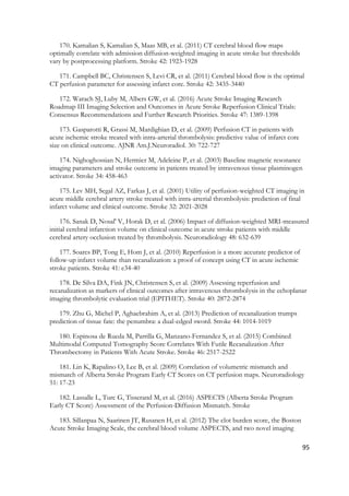
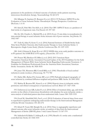
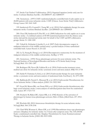
![99
acute ischaemic stroke (the third international stroke trial [IST-3]): a randomised controlled
trial. Lancet 379: 2352-2363
224. IST-3 collaborative group. (2013) Effect of thrombolysis with alteplase within 6 h of
acute ischaemic stroke on long-term outcomes (the third International Stroke Trial [IST-3]): 18-
month follow-up of a randomised controlled trial. Lancet Neurol. 12: 768-776
225. Cohen JE, Gomori JM, Rajz G, et al. (2015) Extracranial carotid artery stenting
followed by intracranial stent-based thrombectomy for acute tandem occlusive disease.
J.Neurointerv Surg. 7: 412-417
226. Ogawa A, Mori E, Minematsu K, et al. (2007) Randomized trial of intraarterial infusion
of urokinase within 6 hours of middle cerebral artery stroke: the middle cerebral artery
embolism local fibrinolytic intervention trial (MELT) Japan. Stroke 38: 2633-2639
227. IMS Study Investigators. (2004) Combined intravenous and intra-arterial recanalization
for acute ischemic stroke: the Interventional Management of Stroke Study. Stroke 35: 904-911
228. IMS II Trial Investigators. (2007) The Interventional Management of Stroke (IMS) II
Study. Stroke 38: 2127-2135
229. Broderick JP, Palesch YY, Demchuk AM, et al. (2013) Endovascular therapy after
intravenous t-PA versus t-PA alone for stroke. N.Engl.J.Med. 368: 893-903
230. Vagal AS, Khatri P, Broderick JP, et al. (2014) Time to angiographic reperfusion in
acute ischemic stroke: decision analysis. Stroke 45: 3625-3630
231. van der Marel K, Chueh JY, Brooks OW, et al. (2016) Quantitative assessment of
device-clot interaction for stent retriever thrombectomy. J.Neurointerv Surg.
232. Schonenberger S, Uhlmann L, Hacke W, et al. (2016) Effect of Conscious Sedation vs
General Anesthesia on Early Neurological Improvement Among Patients With Ischemic Stroke
Undergoing Endovascular Thrombectomy: A Randomized Clinical Trial. JAMA 316: 1986-
1996
233. ALEJANDRO M. SPIOTTA M, KYLE M. FARGEN, MD, MPH, IMRAN
CHAUDRY M, et al. (2016) A Direct AspirationFirst Pass Technique. Endovascular today 15:
68-70
234. Smith WS, Sung G, Starkman S, et al. (2005) Safety and efficacy of mechanical
embolectomy in acute ischemic stroke: results of the MERCI trial. Stroke 36: 1432-1438
235. Smith WS, Sung G, Saver J, et al. (2008) Mechanical thrombectomy for acute ischemic
stroke: final results of the Multi MERCI trial. Stroke 39: 1205-1212
236. Penumbra Pivotal Stroke Trial Investigators. (2009) The penumbra pivotal stroke trial:
safety and effectiveness of a new generation of mechanical devices for clot removal in
intracranial large vessel occlusive disease. Stroke 40: 2761-2768](https://image.slidesharecdn.com/strokethrombectomy-180911063514/85/Stroke-thrombectomy-101-320.jpg)
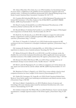
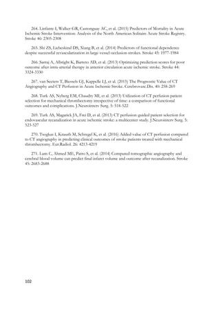

![INTRODUCTION
Mechanical thrombectomy (MT) is a safe and efficient treatment for acute ischemic stroke in
patients with large penumbra and good collateral circulation as established by several recent
randomized studies [1-6].
The success of the intervention depends among other factors on achieving the reperfusion of the
ischemic brain tissue quickly after groin puncture. The technical success is usually measured
using the Thrombolysis in Cerebral Infarction score (TICI) [7, 8].
According to previous studies the location and volume of the clot are critical determinants of the
success in intravenous thrombolysis (IVT). The recanalization results are poor in proximal clot
locations with a higher clot burden [9-11]. The location of the occlusion and the anatomy of the
vessels can influence the recanalization rate and the clinical outcome also in MT. Vessel anatomy
can have an effect on MT with extensive tortuosity and acute angles in the vessel bends being
more difficult to treat [12]. These challenges have been partly addressed by newer generation
retrievable stent-based MT devices which seem to be easier to use and have better technical and
clinical outcomes compared to the older generation devices [13-16].
Our study aimed to evaluate the success of MT in different clot locations using the TICI score
and the duration of the procedure as indicators of technical outcome and the 3-month modified
Rankin Scale (mRs) as a measure of clinical outcome.
METHODS
Overview, participants and variables](https://image.slidesharecdn.com/strokethrombectomy-180911063514/85/Stroke-thrombectomy-108-320.jpg)
![Hemorrhagic complications and postinfarct oedema were classified according to SITS-MOST
criteria [17].
Revascularization was evaluated with TICI, which was scored from the final DSA control runs of
the intervention. The clinical outcome measure was mRS, evaluated three months after the stroke
based on a follow-up visit to a neurologist or a phone interview by a neurologist. The imaging
outcome measure was infarct volume evaluated 24h after the intervention.
Recanalization therapies
MT was performed using a bi-axial system consisting of an 8F or 9F guiding catheter with a tip
balloon and coaxially a 0.021” micro-catheter or a tri-axial system consisting of an 8F guiding
catheter, a distal access catheter through which a micro-catheter was inserted with the aid of a
0.014” micro-guidewire. The micro-catheter was navigated through the occluded segment of the
artery and a suitable stent retriever was positioned trough the micro-catheter to the site of the
thrombus and deployed. The stent was left in place for 4 minutes and then retrieved and at the
same time the guiding catheter or the intermediate catheter was aspirated forcefully. The same
procedure was repeated until satisfactory circulation was restored. Different stent retriever
devices were used based on the preference and judgment of the operator. TREVO® (Stryker
Neurovascular/Concentric Medical, Mountain View, CA, USA) was used in 40 % of cases,
CAPTURE LP™ (eV3/ COVIDIEN/Medtronic, Santa Rosa, CA, USA) in 40%, ERIC®
(MicroVention, Tustin, CA, USA) in 9%, Aperio® (Acandis, Pforzheim, Germany) and
REVIVE® (Codman &Shurtleff, Raynham, MA, USA) in 1% respectively and in 10% of cases
multiple device types were used. The diameters of the devices used varied between 3mm and
6mm with a 4mm device being most commonly used. Intravenous thrombolysis (Actilyse®
0,9mg/kg, Boehringer-Ingelheim, Ingelheim, Germany) was administered as bridging therapy](https://image.slidesharecdn.com/strokethrombectomy-180911063514/85/Stroke-thrombectomy-110-320.jpg)
![5ml/s using 40ml of contrast agent followed by a 40ml saline flush. Digital subtraction
angiographic images were obtained using the Artis Z angiographer (Siemens, Munich, Germany)
using the parameters 102kV, AUTOmA and SMARTmA
Image analysis
NCCT, CTA and CTP examinations were reviewed using dedicated medical imaging
workstations. Parametric perfusion maps – mean transit time (MTT), cerebral blood flow (CBF),
and cerebral blood volume (CBV) – were generated with CT Perfusion 4 software (GE
Healthcare, Milwaukee, WI, USA). CTA images were reviewed by examining both the raw data
and maximum intensity projection images. The Alberta Stroke Program Early CT Score
(ASPECTS) was assessed from admission and follow-up NCCT images, and MTT and CBV
maps as described in our previous article [18]. CTA was used to evaluate the occlusion site, the
Clot Burden Score (CBS) and the Collateral Score (CS) as described in our previous report [19].
The location of the clot was recorded based on the most proximal position of the occlusion. The
examinations were reviewed in the order NCCT, CTA, and finally CTP, paralleling that of the
clinical work flow. Two radiologists assigned ASPECTS, CBS and CS. In cases where the
scoring or the assignment differed, a consensus opinion was agreed on. The reviewers were
blinded to the clinical data apart from the side and nature of the acute symptoms. Final infarct
volumes were measured by one radiologist. The boundaries of the affected areas were determined
visually. Volume was calculated by multiplying the measured area with the slice thickness.
Validation of the measurements including intraclass correlation coefficients (ICC) and Cohen’s
kappa values can be found in the above mentioned previous publications.
Statistics](https://image.slidesharecdn.com/strokethrombectomy-180911063514/85/Stroke-thrombectomy-112-320.jpg)
![outcome (mRS≤1) was seen in 40 patients (39%) and there was a trend towards more patients
having excellent outcome in the ICA and M1 groups (16 patients, 44% vs 19 patients, 41% vs 5
patients, 23%, p=0.22, Table 2) with only 5% of patients having mRS=1 in the M2 group (Figure
3). However, the ASPECTS score at 24h was significantly higher in the M2 group (median
ASPECTS 7 vs 8 vs 9, p=0.02, Table 2). In accordance, patients in the M2 group presented with
smaller infarct volumes in the 24h follow-up NCCT (Table 2, p=0.03). There were 20 patients
(19%) with parenchymal bleeding postoperatively and 21 patients (20%) suffered from post-
infarct oedema graded COED 2 or 3, but no significant differences were found in the occurrence
of these between the three clot locations. Also the mortality rates were similar with an overall
three-month mortality of 14% (p=0.89, Table 2).
DISCUSSION
With the advent of the third generation stent retrievers, superior to the previous generation
devices in efficacy and safety, and the results from recent randomized trials, the treatment of
anterior circulation acute stroke is dramatically changing [1-6, 13-15]. We evaluated in an
observational prospective study setup the technical and clinical outcome of MT, performed with
the new generation devices, in different sites of the occlusion in patients presenting with acute
anterior circulation stroke.
In the recent randomized trials, the majority of the patients treated with MT had occlusion of the
M1 segment whereas far fewer patients had a clot in the M2 segment [1-5]. Only some of these
trials reported their results stratified according to the location of the clot. In the ESCAPE trial the
clinical outcomes in different clot locations were comparable to ours. However, we had slightly
better outcomes with ICA occlusions: the proportion of patients with mRS=0 at three months was](https://image.slidesharecdn.com/strokethrombectomy-180911063514/85/Stroke-thrombectomy-115-320.jpg)
![15% in our study vs 13% in the ESCAPE study, mRS=1: 31% vs 16%, and mRS=2: 17% vs
22%, respectively. The finding was similar also when comparing to the results of the
REVASCAT study. Especially, we had fewer patients that experienced dismal outcome (mRS 5-
6) in the ICA group (23% in our study vs 46% in REVASCAT). However, these differences may
be partly due to longer average onset-to-recanalization times in both of these randomized trials.
Overall, considering the differences in study populations and delays, our results are compatible
with those of these trials.
Whether occlusions distal to the M1 segment should be treated with MT is still an open issue.
Unexpectedly considering the literature, in our study there were no statistically significant
differences in the 3-month clinical outcome between the different clot locations despite
significantly larger thrombus burden, worse collateral circulation and larger perfusion defects in
the more proximal occlusions (Table 1). On the contrary, there was a trend towards worse results
in the M2 group when attaining excellent clinical outcome (mRS≤1) was considered.
Correspondingly, also the recanalization outcomes were somewhat poorer in the M2 group.
These findings may be because patients in the M2 group had more severe strokes (median NIHSS
14) than expected from the literature [20]. In addition, there was higher average age and slight
female predominance in the M2 group. There may be selection of more symptomatic patients,
patients with occlusions of multiple M2 branches and non-responders to IVT into treatment with
MT. Finally, the worse technical outcome may be due to increased tortuosity of the vessels,
involvement of multiple vessel branches, a smaller proportion of patients that had received IVT,
and a thrombus composition less amenable to MT and/or IVT. However, there were no
statistically significant differences in the duration of the intervention between the three groups.
The total infarct volume at 24h was, as expected, significantly smaller in the M2 group. This was
not reflected in the 3-month clinical outcome. This may be due to quite small infarct volumes](https://image.slidesharecdn.com/strokethrombectomy-180911063514/85/Stroke-thrombectomy-116-320.jpg)
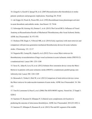
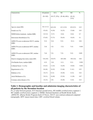
![ORIGINAL RESEARCH
TREVO and Capture LP have equal technical success
rates in mechanical thrombectomy of proximal and
distal anterior circulation occlusions
Sara Protto,1
Juha-Pekka Pienimäki,1
Janne Seppänen,1
Ira Matkaselkä,1
Jyrki Ollikainen,2
Heikki Numminen,2
Niko Sillanpää1
▸ Additional material is
published online only. To view
please visit the journal online
(http://dx.doi.org/10.1136/
neurintsurg-2016-012354).
1
Medical Imaging Center,
Tampere University Hospital,
Tampere, Finland
2
Department of Neurology,
Tampere University Hospital,
Tampere, Finland
Correspondence to
Dr Sara Protto, Medical
Imaging Center, Tampere
University Hospital, PL 2000,
Tampere 33521, Finland;
sara.protto@pshp.fi
Received 1 March 2016
Revised 16 May 2016
Accepted 23 May 2016
To cite: Protto S,
Pienimäki J-P, Seppänen J,
et al. J NeuroIntervent Surg
Published Online First:
[please include Day Month
Year] doi:10.1136/
neurintsurg-2016-012354
ABSTRACT
Purpose Mechanical thrombectomy (MT) is a proven
method to treat large vessel occlusions in acute anterior
circulation stroke. We compared the technical, imaging,
and clinical outcomes of MT performed with either
TREVO or Capture LP devices.
Methods There were 42 and 43 patients in the TREVO
and Capture LP groups, respectively. Baseline variables,
technical outcome (Thrombolysis In Cerebral Infarction,
TICI), 24 hours imaging outcome, and 3-month clinical
outcome (modified Rankin Scale, mRS) were
prospectively recorded. The patients were stratified
according to clot location, groups compared, and logistic
regression models devised to study the effect of device
selection on the clinical outcome.
Results The technical success rates were equal in both
proximal (internal carotid artery and proximal M1
segment) and distal occlusions (distal M1 and M2
segments). The proportion of TICI 2b or 3 was 96% and
87% with TREVO and 87% and 89% with Capture LP
(p=0.25 and p=0.80, respectively). Device selection did
not significantly predict good clinical outcome (mRS ≤2)
in either proximal or distal occlusions. In multivariate
analysis, selecting Capture LP borderline significantly
increased the odds of an excellent outcome close to
sixfold both in proximal and distal occlusions (OR 6.7,
95% CI 0.82 to 53.7, p=0.08 and OR 5.7, 95%
CI 0.88 to 37.8, p=0.07, respectively).
Conclusions TREVO and Capture LP perform equally
well in proximal and distal occlusions in the anterior
circulation when technical and good clinical outcome are
considered. Capture LP may have a small advantage in
reaching mRS ≤1 at 3 months. However, this needs to
be confirmed in a randomized study.
INTRODUCTION
Recent randomized studies have established the effi-
cacy and safety of mechanical thrombectomy (MT)
as treatment for acute anterior circulation stroke in
patients having a proximal vessel occlusion with
substantial penumbra in perfusion imaging, a rea-
sonably small infarct core, and adequate collateral
circulation.1–5
There are a number of newer generation retriev-
able stent-based MT devices in the market. Among
the more popular of these systems are TREVO
ProVue (Stryker Neurovascular/Concentric
Medical, Mountain View, California, USA) and
Capture LP (eV3/ Covidien/Medtronic, Santa Rosa,
California, USA). However, currently only a few
studies have compared the different retrievable
stent systems.
The TREVO Retriever is a thrombectomy device
designed to optimize the integration of the clot,
especially in intracranial proximal large vessel
occlusions. It is a retrievable, hydrophilic coated,
closed cell stent-like system. This device has con-
ventional stent characteristics with optimized orien-
tation of the struts, which aims at better thrombus
incorporation. Capture LP is a laser-cut stent
attached to a nitinol wire. The device is specially
designed to fit in smaller microcatheters to allow
thrombectomy in distal and more tortuous vessels.
Our study aimed to compare the efficacy and
safety of two different newer generation stent
retriever devices with different target vessel profiles
—the TREVO Retriever and Capture LP—in the
treatment of both proximal and distal occlusions in
the anterior circulation.
METHODS
Overview, participants and variables
We prospectively collected and analyzed the clinical
and imaging data of 130 consecutive patients who
were admitted to Tampere University Hospital
between January 2013 and December 2014 with
acute ischemic stroke. Patients underwent clinical
and imaging evaluation and proceeded to digital
subtraction angiography with an intention to
perform MT. The inclusion criteria for this study
were occlusion of the internal carotid artery (ICA)
and/or middle cerebral artery and MT with either
TREVO or Capture LP. These criteria were met by
85 patients and 45 patients were excluded. Eight
were excluded because they had posterior circula-
tion stroke and one patient had an occlusion of the
A3 segment of the anterior cerebral artery. In 15
cases thrombectomy with a stent retriever device
was not performed either because the clot had dis-
solved, there was no access to the thrombus, or
only aspiration thrombectomy was done. In 10
patients more than one device type was used and in
11 cases the device used was neither TREVO nor
Capture LP.
The initial imaging evaluation consisted of
non-contrast-enhanced CT (NCCT), CT angiog-
raphy (CTA), and CT perfusion (CTP). The selec-
tion of patients as candidates for MT was based on
absence of extensive irreversible ischemic changes
and hemorrhage in NCCT, evaluation of the
amount of salvageable tissue in CTP imaging and
Protto S, et al. J NeuroIntervent Surg 2016;0:1–6. doi:10.1136/neurintsurg-2016-012354 1
Ischemic stroke](https://image.slidesharecdn.com/strokethrombectomy-180911063514/85/Stroke-thrombectomy-124-320.jpg)
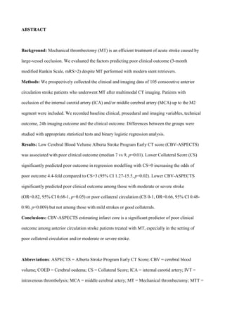
![mean transit time; NIHSS = National Institutes of Health Stroke Scale; TICI = Thrombolysis In
Cerebral Infarction
Keywords: interventional radiology; ischemic stroke; mechanical thrombectomy; perfusion CT.
INTRODUCTION
Mechanical thrombectomy (MT) is an efficient treatment of acute stroke caused by large-vessel
occlusion in patients presenting with favourable results in imaging evaluation [1-7]. Good clinical
outcome, as signified by three-month modified Rankin score (mRS) ≤2, has been reported in
roughly half of patients treated with MT despite high rates of successful reperfusion achieved with
newer generation stent retrievers [3-7]. Thus, identification of patients with poor prognosis despite
reperfusion is of central importance in improving the effectiveness of MT.
Fast and effective recanalization of the occluded vessel with restoration of normal antegrade
circulation has a crucial effect on the clinical outcome of acute stroke. However, it is not the only
important factor [8, 9]. Multiple studies have mapped both clinical and imaging variables that
predict favourable clinical outcome while those that directly address predictors of poor outcome in
different settings are far fewer in number [9, 10, 10-13].
The objective of this investigation was to clarify which clinical and imaging variables predict poor
clinical outcome (mRS>2) in patients with anterior circulation stroke who underwent MT.
METHODS
Participants and variables
The study was approved by the institutional review board. We prospectively collected and analysed
the clinical and imaging data of 130 consecutive patients presenting with stroke symptoms admitted](https://image.slidesharecdn.com/strokethrombectomy-180911063514/85/Stroke-thrombectomy-132-320.jpg)
![between January 2013 and December 2014 to Tampere University Hospital. They underwent
clinical and imaging evaluation and proceeded to digital subtraction angiography (DSA) with an
intention to perform MT. The inclusion criteria for this study were occlusion of the internal carotid
artery (ICA) and/or middle cerebral artery (MCA) up to the M2 segment, and MT with stent
retriever. 105 patients met these criteria and 25 patients were excluded: eight patients because they
had posterior circulation stroke, one patient had an occlusion of the A3 segment of the anterior
cerebral artery and another of the M3 segment. In 15 cases thrombectomy with a stent retriever
device was not performed because the clot had dissolved, there was no access to the thrombus or
only aspiration thrombectomy was done. The baseline clinical characteristics included age, sex,
clinical risk factors for ischemic stroke (hypertension, diabetes, coronary heart disease, atrial
fibrillation), time from symptom onset to imaging and to initiation of IVT. This data was collected
from the patient records. National Institutes of Health Stroke Scale (NIHSS) score at the
presentation, time from symptom onset to imaging and to recanalization of the occluded vessel, the
duration of the procedure, TICI (Thrombolysis in Cerebral Ischemia) grading evaluated with DSA
at the end of the procedure, and procedural complications had been prospectively stored. A follow-
up NCCT was performed 24h after MT. Haemorrhagic complications and post-infarct oedema were
classified according to SITS-MOST criteria [14]. The clinical outcome measure was the modified
Rankin Scale (mRS), evaluated three months after the stroke based on a follow-up visit to
neurologist or a phone interview by neurologist. One patient could not be reached for this control.
Imaging protocol, clinical decision making and image analysis
The initial imaging evaluation consisted of non-contrast-enhanced computed tomography (NCCT),
CT angiography (CTA) and CT perfusion (CTP). The selection of patients as candidates for MT
was based on absence of extensive irreversible ischemic changes (frank hypodensity more than 1/3
of the MCA territory) and haemorrhage in NCCT, evaluation of the amount of salvageable tissue in
CTP imaging and proximal clot position in CTA. The decision to proceed to MT was](https://image.slidesharecdn.com/strokethrombectomy-180911063514/85/Stroke-thrombectomy-133-320.jpg)
![multidisciplinary (stroke neurologist and neurointerventional radiologist). Patients referred to our
institution from other hospitals were re-evaluated with at least NCCT and CTA upon arrival before
proceeding to the angiographic suite to rule out bleeding and extensive irreversible ischemic lesion.
In the case of wake-up strokes, CTP was performed if no large infarct was seen in NCCT. NCCT,
CTA and CTP examinations were reviewed using dedicated medical imaging workstations.
Parametric perfusion maps – mean transit time (MTT), cerebral blood flow (CBF), and cerebral
blood volume (CBV) – were generated with the CT Perfusion 4 software that uses a delay
insensitive deconvolution based algorithm (GE Healthcare). CTA images were reviewed by
examining both the raw data and maximum intensity projection images. The Alberta Stroke
Program Early CT Score (ASPECTS) was assessed from admission and follow-up 24h NCCT
images, and from MTT and CBV maps as described in our previous article [15]. CTA was used to
evaluate the occlusion site, the Clot Burden Score (CBS) and the Collateral Score (CS) as described
in our previous report [16] and Table 1. The location of the clot was recorded based on the most
proximal position of the occlusion. The examinations were reviewed in the order NCCT, CTA, and
finally CTP, paralleling that of the clinical work flow. Two radiologists assigned ASPECTS, CBS
and CS. In cases the scoring or the assignment differed, a consensus opinion was agreed on. The
reviewers were blinded to the clinical data apart from the side and nature of the acute symptoms.
One radiologist measured the final infarct volumes on the 24h NCCT. The boundaries of the
affected areas were determined visually. Volume was calculated by multiplying the measured area
with the slice thickness. Validation of the measurements including intraclass correlation coefficients
(ICC) and Cohen’s kappa values can be found in the above-mentioned previous publications.
Recanalization therapies
MT was performed with different stent retrievers and sometimes with multiple devices based on the
judgment of the operator. We used a bi-axial system consisting of an 8F or 9F guiding catheter with](https://image.slidesharecdn.com/strokethrombectomy-180911063514/85/Stroke-thrombectomy-134-320.jpg)

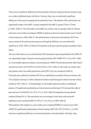
![restricting the analysis to patients with poor collateral circulation (CS 0-1), incrementing one CBV-
ASPECTS point significantly decreased the odds of poor prognosis (OR=0.66, 95% CI 0.48-0.90,
p=0.009) whereas no significant difference was found among those with good collaterals. Patients
with reperfusion result TICI=2a or worse showed a trend to poorer outcome (OR=3.14, 95%
CI=0.9-11. p=0.07, Table 4).
Finally, a binary logistic multivariable model using backwards likelihood method was devised with
poor 3-month clinical outcome as the dependent variable and variables with p<0.1 in the
univariable analysis included as covariates. Lower CBV-ASPECTS (OR=0.77, 95% CI=0.63 to
0.95, p=0.01) and not receiving IVT (OR=3.2, 95% CI=1.1 to 9.4, p=0.04) emerged as the only
statistically significant predictors of poor outcome in the final model.
Imaging outcome
ASPECTS was markedly lower in the 24h-follow-up NCCT of patients with a mRS >2 (7 vs 9,
p<0.001, Table 3). Correspondingly, the total infarct volume was larger among those with poor
outcome (64 ccm vs 11 ccm, p<0.001, Table 3). Furthermore, the number of haemorrhagic
complications and the extent of post-infarct oedema were significantly larger among these patients:
the proportion of those with a haemorrhagic complication at 24 hours 14% vs 6% (p=0.01, Table
3), major space occupying haemorrhage type PH2 or PHr2 5% vs 2% (p=0.05), and post-infarct
oedema grade COED 2 or 3 18% vs 2% (p<0.001).
DISCUSSION
Rapid and effective recanalization of the occluded vessel is a crucial predictor of good clinical
outcome in the treatment of acute anterior circulation ischemic stroke [9, 12]. It is still unclear
which patients with a large-vessel occlusion should be denied of MT because of poor prognosis
regardless of the type of reperfusion therapy. A number of studies have focused on evaluating the](https://image.slidesharecdn.com/strokethrombectomy-180911063514/85/Stroke-thrombectomy-138-320.jpg)
![factors predicting good outcome, whereas fewer studies have primarily addressed the factors related
to poor clinical outcome at 3 months [11, 17-21]. The setup of the majority of these studies has
been the evaluation of factors associated with poor clinical outcome despite successful
recanalization. In contrast, we studied all consecutive patients presenting with anterior circulation
stroke who underwent MT with a goal to understand which factors that are known at the time of the
clinical decision making to perform MT predict poor outcome. However, a subgroup analysis
including only patients with successful recanalization revealed that these factors are essentially the
same as in the entire study population. This finding was somewhat expected because of the high
recanalization rate (88%).
Higher NIHSS score at admission signifying more severe stroke, proximal site of occlusion and
absence of IVT treatment are closely associated with a poor 3-month outcome among patients
undergoing MT [17, 19, 20]. In our study we saw similar trends but these findings were borderline
statistically non-significant apart from the last. Interestingly, according to a meta-analysis of the
five recent randomized studies that established MT as the treatment of choice in large-vessel
occlusions, MT increased the odds of good clinical outcome regardless of IVT [2]. In our study
also the location of the occlusion was not a significant predictor of outcome which can in part be
due to worse recanalization outcomes among those with an M2 occlusion compared to those with
more proximal occlusions [22]. Moreover, patients with an M2 occlusion had more severe strokes
(median NIHSS 14) than expected from the literature [23] and somewhat higher average age (69.8
years vs. 64.8 and 65.3 years among patients with ICA and M1 occlusions, respectively). On the
other hand, age was not significantly related to a poor outcome in our study. This may be because
only a small number of patients in our study were 80 years of age or older, which has been
demonstrated to be related to a poorer outcome. [17, 19]
There was no significant difference in NCCT-ASPECTS scores at admission between those with
good and poor outcome. This needs to be interpreted in the context of relatively short average onset-](https://image.slidesharecdn.com/strokethrombectomy-180911063514/85/Stroke-thrombectomy-139-320.jpg)
![to-imaging times. However, where NCCT-ASPECTS lacks sensitivity, CBV-ASPECTS emerged as
an important imaging parameter with lower scores predicting poor outcome (Table 2, Figure 1).
This is compatible with previous results regarding both IVT and MT [4, 21, 24-29]. As
demonstrated in previous studies on IVT a CBV-ASPECTS threshold of 7 or 8 best
differentiates good and poor outcome. [15, 30] In our study the median CBV-ASPECTS in the
poor outcome group was 7.
The status of the collateral circulation as measured with Collateral Score has been shown to be an
important predictor of clinical outcome [31-33]. In our study having no visible collateral circulation
at all (CS=0) was specifically related to poor outcome.
CBV-ASPECTS did not significantly predict poor clinical outcome among patients with mild
strokes (NIHSS<8) or good collateral circulation (CS 2-4) whereas low CBV-ASPECTS was
significantly associated with poor outcome among those suffering from moderate or severe
stroke and/or having poor collateral circulation. A mild stroke entails less extensive and severe
ischemia which considering the mean onset-to-imaging time of less than 3 hours translates into
a small infarct core especially if an aggregating quantification method like ASPECTS is used.
Similarly, good collateral circulation is related to smaller infarct core in this short timeframe.
In a recent meta-analysis, MT did not significantly improve the clinical outcome of patients
with NIHSS<10 compared to IVT [2]. These findings imply that perfusion imaging in the
context of MT may be targeted to specific subgroups if not performed routinely to all patients
presenting with stroke symptoms. This would also reduce the total radiation dose incurred in
diagnostic imaging.
Our study has limitations. Firstly, as the formation of the MT cohort was clinically driven and thus
non-randomized, there can be selection biases. Further, due to relatively small number of patients,
subgroup analyses may be underpowered to detect small differences between the subgroups.](https://image.slidesharecdn.com/strokethrombectomy-180911063514/85/Stroke-thrombectomy-140-320.jpg)
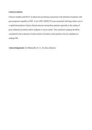
![Figure 1: A 82-year old male with type 2 diabetes mellitus and chronic hypertension was brought
to the emergency department because of right-sided hemiparesis. NCCT revealed minor ischemic
changes (ASPECTS=9, only one level shown, Panel A). There was an occlusion of the left distal
M1 segment in CTA (Panel B). CBV-ASPECTS was scored 7 and MTT-ASPECTS 5 (Panels D
and E. White arrows indicate the boundaries of the lesion). Revascularization was achieved with
MT (TICI=2b, Panel C). In the 24h control NCCT ASPECTS was 7. The 3-month mRS was 3
(Panel F).
Table 1: Collateral Score
Collateral Score (CS) was evaluated from single-phase CTA using a modified score as defined by
Souza et al. (2012). [34]
COLLATERAL SCORE DEFINITION
0 Absent collaterals in > 50% of the MCA territory
1 Diminished collaterals in > 50% of the MCA territory
2 Diminished collaterals in < 50% of the MCA territory](https://image.slidesharecdn.com/strokethrombectomy-180911063514/85/Stroke-thrombectomy-142-320.jpg)

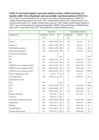
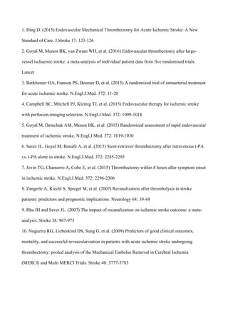
![208Intervent Neurol 2017;6:207–218
DOI: 10.1159/000475606
Sillanpää et al.: Internal Carotid Artery and the Proximal M1 Segment Are Optimal
Targets for Mechanical Thrombectomy
www.karger.com/ine
© 2017 S. Karger AG, Basel
Introduction
Mechanical thrombectomy (MT) is a safe and efficient treatment for acute ischemic
stroke caused by large vessel occlusion (LVO) with sufficiently small infarct volume on
admission imaging as established by several recent randomized studies [1–5]. Based on these
trials there is convincing evidence that MT yields better results compared to revascular-
ization with intravenous thrombolysis (IVT) only if the occlusion is located in the distal
internal carotid artery (ICA) including the different conformations carotid I, L and T, or the
M1 segment of the middle cerebral artery (MCA). It is still unclear if MT outperforms IVT in
the M2 segment because of the small number and heterogeneousness of patients with M2
occlusions in the randomized trials and other previous research [6–8]. Further, according to
a recent recommendation and previous reports, the proximal and distal halves of the M1
segments should be examined separately because these vessel subsegments have differing
properties that seem to influence the potential for good clinical outcome and the efficacy of
revascularization therapies [8–12].
We compared the clinical outcome measured with 3-month modified Rankin Scale (mRS)
in hyperacute (<3 h from the onset of symptoms) ischemic stroke between an MT and an IVT
cohort in different sites of occlusion: ICA, the proximal M1 (M1P), the distal M1 (M1D), and
the M2 segment of the MCA. The first two sites were considered proximal LVOs and the latter
two distal LVOs.
Methods
Overview, Participants, and Variables
We prospectively collected and analyzed the clinical and imaging data of 130 consecutive patients, who
were admitted to Tampere University Hospital from January 2013 to December 2014 because of acute ischemic
stroke symptoms and who underwent clinical and imaging evaluation and proceeded to digital subtraction angi-
ography (DSA) with an intention to perform MT. The inclusion criteria were occlusion of ICA (the intracranial
part with possible extension to the extracranial part) and/or the M1 or M2 segment of the MCA, MT with a stent
retriever, and onset-to-imaging time 3 h or less. Sixty-seven patients met these criteria. An anterior circulation
occlusion IVT-only cohort was used as a control group. This cohort has been extensively characterized in our
previous report [12]. Ninety-eight from 105 patients remained in this group after patients with occlusions distal
to the M2 segment were removed. The IVT-only cohort includes all patients treated with IVT from 2004 to 2007
for anterior circulation stroke and who had a clot visible in CT angiography (CTA). Starting from 2008 with the
advent of the first generation MT devices we began to treat selected patients with MT. By 2012, we had estab-
lished a stroke protocol where all anterior circulation stroke patients being evaluated for IVT were also eval-
uated for MT if there was a clot visible in CTA. The inclusion criteria to treat LVO (ICA and the M1 and M2
segments) with MT paralleled those of IVT apart from the time limit from the onset of symptoms that was <12 h
with MT, and therapy-related differences in contraindications. The prestroke mRS was required to be ≤2. In the
current study, only onset-to-imaging times <3 h were included to maintain congruence with the IVT-only cohort.
The initial imaging evaluation consisted of non-contrast-enhanced computed tomography (NCCT), CTA,
and CT perfusion (CTP). CTP could be dropped to save time at discretion of the attending neurologist. The
selection of patients as candidates for MT was based on absence of extensive irreversible ischemic changes
(frank hypodensity spanning more than one-third the volume of MCA vascular territory) and hemorrhage in
NCCT, presence of a proximal clot in CTA, and evaluation of the amount of salvageable tissue in CTP imaging
(if available).
Baseline clinical characteristics included age, sex, and clinical risk factors for ischemic stroke (hyper-
tension, diabetes, coronary heart disease, atrial fibrillation). These data were collected from the patient
records. National Institutes of Health Stroke Scale (NIHSS) score at the presentation, time from symptom
onset to imaging and recanalization of the occluded vessel, the duration of the procedure, the Thrombolysis
in Cerebral Infarction (TICI) score, and possible complications had been prospectively stored to a specifically
devised questionnaire. A follow-up NCCT was performed 24 h after revascularization. Hemorrhagic compli-
cations and cerebral edema (COED) were classified according to the SITS-MOST criteria.
Downloadedby:P.Sara-436829
85.76.9.17-7/7/20178:28:32AM](https://image.slidesharecdn.com/strokethrombectomy-180911063514/85/Stroke-thrombectomy-152-320.jpg)
![211Intervent Neurol 2017;6:207–218
DOI: 10.1159/000475606
Sillanpää et al.: Internal Carotid Artery and the Proximal M1 Segment Are Optimal
Targets for Mechanical Thrombectomy
www.karger.com/ine
© 2017 S. Karger AG, Basel
There were no statistically significant differences in clinical outcome between the MT
and IVT-only groups when distal LVOs were considered after controlling for confounders
(Tables 2, 3). However, there was a trend toward patients treated with MT for an M2 occlusion
more often experiencing unfavorable clinical outcome (Fig. 3; Tables 2, 3). This difference
was statistically significant (p = 0.03) before confounding was accounted for.
Sixty-three percent (42 of 67) of the patients who underwent MT received IVT before the
procedure. Twenty-six of these (62%) experienced good clinical outcome compared to 12
patients (48%) among those without IVT (p = 0.27).
Discussion
We compared the 3-month clinical outcome of MT performed with third-generation stent
retrievers and IVT only in the treatment of hyperacute ischemic stroke in different sites of
occlusion: ICA, the M1P, the M1D, and the M2 segments of the MCA. Our study confirms the
findings of the recent randomized trials [1–5, 13, 14] that MT is clearly superior to IVT in the
treatmentICAandM1Pocclusions(proximalLVOs).Only12and24%,respectively,ofpatients
who received only IVT experienced good clinical outcome (mRS ≤2) compared to 54 and 80%
among those treated with MT. However, we did not find a significant difference in favor of
either revascularization therapy in the treatment of M1D or M2 occlusions (distal LVOs).
Table 1. Baseline and admission imaging characteristics and the clinical and imaging outcomes of patients with proximal and
distal large vessel occlusions in the MT and IVT-only groups
Characteristic ICA and proximal M1 segment Distal M1 and M2 segments
MT
(n = 35)
IVT only
(n = 38)
p1 MT
(n = 32)
IVT only
(n = 60)
p2
Age, years 67.4±9.4 65.7±15.2 0.64 68.4±10.4 70.7±12.8 0.21
Male sex 18 (51) 27 (71) 0.09 12 (38) 28 (47) 0.51
NIHSS before treatment 14.5 [5] 17.5 [7] 0.12 14 [6] 11 [10] 0.01
ASPECTS score at admission NCCT 9 [2] 9 [3] 0.66 10 [1] 10 [2] 0.68
ASPECTS score at admission MTT 1 [2] 3 [4] 0.001 3.5 [(5] 6 [3] 0.04
ASPECTS score at admission CBV 6.5 [6] 7 [5] 0.43 8.5 [(3] 8 [3] 0.53
Onset to imaging time, min 94 [79] 87 [40] 0.22 75 [58] 92 [31] 0.04
Clot burden score 4 [3] 4 [2] 0.27 6 [2] 8 [3] 0.01
Collateral score 1 [2] 1 [3] 0.91 2 [2] 2 [2] 0.90
Hypertension 20 (57) 22 (58) 0.95 15 (47) 40 (67) 0.08
Diabetes 3 (7) 8 (21) 0.14 7 (22) 8 (13) 0.38
Atrial fibrillation 19 (54) 12 (32) 0.05 18 (56) 25 (42) 0.20
Coronary artery disease 6 (17) 16 (42) 0.01 5 (16) 18 (30) 0.21
ASPECTS score at 24-h CT 8 [4] 4 [5] 0.001 9 [3] 8 [4] 0.17
Total infarct volume at 24 h, cm3 4.5 [45.7] 59.4 [153.1] 0.001 2.5 [24.3] 8.3 [38.7] 0.32
Hemorrhagic complication at 24 h 5 (15) 4 (11) 0.73 4 (13) 3 (5) 0.23
Major space-occupying effect (PH2 or PHr2) 2 (6) 2 (5) 0.93 2 (6) 1 (2) 0.28
Postinfarct edema COED2 or COED3 8 (24) 19 (53) 0.01 6 (19) 10 (17) 0.78
Three-month mortality 4 (12) 12 (32) 0.04 4 (13) 2 (3) 0.18
Data are presented as mean ± SD, n (%), or median [IQR]. P1, P2: p values between groups; ASPECTS: Alberta Stroke Program
Early CT Score; CBV, cerebral blood volume; COED, cerebral edema; MTT, mean transit time; NCCT, non-contrast-enhanced
computed tomography; NIHSS, National Institutes of Health Stroke Scale; PH, parenchymal hemorrhage; PHr, parenchymal
hemorrhage remote.
Downloadedby:P.Sara-436829
85.76.9.17-7/7/20178:28:32AM](https://image.slidesharecdn.com/strokethrombectomy-180911063514/85/Stroke-thrombectomy-155-320.jpg)
![213Intervent Neurol 2017;6:207–218
DOI: 10.1159/000475606
Sillanpää et al.: Internal Carotid Artery and the Proximal M1 Segment Are Optimal
Targets for Mechanical Thrombectomy
www.karger.com/ine
© 2017 S. Karger AG, Basel
A current consensus statement recommends that the M1 segment should be divided into
proximal and distal subsegments [9]. None of the recent randomized trials reported this
distinction in their published analyses. In the earlier IMS 3 trial, 64% of subjects in the
IVT-only arm compared to 55% in the endovascular arm had good clinical outcome of an M1D
occlusion [8]. The difference was not statistically significant. According to a recent retro-
spective study, 61% of subjects treated for M1D occlusion with MT experienced good clinical
outcome and 44% had excellent outcome [10]. These findings are in alignment with our
results (Fig. 3). When we analyzed the whole M1 segment, MT borderline significantly
improved the outcome (OR = 2.6, CI 95% 0.94–7.2, p = 0.07). This is compatible with the
results of recent meta-analyses [14, 15].
The feasibility and efficacy of MT of the M2 segment has been demonstrated in a few
studies [6, 7, 16]. Successful reperfusion rates range from 78.5 to 93.3%, and up to 60% of
patients achieved good clinical outcome. Four of the 5 recent randomized trials reported
results on the M2 segment. In the ESCAPE study, 79% of patients in the control group received
IVT [3]. Only 6 patients in the intervention group had an isolated M2 occlusion and patients
with occlusion of all M2 segment arteries were pooled with M1 occlusions. The authors state
that the treatment effect (showing benefit from MT) was similar in location-based subgroup
Table 2. Good clinical outcome in the MT and IVT-only groups
Clot location MT
mRS ≤2
IVT only
mRS ≤2
p1 Odds ratio 95% CI p2
ICA 13 (54%) 2 (12%) 0.008 4.9 0.77–31.5 0.09
Proximal M1 8 (80%) 5 (24%) 0.006 11.9 1.6–90.1 0.02
Distal M1 10 (56%) 17 (59%) 0.84 1.0 0.26–3.7 0.96
M2 6 (43%) 23 (77%) 0.03 0.30 0.05–1.9 0.20
ICA and proximal M1 21 (62%) 7 (18%) <0.001 6.0 1.9–18.3 0.002
Distal M1 and M2 16 (50%) 40 (69%) 0.12 0.54 0.16–1.5 0.24
P1, p value between groups; P2, significance of the covariate in binary logistic regression model; CI,
confidence interval; ICA, internal carotid artery; M1, middle cerebral artery segment 1; M2, middle cerebral
artery segment 2.
Table 3. Excellent clinical outcome in the MT and IVT-only groups
Clot location MT
mRS ≤1
IVT only
mRS ≤1
p1 Odds ratio 95% CI p2
ICA 12 (50%) 1 (6%) 0.005 9.0 0.9–89.8 0.06
Proximal M1 4 (40%) 0 0.007 n.a. n.a. n.a.
Distal M1 8 (44%) 9 (31%) 0.35 1.9 0.49–7.6 0.37
M2 4 (29%) 11 (37%) 0.73 0.51 0.07–3.7 0.50
ICA and proximal M1 16 (47%) 1 (3%) <0.001 28.0 3.4–233.8 0.002
Distal M1 and M2 12 (38%) 20 (34%) 0.82 1.1 0.37–3.1 0.90
P1, p value between groups; P2, significance of the covariate in binary logistic regression model; CI,
confidence interval; ICA, internal carotid artery; M1, middle cerebral artery segment 1; M2, middle cerebral
artery segment 2.
Downloadedby:P.Sara-436829
85.76.9.17-7/7/20178:28:32AM](https://image.slidesharecdn.com/strokethrombectomy-180911063514/85/Stroke-thrombectomy-157-320.jpg)
![214Intervent Neurol 2017;6:207–218
DOI: 10.1159/000475606
Sillanpää et al.: Internal Carotid Artery and the Proximal M1 Segment Are Optimal
Targets for Mechanical Thrombectomy
www.karger.com/ine
© 2017 S. Karger AG, Basel
analyses but details are not provided. The SWIFT PRIME trial had 18 patients with an M2
occlusion [5]. Thirteen of them were in the MT and IVT group, and 5 in the IVT group with 53
and 40% experiencing good clinical outcome, respectively. The difference was not statisti-
cally significant. In the REVASCAT study, 78% of patients in the control group received IVT
[4]. Ten patients in the MT group and 8 in the control group had an M2 occlusion whose
clinical outcome was not detailed. In the MR CLEAN trial, 91% of patients in the control group
were administered IVT [1]. Eighteen patients in the MT group and 21 patients in the control
group had an M2 occlusion. A subgroup analysis of M2 occlusions was not reported. A meta-
analysis of these trials has been published recently [14]. Pooled together, there were 51
eligible patients with an M2 occlusion in the MT group and 44 in the control group. The
direction of the effect favored MT but the adjusted OR was not significant (OR = 1.28, 95% CI
0.51–3.21). Another meta-analysis of somewhat older studies reported similar results (OR =
1.5, 95% CI 0.8–3.0) [15].
The recanalization rate of an M2 segment occlusion with MT was 79% in our study, which
is comparable to the literature but not optimal. After adjusting for confounders, there was a
nonsignificant trend toward worse clinical outcome in the MT group which was observed also
when the M2 and M1D segments were analyzed together (Table 2). The M2 occlusion patients
in the IVT-only group had milder strokes compared to the MT group (median NIHSS 10 vs.
14). However, the trend remained unchanged when patients with mild strokes (NIHSS <8)
were excluded (data not shown). The reported recanalization rates of M2 occlusions with IVT
are reasonably high at 44–79% and correspondingly the clinical outcomes better compared
to proximal LVOs [8, 17–19]. In our data, 77% of M2 occlusion patients treated with only IVT
experienced good clinical outcome (Fig. 3), a considerably higher proportion than in the IMS
3 trial (44.5%) [8]. This along with the somewhat suboptimal reperfusion rate in our MT
group and selection of moderate and severe strokes to the MT group probably leads to too
pessimistic an estimate of the efficacy of MT. That withstanding, there is no indication in our
data that MT would outperform IVT in the treatment of hyperacute M2 occlusions. A very
recent multicenter retrospective analysis comparing modern endovascular therapy with best
medical management reported a substantial (OR 3.2, CI 2–5.2, p < 0.001) advantage in favor
of endovascular therapy [20]. However, this result is not directly comparable to ours because
not all patients were treated with MT in the endovascular group and IVT in the medical
management group. Further, patients presenting up to 8 h from the onset were included in
the study.
Almost half of the patients in the MT group who had proximal LVO experienced excellent
clinical outcome compared to only 1 patient (3%) in the IVT-only group. Thus, it appears that
surviving an ICA or M1P occlusion without any disability-causing neurological deficits essen-
tially requires MT. This has potential implications especially in the management of patients
with low prestroke mRS (0–1).
We restricted our analysis to hyperacute strokes. The time window of IVT was extended
to 4.5 h in 2008. The efficacy of IVT deteriorates rapidly in the first few hours resulting in a
sizeable number-needed-to-treat figure of 14.9 in the 3- to 4.5-h time window [21]. Consid-
ering the superior reperfusion rates of MT, one would expect the performance of MT to be at
least at par with IVT in the extended time window. Extrapolating from the results of a recent
meta-analysis, this appears to be the case [14].
Our observational study has several limitations: a major limitation is its retrospective
nature,althoughthedataforbothgroupswerecollectedandrecordedprospectivelyaccording
to a preset protocol. There may be a selection of patients with more severe symptoms to MT
especially in distal occlusions, and conversely those with milder symptoms may have been
denied an invasive procedure. Patients that responded to IVT or experienced significant and
sustained spontaneous resolution of symptoms between admission imaging and groin
Downloadedby:P.Sara-436829
85.76.9.17-7/7/20178:28:32AM](https://image.slidesharecdn.com/strokethrombectomy-180911063514/85/Stroke-thrombectomy-158-320.jpg)
![215Intervent Neurol 2017;6:207–218
DOI: 10.1159/000475606
Sillanpää et al.: Internal Carotid Artery and the Proximal M1 Segment Are Optimal
Targets for Mechanical Thrombectomy
www.karger.com/ine
© 2017 S. Karger AG, Basel
puncture were not included in the MT group, whereas those with contraindications to IVT
were obviously not in the IVT-only group. The latter is the reason why the proportion of
patients with atrial fibrillation was larger in the MT group. However, all these biases act to
diminish the perceived efficacy of MT in relation to IVT only. Further, the somewhat small
study population decreases the value of subgroup analyses. Patient level data on prestroke
mRS were available only in the IVT-only group (the median mRS was 1). Several different
stent retrievers or a combination of them were used in varying proportions in different clot
locations based on the preference of the operator, which may lead to a bias between the
proximal and distal LVOs. However, there were no significant differences in technical success
between the two main devices that were used [22]. Finally, the technical details (primary
stent retriever thrombectomy with a balloon-tipped guiding catheter) limit the generaliz-
ability of the results.
In conclusion, our study shows that MT is superior to IVT in achieving good clinical
outcome in the treatment of hyperacute anterior circulation stroke in the most proximal
occlusions, i.e. ICA or the M1P segment. In the distal M1 segment or the M2 segment, neither
of these therapies clearly outperforms the other.
Acknowledgments
The authors are grateful to Jari Hakomäki, MD, for image interpretation and Ira Matkaselkä, BA, for data
collection.
Disclosure Statement
All authors declare that they have no conflicts of interest relevant to this work.
Funding Sources
This study was financially supported by the Competitive State Research Financing of the Expert Respon-
sibility Area of Tampere University Hospital (Grant 9S061).
Appendix
Imaging Parameters
In the MT cohort, the CT scans were obtained using a 64-row multidetector CT scanner (General Electric
LightSpeed VCT, GE Healthcare, Milwaukee, WI, USA). Brain NCCT was performed using the parameters 120
kV with AUTOmA and SMARTmA technic, noise index 3.3, collimation 4 × 5 mm, 40% adaptive statistical
iterative reconstruction (ASIR), and rotation 0.5 s. Images were obtained axially (0.625-mm-thick slices), and
then contiguous axial slices were reconstructed to the thickness of 5 mm and coronal slices to the thickness
of 2 mm. CTA was performed with helical technique using a scanning range from the aortic arch to the vertex
of the skull. The imaging parameters were 100 kV, AUTOmA and SMARTmA, noise index 9, 40% ASIR, colli-
mation 40 × 0.625 mm, rotation 0.5 s, pitch factor 0.984. The contrast agent (iomeprol, 350 mg I/mL;
IOMERON, Bracco, Milan, Italy) was administered via an antecubital vein with 18-gauge cannula using a
double-piston power injector with a flow rate of 5 mL/s using 70 mL contrast agent followed by a 50 mL
saline flush. Automatic bolus triggering from the aortic arch was used. CTP was performed using the param-
eters 80 kV, 250 mA, 50% ASIR, collimation 8 × 5 mm, and rotation 0.4 s. 272 slices covering a range of 80
mm were generated in 46 s using alternating toggle table protocol to increase the z-axis coverage. Contiguous
slices were reconstructed to a thickness of 5 mm at even intervals. The contrast agent (IOMERON 350 mg I/
mL) was administered via an antecubital vein with an 18-gauge cannula using a double-piston power injector
with flow rate of 5 mL/s using 40 mL of contrast agent followed by a 40 mL saline flush. DSA images were
Downloadedby:P.Sara-436829
85.76.9.17-7/7/20178:28:32AM](https://image.slidesharecdn.com/strokethrombectomy-180911063514/85/Stroke-thrombectomy-159-320.jpg)
![216Intervent Neurol 2017;6:207–218
DOI: 10.1159/000475606
Sillanpää et al.: Internal Carotid Artery and the Proximal M1 Segment Are Optimal
Targets for Mechanical Thrombectomy
www.karger.com/ine
© 2017 S. Karger AG, Basel
obtained using the Artis Z angiographer (Siemens, Munich, Germany) using the parameters 102 kV, AUTOmA
and SMARTmA.
In the IVT cohort, imaging was performed using two different multidetector scanners: General Electric
LightSpeed 16-row (GE Healthcare, Milwaukee, WI, USA) and Philips Brilliance 64-row (Philips, Cleveland,
OH, USA). Brain NCCT was performed using the parameters 120 kV, 430 mAs, collimation 12 × 1.25 mm,
rotation 1.5 s (64-row) or 120 kV, 320 mAs, collimation 16 × 1.25 mm, rotation 1 s (16-row). Contiguous
slices were reconstructed to the thickness of 5 mm in the whole scanning range (64-row) or to the thickness
of 5 mm in the skull base and 7.5 mm in the supratentorial region (16-row). CTA was performed using a
scanning range extending from the C2-vertebra to the vertex of the skull. The imaging parameters were 120
kV, 212 mAs (using dynamic tube current modulation), collimation 64 × 0.625 mm, rotation 0.75 s, pitch
factor 0.923 (64-row) or 120 kV, 160 mAs, collimation 16 × 0.625 mm, rotation 0.8 s, pitch factor 0.938
(16-row). Contiguous slices were reconstructed to the thickness of 0.9 mm with a 0.45 mm overlap (64-row)
or to the thickness of 1.25 mm (16-row). The contrast agent (iobitridol, Xenetix 350 mg I/mL; Aulnay-sous-
Bois, France) was administered through an antecubital 18-gauge cannula using a double-piston power
injector with a flow rate of 4 mL/s using 70 mL of contrast agent followed by a 50 mL saline flush. Manual
bolus triggering was used. CTP was performed using the parameters 80 kV, 200 mAs (effective), collimation
32 × 1.25 mm, rotation 0.4 s (64-row) or 80 kV, 200 mAs, collimation 8 × 2.5 mm, rotation 1 s (16-row). 120
slices covering a range of 80 mm were generated in 55 s using an alternating toggle table protocol (64-row),
or 200 slices covering a range of 20 mm were generated in 50 s with a stationary gantry position (16-row).
Contiguous slices were reconstructed to the thickness of 10 mm (64-row) or to the thickness of 5 mm
(16-row) at even time intervals. The imaging range was positioned so that the ASPECTS levels (the level of
the basal ganglia and the supraganglionic level) were always covered. The rest of the 80 mm range (64-row)
was positioned both cranial and caudal to the ASPECTS planes with the exact balancing depending on the
clinical presentation. The contrast agent (Xenetix 350 mg I/mL) was administered through an antecubital
18-gauge cannula using a double-piston power injector with a flow rate of 5 mL/s using 60 mL of contrast
agent followed by a 40 mL saline flush.
Image Analysis
NCCT, CTA, and CTP examinations were reviewed using dedicated medical imaging workstations. Para-
metric perfusion maps – mean transit time (MTT), cerebral blood flow (CBF), and cerebral blood volume
(CBV) – were generated with the CT Perfusion 4 software that uses a delay insensitive deconvolution-based
algorithm (GE Healthcare). CTA images were reviewed by examining both the raw data and maximum
intensity projection images. The Alberta Stroke Program Early CT Score (ASPECTS) was assessed from
admission and follow-up 24-h NCCT images and from MTT and CBV maps, and CTA was used to evaluate the
occlusion site, the Clot Burden Score (CBS) and the Collateral Score (CS) as described in previous reports [23,
24]. The location of the clot was recorded based on the most proximal position of the occlusion. The M1
segment of the MCA was divided in two parts of equal length: the proximal and the distal half as described
previously (M1P and M1D, respectively) [25]. The examinations were reviewed in the order NCCT, CTA, and
finally CTP, paralleling that of the clinical work flow. Two radiologists assigned ASPECTS, CBS, and CS. In
cases where the scoring or the assignment differed, a consensus opinion was agreed on. The reviewers were
blinded to the clinical data apart from the side and nature of the acute symptoms. One radiologist measured
the final infarct volumes. The boundaries of the affected areas were determined visually. Volume was calcu-
lated by multiplying the measured area with the slice thickness. TICI was evaluated and recorded prospec-
tively to a questionnaire by the interventional neuroradiologist that performed the procedure. Intraclass
correlation coefficients (ICC) between staff radiologists and an experienced neuroradiologist were calcu-
lated for ASPECTS assignments in a test sample (n = 20): ICCNCCT = 0.86, ICCMTT = 0.79, ICCCBV = 0.73, and
ICCNCCT 24 h = 0.93. Median interobserver agreement indices for areas and volumes were AREAMTT: 68%,
AREACBV: 90% and VOLUMEINFARCT: 80%. ICC for CS was 0.87. Cohen’s kappa was 0.94 for the location of the
clot and 0.68 for the collateral score (0.90 after dichotomization 0–1 vs. 2–4).
Downloadedby:P.Sara-436829
85.76.9.17-7/7/20178:28:32AM](https://image.slidesharecdn.com/strokethrombectomy-180911063514/85/Stroke-thrombectomy-160-320.jpg)
![217Intervent Neurol 2017;6:207–218
DOI: 10.1159/000475606
Sillanpää et al.: Internal Carotid Artery and the Proximal M1 Segment Are Optimal
Targets for Mechanical Thrombectomy
www.karger.com/ine
© 2017 S. Karger AG, Basel
Results
Baseline and admission imaging characteristics and the clinical and imaging outcomes of all patients and
patients in the MT and IVT-only groups
Characteristic All patients
(n = 165)
MT
(n = 67)
IVT only
(n = 98)
p1
Age, years 68.5±12.4 67.9±9.7 68.9±13.8 0.60
Male sex 85 (52) 30 (45) 55 (56) 0.18
NIHSS before treatment 13.5 [9] 14 (5) 14 (9) 0.26
ASPECTS score at admission NCCT 10 [2] 9 (2) 10 (2) 0.34
ASPECTS score at admission MTT 4 [4] 2 (4) 5 (4) <0.001
ASPECTS score at admission CBV 8 [3] 7 (4) 8 (4) 0.10
Onset to imaging time, min 90±34 86±64 90±34 0.58
Clot burden score 6 [4] 6 (4) 6 (4) 0.03
Collateral score 1 [3] 1 (2) 2 (3) 0.58
Hypertension 97 (59) 35 (52) 62 (64) 0.16
Diabetes 26 (16) 10 (15) 16 (16) 0.81
Atrial fibrillation 74 (45) 37 (55) 37 (38) 0.04
Coronary artery disease 45 (27) 11 (16) 34 (35) 0.01
Three-month modified Rankin scale 0–2 84 (52) 37 (56) 47 (49) 0.34
Three-month modified Rankin scale 0–1 49 (30) 28 (42) 21 (22) 0.005
Three-month mortality 22 (13) 8 (12) 14 (14) 0.67
ASPECTS score at 24-h CT 7.5 [5] 8 (3) 7 (5) 0.007
Total infarct volume at 24 h, cm3
45±75 27±52 57±85 0.005
Hemorrhagic complication at 24 h 16 (19) 9 (14) 7 (7) 0.18
Major space-occupying effect (PH2 or PHr2) 7 (4) 4 (6) 3 (3) 0.44
Postinfarct edema COED2 or COED3 43 (26) 14 (21) 29 (30) 0.20
Data are presented as mean ± SD, n (%), or median [IQR]. p1, p value between groups; ASPECTS, Alberta
Stroke Program Early CT Score; CBV, cerebral blood volume; COED, cerebral edema; MTT, mean transit time;
NCCT, non-contrast-enhanced computed tomography; NIHSS, National Institutes of Health Stroke Scale; PH,
parenchymal hemorrhage; PHr, parenchymal hemorrhage remote.
References
1 Berkhemer OA, Fransen PS, Beumer D, van den Berg LA, et al: A randomized trial of intraarterial treatment for
acute ischemic stroke. N Engl J Med 2015;372:11–20.
2 CampbellBC,MitchellPJ,KleinigTJ,DeweyHM,etal:Endovasculartherapyforischemicstrokewithperfusion-
imaging selection. N Engl J Med 2015;372:1009–1018.
3 Goyal M, Demchuk AM, Menon BK, Eesa M, et al: Randomized assessment of rapid endovascular treatment of
ischemic stroke. N Engl J Med 2015;372:1019–1030.
4 Jovin TG, Chamorro A, Cobo E, de Miquel MA, et al: Thrombectomy within 8 hours after symptom onset in
ischemic stroke. N Engl J Med 2015;372:2296–2306.
5 Saver JL, Goyal M, Bonafe A, Diener HC, et al: Stent-retriever thrombectomy after intravenous t-PA vs. t-PA
alone in stroke. N Engl J Med 2015;372:2285–2295.
6 Dorn F, Lockau H, Stetefeld H, Kabbasch C, Kraus B, Dohmen C, Henning T, Mpotsaris A, Liebig T: Mechanical
thrombectomy of M2-occlusion. J Stroke Cerebrovasc Dis 2015;24:1465–1470.
7 Flores A, Tomasello A, Cardona P, de Miquel MA, Gomis M, Garcia Bermejo P, Obach V, Urra X, Marti-Fabregas
J, Canovas D, Roquer J, Abilleira S, Ribo M; Catalan Stroke Code and Reperfusion Consortium Cat-SCR: Endo-
vascular treatment for M2 occlusions in the era of stentrievers: a descriptive multicenter experience. J Neuro-
interv Surg 2015;7:234–237.
8 Demchuk AM, Goyal M, Yeatts SD, Carrozzella J, Foster LD, Qazi E, Hill MD, Jovin TG, Ribo M, Yan B, Zaidat OO,
Frei D, von Kummer R, Cockroft KM, Khatri P, Liebeskind DS, Tomsick TA, Palesch YY, Broderick JP; IMS III
Investigators: Recanalization and clinical outcome of occlusion sites at baseline CT angiography in the Inter-
ventional Management of Stroke III trial. Radiology 2014;273:202–210.
Downloadedby:P.Sara-436829
85.76.9.17-7/7/20178:28:32AM](https://image.slidesharecdn.com/strokethrombectomy-180911063514/85/Stroke-thrombectomy-161-320.jpg)

This thesis examined factors influencing outcomes of mechanical thrombectomy (MT) for acute anterior circulation stroke. The study analyzed a prospectively collected cohort of MT patients from 2013-2014. The location of clot on CTA, device used for MT, and CTP parameters including CBV-ASPECTS were evaluated for their effects on technical success (TICI score) and clinical outcome (mRS at 3 months). MT was superior to IVT for proximal large vessel occlusions but neither was clearly better for distal occlusions. Site of occlusion did not affect MT success rates. CBV-ASPECTS predicted infarct core on follow-up and poor outcome, especially with poor collaterals or moderate-severe strokes. Low


![Mechanical thrombectomy in acute stroke [Autosaved].pptx](https://cdn.slidesharecdn.com/ss_thumbnails/mechanicalthrombectomyinacutestrokeautosaved-230102142009-fe15766f-thumbnail.jpg?width=640&height=640&fit=bounds)


![CTEV [ clubfoot] DR ARUN LAL ,DR MOHAMED ASHRAF travancore medical college k...](https://cdn.slidesharecdn.com/ss_thumbnails/ctevclubfootdrarunlaldrmohamedashraftravancoremedicalcollegekollamkeralaindia-260208063247-18fc466c-thumbnail.jpg?width=640&height=640&fit=bounds)


