The 2024 ESC guidelines for atrial fibrillation management, developed by the European Society of Cardiology in collaboration with several cardiology associations, provide comprehensive recommendations on diagnosis, treatment strategies, and patient management. The guidelines emphasize patient-centered care, addressing comorbidities, and optimizing both rate and rhythm control, while also outlining strategies to reduce stroke risk. Health professionals are urged to use these guidelines as a supportive reference while exercising their clinical judgment for individual patient cases.
![Table of contents
1. Preamble .............................................................................................................. 3319
2. Introduction ....................................................................................................... 3321
2.1. What is new ............................................................................................. 3322
3. Definitions and clinical impact .................................................................... 3326
3.1. Definition and classification of AF ................................................... 3326
3.2. Diagnostic criteria for AF ................................................................... 3327
3.3. Symptoms attributable to AF ........................................................... 3328
3.4. Diagnostic evaluation of new AF ..................................................... 3328
3.5. Adverse events associated with AF ................................................ 3329
3.6. Atrial flutter .............................................................................................. 3330
4. Patient pathways and management of AF ............................................. 3330
4.1. Patient-centred, multidisciplinary AF management ................. 3330
4.1.1. The patient at the heart of care .............................................. 3330
4.1.2. Education and shared decision-making ................................. 3331
4.1.3. Education of healthcare professionals ................................... 3332
4.1.4. Inclusive management of AF ...................................................... 3332
4.2. Principles of AF-CARE ......................................................................... 3332
5. [C] Comorbidity and risk factor management ................................... 3338
5.1. Hypertension ............................................................................................ 3339
5.2. Heart failure .............................................................................................. 3339
5.3. Type 2 diabetes mellitus ...................................................................... 3340
5.4. Obesity ....................................................................................................... 3340
5.5. Obstructive sleep apnoea ................................................................... 3340
5.6. Physical inactivity .................................................................................... 3340
5.7. Alcohol excess ......................................................................................... 3341
6. [A] Avoid stroke and thromboembolism .............................................. 3341
6.1. Initiating oral anticoagulation ............................................................. 3341
6.1.1. Decision support for anticoagulation in AF ........................ 3341
6.2. Oral anticoagulants ................................................................................ 3343
6.2.1. Direct oral anticoagulants ........................................................... 3344
6.2.2. Vitamin K antagonists ................................................................... 3345
6.2.3. Clinical vs. device-detected subclinical AF ........................... 3345
6.3. Antiplatelet drugs and combinations with anticoagulants .... 3346
6.4. Residual ischaemic stroke risk despite anticoagulation .......... 3346
6.5. Percutaneous left atrial appendage occlusion ............................ 3346
6.6. Surgical left atrial appendage occlusion ........................................ 3347
6.7. Bleeding risk .............................................................................................. 3348
6.7.1. Assessment of bleeding risk ....................................................... 3348
6.7.2. Management of bleeding on anticoagulant therapy ......... 3348
7. [R] Reduce symptoms by rate and rhythm control ......................... 3351
7.1. Management of heart rate in patients with AF ......................... 3351
7.1.1. Indications and target heart rate .............................................. 3352
7.1.2. Heart rate control in the acute setting ................................. 3352
7.1.3. Long-term heart rate control .................................................... 3352
7.1.4. Atrioventricular node ablation and pacemaker
implantation ................................................................................................... 3353
7.2. Rhythm control strategies in patients with AF .......................... 3353
7.2.1. General principles and anticoagulation ................................. 3353
7.2.2. Electrical cardioversion ................................................................ 3356
7.2.3. Pharmacological cardioversion .................................................. 3356
7.2.4. Antiarrhythmic drugs .................................................................... 3357
7.2.5. Catheter ablation ............................................................................ 3358
7.2.6. Anticoagulation in patients undergoing catheter ablation 3359
7.2.7. Endoscopic and hybrid AF ablation ........................................ 3360
7.2.8. AF ablation during cardiac surgery .......................................... 3361
7.2.9. Atrial tachycardia after pulmonary vein isolation ............. 3361
8. [E] Evaluation and dynamic reassessment ............................................. 3361
8.1. Implementation of dynamic care ..................................................... 3362
8.2. Improving treatment adherence ...................................................... 3362
8.3. Cardiac imaging ....................................................................................... 3362
8.4. Patient-reported outcome measures ............................................ 3363
9. The AF-CARE pathway in specific clinical settings ............................ 3364
9.1. AF-CARE in unstable patients .......................................................... 3364
9.2. AF-CARE in acute and chronic coronary syndromes ............ 3364
9.3. AF-CARE in vascular disease ............................................................. 3366
9.4. AF-CARE in acute stroke or intracranial haemorrhage ........ 3366
9.4.1. Management of acute ischaemic stroke ................................ 3366
9.4.2. Introduction or re-introduction of anticoagulation after
ischaemic stroke .......................................................................................... 3367
9.4.3. Introduction or re-introduction of anticoagulation after
haemorrhagic stroke .................................................................................. 3367
9.5. AF-CARE for trigger-induced AF .................................................... 3367
9.6. AF-CARE in post-operative patients ............................................. 3368
9.7. AF-CARE in embolic stroke of unknown source ..................... 3368
9.8. AF-CARE during pregnancy ............................................................... 3369
9.9. AF-CARE in congenital heart disease ............................................ 3370
9.10. AF-CARE in endocrine disorders ................................................. 3370
9.11. AF-CARE in inherited cardiomyopathies and primary
arrhythmia syndromes .................................................................................. 3370
9.12. AF-CARE in cancer ............................................................................. 3371
9.13. AF-CARE in older, multimorbid, or frail patients .................. 3371
9.14. AF-CARE in atrial flutter .................................................................. 3371
10. Screening and prevention of AF ............................................................. 3371
10.1. Epidemiology of AF ............................................................................. 3371
10.2. Screening tools for AF ....................................................................... 3372
10.3. Screening strategies for AF .............................................................. 3373
10.3.1. Single timepoint screening ‘snapshot’ .................................. 3374
10.3.2. Prolonged screening ................................................................... 3374
10.4. Factors associated with incident AF ............................................ 3375
10.5. Primary prevention of AF ................................................................ 3375
10.5.1. Hypertension ................................................................................. 3376
10.5.2. Heart failure ................................................................................... 3376
10.5.3. Type 2 diabetes mellitus ........................................................... 3376
10.5.4. Obesity ............................................................................................. 3376
10.5.5. Sleep apnoea syndrome ............................................................ 3376
10.5.6. Physical activity .............................................................................. 3376
10.5.7. Alcohol intake ................................................................................ 3377
11. Key messages .................................................................................................. 3377
12. Gaps in evidence ............................................................................................ 3377
13. ‘What to do’ and ‘What not to do’ messages from the
guidelines .................................................................................................................. 3379
14. Evidence tables ............................................................................................... 3382
15. Data availability statement ......................................................................... 3382
16. Author information ...................................................................................... 3382
17. Appendix ........................................................................................................... 3383
18. References ........................................................................................................ 3384
3316 ESC Guidelines
Downloaded
from
https://academic.oup.com/eurheartj/article/45/36/3314/7738779
by
guest
on
21
October
2024](https://image.slidesharecdn.com/atriafibr24-241118112337-7c4f5251/85/ATRIAL-FIBRILLATION-2024-Guidelines-ESC-3-320.jpg)
![Tables of Recommendations
Recommendation Table 1 — Recommendations for the diagnosis of
AF (see also Evidence Table 1) ....................................................................... 3328
Recommendation Table 2 — Recommendations for symptom
evaluation in patients with AF (see also Evidence Table 2) ................ 3328
Recommendation Table 3 — Recommendations for diagnostic
evaluation in patients with new AF (see also Evidence Table 3) ..... 3328
Recommendation Table 4 — Recommendations for patient-centred
care and education (see also Evidence Table 4) ...................................... 3332
Recommendation Table 5 — Recommendations for comorbidity
and risk factor management in AF (see also Evidence Table 5) ....... 3339
Recommendation Table 6 — Recommendations to assess and
manage thromboembolic risk in AF (see also Evidence Table 6) .... 3342
Recommendation Table 7 — Recommendations for oral
anticoagulation in AF (see also Evidence Table 7) .................................. 3344
Recommendation Table 8 — Recommendations for combining
antiplatelet drugs with anticoagulants for stroke prevention (see also
Evidence Table 8) .................................................................................................. 3346
Recommendation Table 9 — Recommendations for
thromboembolism despite anticoagulation (see also Evidence
Table 9) ..................................................................................................................... 3346
Recommendation Table 10 — Recommendations for percutaneous
left atrial appendage occlusion (see also Evidence Table 10) ............ 3347
Recommendation Table 11 — Recommendations for surgical left
atrial appendage occlusion (see also Evidence Table 11) .................... 3348
Recommendation Table 12 — Recommendations for assessment of
bleeding risk (see also Evidence Table 12) ................................................. 3348
Recommendation Table 13 — Recommendations for management
of bleeding in anticoagulated patients (see also Evidence Table 13) 3351
Recommendation Table 14 — Recommendations for heart rate
control in patients with AF (see also Evidence Table 14) ................... 3351
Recommendation Table 15 — Recommendations for general
concepts in rhythm control (see also Evidence Table 15) .................. 3355
Recommendation Table 16 — Recommendations for electrical
cardioversion of AF (see also Evidence Table 16) .................................. 3356
Recommendation Table 17 — Recommendations for
pharmacological cardioversion of AF (see also Evidence Table 17) 3356
Recommendation Table 18 — Recommendations for
antiarrhythmic drugs for long-term maintenance of sinus rhythm
(see also Evidence Table 18) ............................................................................ 3358
Recommendation Table 19 — Recommendations for catheter
ablation of AF (see also Evidence Table 19) ............................................. 3359
Recommendation Table 20 — Recommendations for
anticoagulation in patients undergoing catheter ablation (see also
Evidence Table 20) ............................................................................................... 3360
Recommendation Table 21 — Recommendations for endoscopic
and hybrid AF ablation (see also Evidence Table 21) ........................... 3360
Recommendation Table 22 — Recommendations for AF ablation
during cardiac surgery (see also Evidence Table 22) ............................. 3361
Recommendation Table 23 — Recommendations to improve
patient experience (see also Evidence Table 23) .................................... 3364
Recommendation Table 24 — Recommendations for patients with
acute coronary syndromes or undergoing percutaneous
intervention (see also Evidence Table 24) ................................................. 3366
Recommendation Table 25 — Recommendations for
trigger-induced AF (see also Evidence Table 25) .................................... 3368
Recommendation Table 26 — Recommendations for management
of post-operative AF (see also Evidence Table 26) ............................... 3368
Recommendation Table 27 — Recommendations for patients with
embolic stroke of unknown source (see also Evidence Table 27) .. 3369
Recommendation Table 28 — Recommendations for patients with
AF during pregnancy (see also Evidence Table 28) ................................ 3369
Recommendation Table 29 — Recommendations for patients with
AF and congenital heart disease (see also Evidence Table 29) ......... 3370
Recommendation Table 30 — Recommendations for prevention of
thromboembolism in atrial flutter (see also Evidence Table 30) ..... 3371
Recommendation Table 31 — Recommendations for screening for
AF (see also Evidence Table 31) .................................................................... 3374
Recommendation Table 32 — Recommendations for primary
prevention of AF (see also Evidence Table 32) ....................................... 3376
List of tables
Table 1 Classes of recommendations .......................................................... 3320
Table 2 Levels of evidence ................................................................................ 3320
Table 3 New recommendations .................................................................... 3322
Table 4 Revised recommendations ............................................................... 3325
Table 5 Definitions and classifications for the temporal pattern of
AF ................................................................................................................................. 3327
Table 6 Other clinical concepts relevant to AF ....................................... 3327
Table 7 The modified European Heart Rhythm Association
(mEHRA) symptom classification ................................................................... 3329
Table 8 Diagnostic work-up for patients with AF .................................. 3330
Table 9 Achieving patient-centred AF management ............................. 3331
Table 10 Updated definitions for the CHA2DS2-VA score ............... 3342
Table 11 Recommended doses for direct oral anticoagulant
therapy ....................................................................................................................... 3345
Table 12 Drugs for rate control in AF ........................................................ 3352
Table 13 Antiarrhythmic drugs for sinus rhythm restoration ........... 3357
Table 14 Non-cardiac conditions associated with trigger-induced
AF ................................................................................................................................. 3367
Table 15 Tools for AF screening .................................................................... 3373
Table 16 Factors associated with incident AF .......................................... 3375
Table 17 ‘What to do’ and ‘what not to do’ ............................................ 3379
List of figures
Figure 1 Impacts and outcomes associated with clinical AF. AF, atrial
fibrillation .................................................................................................................. 3329
Figure 2 Multidisciplinary approach to AF management ...................... 3331
Figure 3 Central illustration. Patient pathway for AF-CARE (see Figures
4, 5, 6, and 7 for the [R] pathways for first-diagnosed, paroxysmal,
persistent and permanent AF) ................................................................................ 3333
Figure 4 [R] Pathway for patients with first-diagnosed AF ................. 3334
Figure 5 [R] Pathway for patients with paroxysmal AF ....................... 3335
Figure 6 [R] Pathway for patients with persistent AF .......................... 3336
Figure 7 [R] Pathway for patients with permanent AF ........................ 3337
Figure 8 Management of key comorbidities to reduce AF
recurrence ................................................................................................................ 3338
Figure 9 Common drug interactions with oral anticoagulants .......... 3343
Figure 10 Modifying the risk of bleeding associated with OAC ....... 3349
Figure 11 Management of oral anticoagulant-related bleeding in
patients with AF .................................................................................................... 3350
Figure 12 Approaches for cardioversion in patients with AF ............ 3354
Figure 13 Relevance of echocardiography in the AF-CARE pathway 3363
Figure 14 Antithrombotic therapy in patients with AF and acute or
chronic coronary syndromes ........................................................................... 3365
ESC Guidelines 3317
Downloaded
from
https://academic.oup.com/eurheartj/article/45/36/3314/7738779
by
guest
on
21
October
2024](https://image.slidesharecdn.com/atriafibr24-241118112337-7c4f5251/85/ATRIAL-FIBRILLATION-2024-Guidelines-ESC-4-320.jpg)
![Figure 15 Non-invasive diagnostic methods for AF screening .......... 3372
Figure 16 Approaches to screening for AF ............................................... 3374
Abbreviations and acronyms
AAD Antiarrhythmic drugs
ACE Angiotensin-converting enzyme
ACEi Angiotensin-converting enzyme inhibitor
ACS Acute coronary syndromes
ACTIVE W Atrial fibrillation Clopidogrel Trial with Irbesartan
for prevention of Vascular Events (trial)
AF Atrial fibrillation
AF-CARE Atrial fibrillation—[C] Comorbidity and risk factor
management, [A] Avoid stroke and
thromboembolism,[R]Reducesymptomsbyrateand
rhythm control, [E] Evaluation and dynamic
reassessment
AFEQT Atrial Fibrillation Effect on QualiTy-of-Life
(questionnaire)
AFFIRM Atrial Fibrillation Follow-up Investigation of
Rhythm Management (trial)
AFL Atrial flutter
AFQLQ Atrial Fibrillation Quality of Life Questionnaire
AF-QoL Atrial Fibrillation Quality of Life (questionnaire)
AFSS Atrial Fibrillation Severity Scale
AI Artificial intelligence
APACHE-AF Apixaban After Anticoagulation-associated
Intracerebral Haemorrhage in Patients With Atrial
Fibrillation (trial)
APAF-CRT Ablate and Pace for Atrial Fibrillation—cardiac
resynchronization therapy
ARB Angiotensin receptor blocker
ARTESiA Apixaban for the Reduction of Thromboembolism
in Patients With Device-Detected Sub-Clinical
Atrial Fibrillation (trial)
AT Atrial tachycardia
ATHENA A Placebo-Controlled, Double-Blind, Parallel Arm
Trial to Assess the Efficacy of Dronedarone 400 mg
twice daily for the Prevention of Cardiovascular
Hospitalization or Death from Any Cause in
Patients with Atrial Fibrillation/Atrial Flutter (trial)
AUGUSTUS Anopen-label, 2 ×2 factorial, randomized controlled,
clinical trial to evaluate the safety of apixaban vs.
vitamin k antagonist and aspirin vs. aspirin placebo in
patients with atrial fibrillation and acute coronary
syndrome or percutaneous coronary intervention
AVERROES Apixaban Versus Acetylsalicylic Acid to Prevent
Stroke in Atrial Fibrillation Patients Who Have
Failed or Are Unsuitable for Vitamin K Antagonist
Treatment (trial)
AVN Atrioventricular node
b.p.m. Beats per minute
BMI Body mass index
BNP B-type natriuretic peptide
BP Blood pressure
C2HEST Coronary artery disease or chronic obstructive
pulmonary disease (1 point each); hypertension
(1 point); elderly (age ≥75 years, 2 points); systolic
heart failure (2 points); thyroid disease
(hyperthyroidism, 1 point)
CABANA Catheter Ablation versus Anti-arrhythmic Drug
Therapy for Atrial Fibrillation (trial)
CAD Coronary artery disease
CASTLE-AF Catheter Ablation versus Standard Conventional
Treatment in Patients With Left Ventricle (LV)
Dysfunction and AF (trial)
CASTLE-HTx Catheter Ablation for Atrial Fibrillation in Patients
With End-Stage Heart Failure and Eligibility for
Heart Transplantation (trial)
CCS Chronic coronary syndrome
CHADS2 Congestive heart failure, hypertension, age >75
years, diabetes; previous stroke (2 points)
CHA2DS2-VA Congestive heart failure, hypertension, age ≥75 years
(2 points), diabetes mellitus, prior stroke/transient
ischaemic attack/arterial thromboembolism (2
points), vascular disease, age 65–74 years (score)
CHA2DS2-VASc Congestive heart failure, hypertension, age ≥75
years (2 points), diabetes mellitus, prior stroke or
TIA or thromboembolism (2 points), vascular
disease, age 65–74 years, sex category
CKD Chronic kidney disease
CMR Cardiac magnetic resonance
COMPASS Cardiovascular Outcomes for People Using
Anticoagulation Strategies (trial)
CPAP Continuous positive airway pressure
CrCl Creatinine clearance
CRT Cardiac resynchronization therapy
CT Computed tomography
CTA Computed tomography angiography
CTI Cavo-tricuspid isthmus
DAPT Dual antiplatelet therapy
DOAC Direct oral anticoagulant
EAST-AFNET 4 Early treatment of Atrial fibrillation for Stroke
prevention Trial
ECG Electrocardiogram
ECV Electrical cardioversion
EHRA European Heart Rhythm Association
ELAN Early versus Late initiation of direct oral
Anticoagulants in post-ischaemic stroke patients
with atrial fibrillatioN (trial)
ESUS Embolic stroke of undetermined source
FFP Fresh frozen plasma
GI Gastrointestinal
GWAS Genome-wide association studies
HAS-BLED Hypertension, Abnormal renal/liver function,
Stroke, Bleeding history or predisposition, Labile
international normalized ratio, Elderly (>65 years),
Drugs/alcohol concomitantly (score)
HAVOC Hypertension, age, valvular heart disease,
peripheral vascular disease, obesity, congestive
heart failure, and coronary artery disease
HbA1c Haemoglobin A1c (glycated or glycosylated
haemoglobin)
HCM Hypertrophic cardiomyopathy
HF Heart failure
HFmrEF Heart failure with mildly reduced ejection fraction
HFpEF Heart failure with preserved ejection fraction
HFrEF Heart failure with reduced ejection fraction
HR Hazard ratio
i.v. Intravenous
3318 ESC Guidelines
Downloaded
from
https://academic.oup.com/eurheartj/article/45/36/3314/7738779
by
guest
on
21
October
2024](https://image.slidesharecdn.com/atriafibr24-241118112337-7c4f5251/85/ATRIAL-FIBRILLATION-2024-Guidelines-ESC-5-320.jpg)
![Theexperts ofthewritingandreviewingpanelsprovideddeclarationof
interest forms for all relationships that might be perceived as real or po
tential sources of conflicts of interest. Their declarations of interest were
reviewed according to the ESC declaration of interest rules which can be
found on the ESC website (http://www.escardio.org/guidelines) and have
been compiled in a report published in a supplementary document with
the guidelines. Funding for the development of ESC Guidelines is derived
entirely from the ESC with no involvement of the healthcare industry.
The ESC Clinical Practice Guidelines (CPG) Committee supervises and
co-ordinates the preparation of new guidelines and is responsible for the
approval process. In addition to review by the CPG Committee, ESC
Guidelines undergo multiple rounds of double-blind peer review by exter
nalexperts,includingmembersfromacrossthewholeoftheESCregion,all
National Cardiac Societies of the ESC and from relevant ESC Subspecialty
Communities. After appropriate revisions, the guidelines are signed off by
all the experts in the task force. The finalized document is signed off by the
CPG Committee for publication in the European Heart Journal.
ESC Guidelines are based on analyses of published evidence, chiefly on
clinical trials and meta-analyses of trials, but potentially including other
types of studies. Evidence tables summarizing key information from rele
vant studies are generated early in the guideline development process to
facilitate the formulation of recommendations, to enhance comprehension
of recommendations after publication, and reinforce transparency in the
guidelines development process. The tables are published in their
own section of ESC Guidelines and reference specific recommenda
tion tables.
Off-label use of medication may be presented in this guideline if a suf
ficient level of evidence shows that it can be considered medically
appropriate for a given condition. However, the final decisions con
cerning an individual patient must be made by the responsible health
professional giving special consideration to:
• The specific situation of the patient. Unless otherwise provided for
by national regulations, off-label use of medication should be limited
to situations where it is in the patient’s interest with regard to the
quality, safety, and efficacy of care, and only after the patient has
been informed and has provided consent.
• Country-specific health regulations, indications by governmental
drug regulatory agencies, and the ethical rules to which health profes
sionals are subject, where applicable.
2. Introduction
Atrial fibrillation (AF) is one of the most commonly encountered heart
conditions, with a broad impact on all health services across primary
and secondary care. The prevalence of AF is expected to double in
the next few decades as a consequence of the ageing population, an in
creasing burden of comorbidities, improved awareness, and new tech
nologies for detection.
The effects of AF are variable across individual patients; however, mor
bidity from AF remains highly concerning. Patients with AF can suffer
from a variety of symptoms and poor quality of life. Stroke and heart
failure as consequences of AF are now well appreciated by healthcare
professionals, but AF is also linked to a range of other thromboembolic
outcomes. These include subclinical cerebral damage (potentially leading
to vascular dementia), and thromboembolism to every other organ, all of
which contribute to the higher risk of mortality associated with AF.
The typical drivers of AF onset and progression are a range of co
morbidities and associated risk factors. To achieve optimal care for pa
tients with AF, it is now widely accepted that these comorbidities and
risk factors must be managed early and in a dynamic way. Failure to do
so contributes to recurrent cycles of AF, treatment failure, poor patient
outcomes, and a waste of healthcare resources. In this iteration of the
European Society of Cardiology (ESC) practice guidelines on AF, the
task force has consolidated and evolved past approaches to develop
the AF-CARE framework (Atrial Fibrillation—[C] Comorbidity and
risk factor management, [A] Avoid stroke and thromboembolism, [R]
Reduce symptoms by rate and rhythm control, [E] Evaluation and dy
namic reassessment). Comorbidities and risk factors is placed as the ini
tial and central component of patient management. This should be
considered first as it applies to all patients with AF, regardless of their
thromboembolic risk factors or any symptoms that might warrant
intervention. This is followed by considering how best to [A] avoid
stroke and thromboembolism, and then the options available to reduce
symptoms, and in some cases improve prognosis, through [R] rate and
rhythm control. [E] Evaluation and reassessment should be individua
lized for every patient, with a dynamic approach that accounts for
how AF and its associated conditions change over time.
Patient empowerment is critical in any long-term medical problem
to achieve better outcomes, encourage adherence, and to seek timely
guidance on changes in clinical status. A patient-centred, shared
decision-making approach will facilitate the choice of management
that suits each individual patient, particularly in AF where some ther
apies and interventions improve clinical outcomes, and others are
focused on addressing symptoms and quality of life. Education and
awareness are essential, not only for patients but also healthcare pro
fessionals in order to constrain the impact of AF on patients and
healthcare services.
With this in mind, the task force have created a range of patient
pathways that cover the major aspects of AF-CARE. At present, these re
main based on the time-orientated classification of AF (first-diagnosed,
paroxysmal, persistent, and permanent), but ongoing research may allow
for pathology-based classifications and a future of personalized medicine.
Clinical practice guidelines can only cover common scenarios with an evi
dence base, and so there remains a need for healthcare professionals to
care for patients within a local multidisciplinary team. While guideline-
adherent care has repeatedly been shown to improve patient outcomes,
the actual implementation of guidelines is often poor in many healthcare
settings. This has been demonstrated in the ESC’s first randomized con
trolled trial (RCT), STEEER-AF (Stroke prevention and rhythm control
Therapy: Evaluation of an Educational programme of the European
Society of Cardiology in a cluster-Randomised trial in patients with Atrial
Fibrillation), which has sought to improve guideline adherence in parallel
to guideline production. The task force developing the 2024 AF
Guidelineshave madeimplementation a key goal by focusing on theunder
pinning evidence and using a consistent writing style for each recommen
dation (the intervention proposed, the population it should be applied to,
andthepotentialvaluetothepatient,followedbyanyexceptions).Tables3
and 4 below outline new recommendations and those with important re
visions. These initiatives have been designed to make the 2024 ESC
Guidelines for the management of AF easier to read, follow, and implement,
with the aim of improving the lives of patients with AF. A patient version of
these guidelines is also available at http://www.escardio.org/Guidelines/
guidelines-for-patients.
ESC Guidelines 3321
Downloaded
from
https://academic.oup.com/eurheartj/article/45/36/3314/7738779
by
guest
on
21
October
2024](https://image.slidesharecdn.com/atriafibr24-241118112337-7c4f5251/85/ATRIAL-FIBRILLATION-2024-Guidelines-ESC-8-320.jpg)
![2.1. What is new
Table 3 New recommendations
Classa
Levelb
Diagnostic evaluation of new AF—Section 3.4
A transthoracic echocardiogram is recommended in patients with an AF diagnosis where this will guide treatment decisions. I C
Principles of AF-CARE—Section 4.2
Education directed to patients, family members, caregivers, and healthcare professionals is recommended to optimize shared
decision-making, facilitating open discussion of both the benefit and risk associated with each treatment option.
I C
Access to patient-centred management according to the AF-CARE principles is recommended in all patients with AF, regardless of gender,
ethnicity, and socioeconomic status, to ensure equality in healthcare provision and improve outcomes.
I C
Patient-centred AF management with a multidisciplinary approach should be considered in all patients with AF to optimize management and
improve outcomes.
IIa B
[C] Comorbidity and risk factor management—Section 5
Diuretics are recommended in patients with AF, HF, and congestion to alleviate symptoms and facilitate better AF management. I C
Appropriate medical therapy for HF is recommended in AF patients with HF and impaired LVEF to reduce symptoms and/or HF
hospitalization and prevent AF recurrence.
I B
Sodium-glucose cotransporter-2 inhibitors are recommended for patients with HF and AF regardless of left ventricular ejection fraction to
reduce the risk of HF hospitalization and cardiovascular death.
I A
Effective glycaemic control is recommended as part of comprehensive risk factor management in individuals with diabetes mellitus and AF,
to reduce burden, recurrence, and progression of AF.
I C
Bariatric surgery may be considered in conjunction with lifestyle changes and medical management in individuals with AF and body mass
index ≥40 kg/m2 c
where a rhythm control strategy is planned, to reduce recurrence and progression of AF.
IIb C
Management of obstructive sleep apnoea may be considered as part of a comprehensive management of risk factors in individuals with AF to
reduce recurrence and progression.
IIb B
When screening for obstructive sleep apnoea in individuals with AF, using only symptom-based questionnaires is not recommended. III B
Initiating oral anticoagulation—Section 6.1
Oral anticoagulation is recommended in patients with clinical AF at elevated thromboembolic risk to prevent ischaemic stroke and
thromboembolism.
I A
A CHA2DS2-VA score of 2 or more is recommended as an indicator of elevated thromboembolic risk for decisions on initiating oral
anticoagulation.
I C
A CHA2DS2-VA score of 1 should be considered an indicator of elevated thromboembolic risk for decisions on initiating oral
anticoagulation.
IIa C
Oral anticoagulation is recommended in all patients with AF and hypertrophic cardiomyopathy or cardiac amyloidosis, regardless of
CHA2DS2-VA score, to prevent ischaemic stroke and thromboembolism.
I B
Individualized reassessment of thromboembolic risk is recommended at periodic intervals in patients with AF to ensure anticoagulation is
started in appropriate patients.
I B
Direct oral anticoagulant therapy may be considered in patients with asymptomatic device-detected subclinical AF and elevated
thromboembolic risk to prevent ischaemic stroke and thromboembolism, excluding patients at high risk of bleeding.
IIb B
Oral anticoagulants—Section 6.2
A reduced dose of DOAC therapy is not recommended, unless patients meet DOAC-specific criteria, to prevent underdosing and
avoidable thromboembolic events.
III B
Maintaining VKA treatment rather than switching to a DOAC may be considered in patients aged ≥75 years on clinically stable therapeutic
VKA with polypharmacy to prevent excess bleeding risk.
IIb B
Antiplatelet drugs and combinations with anticoagulants—Section 6.3
Adding antiplatelet treatment to oral anticoagulation is not recommended in AF patients for the goal of preventing ischaemic stroke or
thromboembolism.
III B
Continued
3322 ESC Guidelines
Downloaded
from
https://academic.oup.com/eurheartj/article/45/36/3314/7738779
by
guest
on
21
October
2024](https://image.slidesharecdn.com/atriafibr24-241118112337-7c4f5251/85/ATRIAL-FIBRILLATION-2024-Guidelines-ESC-9-320.jpg)
![Endoscopic and hybrid AF ablation—Section 7.2.7
Continuation of oral anticoagulation is recommended in patients with AF at elevated thromboembolic risk after concomitant, endoscopic,
or hybrid AF ablation, independent of rhythm outcome or LAA exclusion, to prevent ischaemic stroke and thromboembolism.
I C
Endoscopic and hybrid ablation procedures should be considered in patients with symptomatic persistent AF refractory to AAD therapy to
prevent symptoms, recurrence, and progression of AF, within a shared decision-making rhythm control team of electrophysiologists and
surgeons.
IIa A
AF ablation during cardiac surgery—Section 7.2.8
Intraprocedural imaging for detection of left atrial thrombus in patients undergoing surgical ablation is recommended to guide surgical
strategy, independent of oral anticoagulant use, to prevent peri-procedural ischaemic stroke and thromboembolism.
I C
Concomitant surgical ablation should be considered in patients undergoing non-mitral valve cardiac surgery and AF suitable for a rhythm
control strategy to prevent symptoms and recurrence of AF, with shared decision-making supported by an experienced team of
electrophysiologists and arrhythmia surgeons.
IIa B
Patient-reported outcome measures—Section 8.4
Evaluating quality of care and identifying opportunities for improved treatment of AF should be considered by practitioners and institutions
to improve patient experiences.
IIa B
Acute and chronic coronary syndromes in patients with AF—Section 9.2
Recommendations for AF patients with chronic coronary or vascular disease
Antiplatelet therapy beyond 12 months is not recommended in stable patients with chronic coronary or vascular disease treated with oral
anticoagulation, due to lack of efficacy and to avoid major bleeding.
III B
Trigger-induced AF—Section 9.5
Long-term oral anticoagulation should be considered in suitable patients with trigger-induced AF at elevated thromboembolic risk to
prevent ischaemic stroke and systemic thromboembolism.
IIa C
Post-operative AF—Section 9.6
Peri-operative amiodarone therapy is recommended where drug therapy is desired to prevent post-operative AF after cardiac surgery. I A
Concomitant posterior peri-cardiotomy should be considered in patients undergoing cardiac surgery to prevent post-operative AF. IIa B
Patients with embolic stroke of unknown source (ESUS)—Section 9.7
Initiation of oral anticoagulation in ESUS patients without documented AF is not recommended due to lack of efficacy in preventing
ischaemic stroke and thromboembolism.
III A
Atrial flutter—Section 9.14
Oral anticoagulation is recommended in patients with atrial flutter at elevated thromboembolic risk to prevent ischaemic stroke and
thromboembolism.
I B
Screening strategies for AF—Section 10.3
Review of an ECG (12-lead, single, or multiple leads) by a physician is recommended to provide a definite diagnosis of AF and commence
appropriate management.
I B
Population-based screening for AF using a prolonged non-invasive ECG-based approach should be considered in individuals aged ≥75 years,
or ≥65 years with additional CHA2DS2-VA risk factors to ensure earlier detection of AF.
IIa B
Primary prevention of AF—Section 10.5
Maintaining optimal blood pressure is recommended in the general population to prevent AF, with ACE inhibitors or ARBs as first-line
therapy.
I B
Appropriate medical HF therapy is recommended in individuals with HFrEF to prevent AF. I B
Maintaining normal weight (BMI 20–25 kg/m2
) is recommended for the general population to prevent AF. I B
Maintaining an active lifestyle is recommended to prevent AF, with the equivalent of 150–300 min per week of moderate intensity or 75–
150 min per week of vigorous intensity aerobic physical activity.
I B
Avoidance of binge drinking and alcohol excess is recommended in the general population to prevent AF. I B
Metformin or SGLT2 inhibitors should be considered for individuals needing pharmacological management of diabetes mellitus to prevent
AF.
IIa B
Weight reduction should be considered in obese individuals to prevent AF. IIa B
©
ESC
2024
AAD, antiarrhythmic drugs; ACEi, angiotensin-converting enzyme inhibitor; AF, atrial fibrillation; AF-CARE, atrial fibrillation—[C] Comorbidity and risk factor management, [A] Avoid stroke
and thromboembolism, [R] Reduce symptoms by rate and rhythm control, [E] Evaluation and dynamic reassessment; ARB, angiotensin receptor blocker; BMI, body mass index; CHA2DS2-VA,
congestive heart failure, hypertension, age ≥75 years (2 points), diabetes mellitus, prior stroke/transient ischaemic attack/arterial thromboembolism (2 points), vascular disease, age 65–74
years; DOAC, direct oral anticoagulant; ECG, electrocardiogram; ESUS, embolic stroke of undetermined source; HF, heart failure; HFrEF, heart failure with reduced ejection fraction; LAA, left
atrial appendage; LVEF, left ventricular ejection fraction; PVI, pulmonary vein isolation; SGLT2, sodium-glucose cotransporter-2; VKA, vitamin K antagonist.
a
Class of recommendation.
b
Level of evidence.
c
Or body mass index ≥35 kg/m2
with obesity-related complications.
3324 ESC Guidelines
Downloaded
from
https://academic.oup.com/eurheartj/article/45/36/3314/7738779
by
guest
on
21
October
2024](https://image.slidesharecdn.com/atriafibr24-241118112337-7c4f5251/85/ATRIAL-FIBRILLATION-2024-Guidelines-ESC-11-320.jpg)
![Table 4 Revised recommendations
Recommendations in 2020 version Classa
Levelb
Recommendations in 2024 version Classa
Levelb
Section 3.2—Diagnostic criteria for AF
ECG documentation is required to establish the
diagnosis of AF. A standard 12-lead ECG recording or a
single-lead ECG tracing of ≥30 s showing heart rhythm
with no discernible repeating P waves and irregular RR
intervals (when atrioventricular conduction is not
impaired) is diagnostic of clinical AF.
I B
Confirmation by an electrocardiogram (12-lead,
multiple, or single leads) is recommended to establish
the diagnosis of clinical AF and commence risk
stratification and treatment.
I A
In patients with AF, it is recommended to:
• Evaluate AF-related symptoms (including fatigue,
tiredness, exertional shortness of breath, palpitations,
and chest pain) and quantify the patient symptom
status using the modified EHRA symptom scale
before and after initiation of treatment.
• Evaluate AF-related symptoms before and after
cardioversion of persistent AF to aid rhythm control
treatment decisions.
I C
Evaluating the impact of AF-related symptoms is
recommended before and after major changes in
treatment to inform shared decision-making and guide
treatment choices.
I B
Section 5—[C] Comorbidity and risk factor management
Attention to good BP control is recommended in AF
patients with hypertension to reduce AF recurrences
and risk of stroke and bleeding.
I B
Blood pressure lowering treatment is recommended in
patients with AF and hypertension to reduce
recurrence and progression of AF and prevent adverse
cardiovascular events.
I B
In obese patients with AF, weight loss together with
management of other risk factors should be considered
to reduce AF incidence, AF progression, AF
recurrences, and symptoms.
IIa B
Weight loss is recommended as part of comprehensive
risk factor management in overweight and obese
individuals with AF to reduce symptoms and AF burden,
with a target of 10% or more reduction in body weight.
I B
Physical activity should be considered to help prevent
AF incidence or recurrence, with the exception of
excessive endurance exercise, which may promote AF.
IIa C
A tailored exercise programme is recommended in
individuals with paroxysmal or persistent AF to improve
cardiorespiratory fitness and reduce AF recurrence.
I B
Advice and management to avoid alcohol excess should
be considered for AF prevention and in AF patients
considered for OAC therapy.
IIa B
Reducing alcohol consumption to ≤3 standard drinks
(≤30 grams of alcohol) per week is recommended as
part of comprehensive risk factor management to
reduce AF recurrence.
I B
Section 6.6—Surgical left atrial appendage occlusion
Surgical occlusion or exclusion of the LAA may be
considered for stroke prevention in patients with AF
undergoing cardiac surgery.
IIb C
Surgical closure of the left atrial appendage is
recommended as an adjunct to oral anticoagulation in
patients with AF undergoing cardiac surgery to prevent
ischaemic stroke and thromboembolism.
I B
Section 6.7—Bleeding risk
For a formal risk-score-based assessment of bleeding
risk, the HAS-BLED score should be considered to help
address modifiable bleeding risk factors, and to identify
patients at high risk of bleeding (HAS-BLED score ≥3)
for early and more frequent clinical review and
follow-up.
IIa B
Assessment and management of modifiable bleeding
risk factors is recommended in all patients eligible for
oral anticoagulation, as part of shared decision-making
to ensure safety and prevent bleeding.
I B
Continued
ESC Guidelines 3325
Downloaded
from
https://academic.oup.com/eurheartj/article/45/36/3314/7738779
by
guest
on
21
October
2024](https://image.slidesharecdn.com/atriafibr24-241118112337-7c4f5251/85/ATRIAL-FIBRILLATION-2024-Guidelines-ESC-12-320.jpg)

![not provide an ECG (see Section 10). To guard against inappropriate
diagnosis of AF, this task force continues to recommend that ECG
documentation is required to initiate risk stratification and AF manage
ment. In current practice, ECG confirmation can include multiple op
tions: not only where AF persists across a standard 12-lead ECG, but
also single- and multiple-lead devices that provide an ECG (see
Supplementary data online, Additional Evidence Table S1). This does
not include non-ECG wearables and other devices that typically use
photoplethysmography. Note that many pivotal AF trials required
two or more ECGs documenting AF, or an established AF diagnosis be
fore randomization.25–29
The time period of AF required for diagnosis
on monitoring devices is not clear cut. A standard 12-lead ECG mea
sures 10 s, while 30 s or more on single-lead or multiple-lead ECG de
vices has generally been the consensus opinion, albeit with limited
evidence.
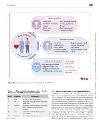
3.3. Symptoms attributable to AF
Symptoms related to episodes of AF are variable and broad, and not
just typical palpitations (Figure 1). Asymptomatic episodes of AF can
occur,30
although 90% of patients with AF describe symptoms with
variable severity.31
Even in symptomatic patients, some episodes of
AF may remain asymptomatic.32,33
The presence or absence of symp
toms is not related to incident stroke, systemic embolism, or mortal
ity.34
However, symptoms do impact on patient quality of life.35,36
Cardiac-specific AF symptoms such as palpitations are less common
than non-specific symptoms such as fatigue, but they significantly
impair quality of life.36,37
Although women are often underrepresented
in clinical trials of AF,38–40
the available literature suggests that
women with AF appear to be more symptomatic and have poorer
quality of life.41,42
Patients with AF report a higher burden of anxiety
and severity of depression (odds ratio [OR], 1.08; 95% confidence
interval [CI], 1.02–1.15; P = .009) as compared with the general
population,43,44
with higher prevalence of these symptoms in women
with AF.45
Assessment of AF-related symptoms should be recorded initially,
after a change in treatment, or before and after intervention. The
modified European Heart Rhythm Association score (mEHRA)
symptom classification (Table 7) is similar to the New York
Heart Association (NYHA) functional class for heart failure. It cor
relates with quality of life scores in clinical trials, is associated with
clinical progress and events, and may be a valuable starting point
in routine practice to assess the burden and impact of symptoms to
gether with the patient.46–48
Note that symptoms may also relate to
associated comorbidities and not just the AF component. The
patient-related effects of symptoms from AF over time can alterna
tively be evaluated using patient-reported outcome measures (see
Section 8.4).
3.4. Diagnostic evaluation of new AF
All patients with AF should be offered a comprehensive diagnostic as
sessment and review of medical history to identify risk factors and/or
comorbidities needing active treatment. Table 8 displays the essential
diagnostic work-up for a patient with AF.
A 12-lead ECG is warranted in all AF patients to confirm rhythm, de
termine ventricular rate, and look for signs of structural heart disease,
conduction defects, or ischaemia.56
Blood tests should be carried out
(kidney function, serum electrolytes, liver function, full blood count, glu
cose/glycated haemoglobin [HbA1c], and thyroid tests) to detect any
concomitant conditions that may exacerbate AF or increase the risk
of bleeding and/or thromboembolism.57,58
Other investigations will depend on individualized assessment and
the planned treatment strategy.59–65
A transthoracic echocardiogram
(TTE) should be carried out in the initial work-up, where this will guide
management decisions, or in patients where there is a change in cardio
vascular signs or symptoms. The task force recognizes that accessibility
to TTE might be limited or delayed in the primary care setting, but this
should not delay initiation of oral anticoagulation (OAC) or other com
ponents of AF-CARE where indicated.66
Further details on TTE and re
assessment (e.g. if elevated heart rate limits diagnostic imaging, or
where there is a change in clinical status) are presented in Section 8.3.
Additional imaging using different modalities may be required to assist
with comorbidity and AF-related management (see Supplementary
data online, Figure S1).
Recommendation Table 1 — Recommendations for the
diagnosis of AF (see also Evidence Table 1)
Recommendations Classa
Levelb
Confirmation by an electrocardiogram (12-lead,
multiple, or single leads) is recommended to establish
the diagnosis of clinical AF and commence risk
stratification and treatment.25–29
I A
©
ESC
2024
AF, atrial fibrillation.
a
Class of recommendation.
b
Level of evidence.
Recommendation Table 2 — Recommendations for
symptom evaluation in patients with AF (see also
Evidence Table 2)
Recommendations Classa
Levelb
Evaluating the impact of AF-related symptoms is
recommended before and after major changes in
treatment to inform shared decision-making and
guide treatment choices.17,36,46–55
I B
©
ESC
2024
AF, atrial fibrillation.
a
Class of recommendation.
b
Level of evidence.
Recommendation Table 3 — Recommendations for
diagnostic evaluation in patients with new AF (see also
Evidence Table 3)
Recommendations Classa
Levelb
A transthoracic echocardiogram is recommended in
patients with an AF diagnosis where this will guide
treatment decisions.59,65,67
I C
©
ESC
2024
AF, atrial fibrillation.
a
Class of recommendation.
b
Level of evidence.
3328 ESC Guidelines
Downloaded
from
https://academic.oup.com/eurheartj/article/45/36/3314/7738779
by
guest
on
21
October
2024](https://image.slidesharecdn.com/atriafibr24-241118112337-7c4f5251/85/ATRIAL-FIBRILLATION-2024-Guidelines-ESC-15-320.jpg)
![with AF.72,73
Patients with AF also have an increased risk of cognitive
impairment (adjusted hazard ratio [HR], 1.39; 95% CI, 1.25–1.53)74
and dementia (OR, 1.6; 95% CI, 1.3–2.0).75–77
It should be noted
that most of the observational studies on adverse events have a mix
of patients taking and not taking OAC. When carefully controlling
for the confounding effects of stroke, comorbidities, and OAC, AF ex
posure was still significantly associated with vascular dementia (HR,
1.68; 95% CI, 1.33–2.12; P < .001), but not Alzheimer’s disease (HR,
0.85; 95% CI, 0.70–1.03; P = .09).78
Hospital admission rates due to AF vary widely depending on the
population studied, and may be skewed by selection bias. In a Dutch
RCT including first-diagnosed AF patients (mean age 64 years), car
diovascular hospitalization rates were 7.0% to 9.4% per year.79
An
Australian study identified 473 501 hospitalizations for AF during
15 years of follow-up (300 million person-years), with a relative in
crease in AF hospitalizations of 203% over the study period, in con
trast to an increase for all hospitalizations of 71%. The age-specific
incidence of hospital admission increased particularly in the older
age groups.80
Atrial fibrillation is also associated with increased mortality. In
2017, AF contributed to over 250 000 deaths globally, with an
age-standardized mortality rate of 4.0 per 100 000 people (95% un
certainty interval 3.9–4.2).81
The most frequent cause of death in pa
tients with AF is heart failure related,70
with complex relationships to
cardiovascular and non-cardiovascular disease.82
There is up to a
two-fold increased risk of all-cause mortality (RR, 1.95; 95% CI,
1.50–2.54),68
and cardiovascular mortality (RR, 2.03; 95% CI, 1.79–
2.30)69
in AF compared with sinus rhythm. Even in the absence of
major thromboembolic risk factors, the incidence of death is 15.5
per 1000 person-years in those with AF exposure, compared
with 9.4 per 1000 person-years without (adjusted HR, 1.44; 95% CI,
1.38–1.50; P < .001).78
Patients with OAC-related bleeding have
higher mortality, including both minor and major bleeding (as
defined by the International Society on Thrombosis and
Haemostasis scale).83
Despite OAC, patients with AF remain at high
residual risk of death, highlighting the importance of attention to con
comitant disease.84
3.6. Atrial flutter
Atrial flutter (AFL) is the among the most common atrial tachyarrhyth
mias, with an overall incidence rate of 88 per 100 000 person-years, ris
ing to 317 per 100 000 person-years in people over 50 years of age.85
Risk factors for AFL and AF are similar, and more than half of all patients
with AFL will develop AF.85
Observational studies suggest that
thromboembolic risk is elevated in AFL.86
In direct comparison of
AFL with AF, some studies suggest a similar risk of stroke and others
a lower risk in AFL,87–90
possibly due to different comorbidity burdens
and the impact of confounders such as AFL/AF ablation and anticoagu
lation (more frequently stopped in AFL).91
4. Patient pathways and
management of AF
4.1. Patient-centred, multidisciplinary AF
management
4.1.1. The patient at the heart of care
A patient-centred and integrated approach to AF management means
working with a model of care that respects the patient’s experience,
values, needs, and preferences for planning, co-ordination, and delivery
of care. A central component of this model is the therapeutic relationship
between the patient and the multidisciplinary team of healthcare profes
sionals (Figure 2). In patient-centred AF management, patients are seen
not as passive recipients of health services, but as active participants
who work as partners alongside healthcare professionals. Patient-
centred AF management requires integration of all aspects of AF man
agement. This includes symptom control, lifestyle recommendations,
psychosocial support, and management of comorbidities, alongside op
timal medical treatment consisting of pharmacotherapy, cardioversion,
and interventional or surgical ablation (Table 9). Services should be de
signed to ensure that all patients have access to an organized model of
AF management, including tertiary care specialist services when indi
cated (see Supplementary data online, Table S1, Evidence Table 4 and
Additional Evidence Table S3). It is equally important to maintain path
ways for patients to promptly re-engage with specialist services when
their condition alters.
Table 8 Diagnostic work-up for patients with AF
All patients Selected patients
• Medical history to determine AF
pattern, relevant family history,
and comorbidities, and to assess
risk factors for thromboembolism
and bleeding
• Ambulatory ECG monitoring for
assessing AF burden and
ventricular rate control
• Exercise ECG to evaluate rate
control or effects of class IC
antiarrhythmic drugs
• 12-lead ECG • Further blood tests for
investigation of cardiovascular
disease and refinement of stroke/
bleeding risk (e.g. NT-proBNP,
troponin)
• Assess symptoms and functional
impairment
• Transoesophageal
echocardiography for left atrial
thrombus and valvular disease
assessment
• Collect generic or AF-specific
patient-reported outcome
measures
• Coronary CT, angiography, or
ischaemia imaging for suspected
CAD
• Blood tests (full blood count,
kidney function, serum
electrolytes, liver function,
glucose/HbA1c, and thyroid
function)
• CMR for evaluation of atrial and
ventricular cardiomyopathies,
and to plan interventional
procedures
• Transthoracic echocardiography
where this will guide AF-CARE
management decisions
• Brain imaging and cognitive
function assessment for
cerebrovascular disease and
dementia risk
©
ESC
2024
AF, atrial fibrillation; AF-CARE, atrial fibrillation—[C] Comorbidity and risk factor
management, [A] Avoid stroke and thromboembolism, [R] Reduce symptoms by rate
and rhythm control, [E] Evaluation and dynamic reassessment; CAD, coronary artery
disease; CMR, cardiac magnetic resonance; CT, computed tomography; CTA, computed
tomography angiography; ECG, electrocardiogram; HbA1c, glycated haemoglobin;
NT-proBNP, N-terminal pro-B-type natriuretic peptide.
3330 ESC Guidelines
Downloaded
from
https://academic.oup.com/eurheartj/article/45/36/3314/7738779
by
guest
on
21
October
2024](https://image.slidesharecdn.com/atriafibr24-241118112337-7c4f5251/85/ATRIAL-FIBRILLATION-2024-Guidelines-ESC-17-320.jpg)
![4.1.2. Education and shared decision-making
Clear advice about the rationale for treatments, the possibility of
treatment modification, and shared decision-making can help patients
live with AF (see Supplementary data online, Table S2).92
An open and
effective relationship between the patient and the healthcare profes
sional is critical, with shared decision-making found to improve
outcomes for OAC and arrhythmia management.93,94
In using a
shared approach, both the clinician and patient are involved in the
decision-making process (to the extent that the patient prefers).
Information is shared in both directions. Furthermore, both the
clinician and the patient express their preferences and discuss the
options. Of the potential treatment decisions, no treatment is
also a possibility.95
There are several toolkits available to facilitate
this, although most are focused on anticoagulation decisions. For ex
ample, the Shared Decision-Making Toolkit (http://afibguide.com,
http://afibguide.com/clinician) and the Successful Intravenous
Cardioversion for Atrial Fibrillation (SIC-AF) score have been shown
to reduce decisional conflict compared with usual care in patients
with AF.93,94
Patient-support organizations can also make an import
ant contribution to providing understandable and actionable knowl
edge about AF and its treatments (e.g. local support groups and
international charities, such as http://afa-international.org). As AF is a
chronic or recurrent disease in most patients, education is central
to empower patients, their families, and caregivers.
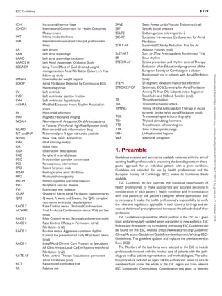
Atrial fibrillation
Patient-centred
C
A R
E
Comorbidity
and risk factor
management
Lifestyle help
Primary care
Cardiology
Internal medicine
Nursing care
Other
Avoid stroke and
thromboembolism
Primary care
Cardiology
Neurology
Nursing care
Anticoagulation
services
e-Health
Reduce symptoms
by rate and
rhythm control
Evaluation and
dynamic
reassessment
Primary care
Cardiology
Pharmacy
Nursing
Family/carers
e-Health
Primary care
Cardiology
Electrophysiology
Cardiac surgeons
e-Health
integrated AF-CARE
Figure 2 Multidisciplinary approach to AF management. Principal caregivers are involved in the community and hospital settings to provide optimal,
patient-centred care for patients living with AF. AF-CARE, atrial fibrillation—[C] Comorbidity and risk factor management, [A] Avoid stroke and
thromboembolism, [R] Reduce symptoms by rate and rhythm control, [E] Evaluation and dynamic reassessment.
Table 9 Achieving patient-centred AF management
Components of patient-centred AF management:
• Optimal treatment according to the AF-CARE pathway, which includes:
∘ [C] Comorbidity and risk factor management
∘ [A] Avoid stroke and thromboembolism
∘ [R] Reduce symptoms by rate and rhythm control
∘ [E] Evaluation and dynamic reassessment
• Lifestyle recommendations
• Psychosocial support
• Education and awareness for patients, family members, and caregivers
• Seamless co-ordination between primary care and specialized AF care
How to implement patient-centred AF management:
• Shared decision-making
• Multidisciplinary team approach
• Patient education and empowerment, with emphasis on self-care
• Structured educational programmes for healthcare professionals
• Technology support (e-Health, m-Health, telemedicine)a
©
ESC
2024
AF, atrial fibrillation; AF-CARE, atrial fibrillation—[C] Comorbidity and risk factor
management, [A] Avoid stroke and thromboembolism, [R] Reduce symptoms by rate
and rhythm control, [E] Evaluation and dynamic reassessment.
a
e-Health refers to healthcare services provided using electronic methods; m-Health,
refers to healthcare services supported by mobile devices; and telemedicine refers to
remote diagnosis or treatment supported by telecommunications technology.
ESC Guidelines 3331
Downloaded
from
https://academic.oup.com/eurheartj/article/45/36/3314/7738779
by
guest
on
21
October
2024](https://image.slidesharecdn.com/atriafibr24-241118112337-7c4f5251/85/ATRIAL-FIBRILLATION-2024-Guidelines-ESC-18-320.jpg)
![4.1.3. Education of healthcare professionals
Gaps in knowledge and skills across all domains of AF care are consist
ently described among cardiologists, neurologists, internal medicine
specialists, emergency physicians, general practitioners, nurses, and al
lied health practitioners.96–98
Healthcare professionals involved in
multidisciplinary AF management should have a knowledge of all avail
able options for diagnosis and treatment.99–101
In the STEEER-AF
trial,99
real-world adherence to clinical practice guidelines for AF
across six ESC countries was poor. These findings highlight the critical
need for appropriate training and education of healthcare
professionals.102
Specifically targeted education for healthcare professionals can in
crease knowledge and lead to more appropriate use of OAC for
prevention of thromboembolism.103
However, educational interventions
for healthcare providers are often not enough to sustainably impact be
haviour.104
Other tools may be needed, such as active feedback,103
clinical decision support tools,105
expert consultation,106
or e-Health
learning.107
4.1.4. Inclusive management of AF
Evidence is growing on differences in AF incidence, prevalence, risk fac
tors, comorbidities, and outcomes according to gender.108
Women di
agnosed with AF are generally older, have more hypertension and heart
failure with preserved ejection fraction (HFpEF), and have less diag
nosed coronary artery disease (CAD).109
Registry studies have re
ported differences in outcomes, with higher morbidity and mortality
in women, although these may be confounded by age and comorbidity
burden.110–112
Women with AF may be more symptomatic, and report
a lower quality of life.41,113
It is unclear whether this is related to de
layed medical assessment in women, or whether there are genuine
sex differences. Despite a higher symptom load, women are less likely
to undergo AF ablation than men, even though antiarrhythmic drug
therapy seems to be associated with more proarrhythmic events in
women.109
These observations call for more research on gender
differences in order to prevent disparities and inequality in care.
Other diversity aspects such as age, race, ethnicity, and transgender
issues, as well as social determinants (including socioeconomic status,
disability, education level, health literacy, and rural/urban location) are
important contributors to inequality that should be actively considered
to improve patient outcomes.114
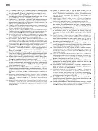
4.2. Principles of AF-CARE
The 2024 ESC Guidelines for the management of AF have compiled and
evolved past approaches to create principles of management to aid im
plementation of these guidelines, and hence improve patient care and
outcomes. There is growing evidence that clinical support tools115–118
can aid best-practice management, with the caveat that any tool is a
guide only, and that all patients require personalized attention. The
AF-CARE approach covers many established principles in the manage
ment of AF, but does so in a systematic, time-orientated format with
four essential treatment pillars (Figure 3; central illustration). Joint man
agement with each patient forms the starting point of the AF-CARE ap
proach. Notably, it takes account of the growing evidence base that
therapies for AF are most effective when associated health conditions
are addressed. A careful search for these comorbidities and risk factors
[C] is critical and should be applied in all patients with a diagnosis of AF.
Avoidance of stroke and thromboembolism [A] in patients with risk
factors is considered next, focused on appropriate use of anticoagu
lant therapy. Reducing AF-related symptoms and morbidity by effect
ive use of heart rate and rhythm control [R] is then applied, which in
selected patients may also reduce hospitalization or improve progno
sis. The potential benefit of rhythm control, accompanied by consid
eration of all risks involved, should be considered in all patients at each
contact point with healthcare professionals. As AF, and its related co
morbidities, changes over time, different levels of evaluation [E] and
re-evaluation are required in each patient, and these approaches
should be dynamic. Due to the wide variability in response to therapy,
and the changing pathophysiology of AF as age and comorbidities ad
vance, reassessment should be built into the standard care pathway to
prevent adverse outcomes for patients and improve population
health.
AF-CARE builds upon prior ESC Guidelines, e.g. the five-step
outcome-focused integrated approach in the 2016 ESC Guidelines for
the management of AF,119
and the AF Better Care (ABC) pathway in
the 2020 ESC Guidelines for the diagnosis and management of AF.120
The reorganization into AF-CARE was based on the parallel develop
ments in new approaches and technologies (in particular for rhythm
control), with new evidence consistently suggesting that all aspects of
AF management are more effective when comorbidities and risk factors
have been considered. This includes management relating to symptom
benefit, improving prognosis, prevention of thromboembolism, and the
response to rate and rhythm control strategies. AF-CARE makes expli
cit the need for individualized evaluation and follow-up in every patient,
with an active approach that accounts for how patients, their AF, and
associated comorbidities change over time. The AF-CARE principles
have been applied to different patient pathways for ease of implemen
tation into routine clinical care. This includes the management of first-
diagnosed AF (Figure 4), paroxysmal AF (Figure 5), persistent AF
(Figure 6), and permanent AF (Figure 7).
Recommendation Table 4 — Recommendations for
patient-centred care and education (see also Evidence
Table 4)
Recommendation Classa
Levelb
Education directed to patients, family members,
caregivers, and healthcare professionals is
recommended to optimize shared decision-making,
facilitating open discussion of both the benefit and
risk associated with each treatment option.94,103
I C
Access to patient-centred management according to
the AF-CARE principles is recommended in all
patients with AF, regardless of gender, ethnicity, and
socioeconomic status, to ensure equality in
healthcare provision and improve outcomes.
I C
Patient-centred AF management with a
multidisciplinary approach should be considered in all
patients with AF to optimize management and
improve outcomes.79,121–124
IIa B
©
ESC
2024
AF, atrial fibrillation; AF-CARE, Atrial fibrillation—[C] Comorbidity and risk factor
management, [A] Avoid stroke and thromboembolism, [R] Reduce symptoms by rate
and rhythm control, [E] Evaluation and dynamic reassessment.
a
Class of recommendation.
b
Level of evidence.
3332 ESC Guidelines
Downloaded
from
https://academic.oup.com/eurheartj/article/45/36/3314/7738779
by
guest
on
21
October
2024](https://image.slidesharecdn.com/atriafibr24-241118112337-7c4f5251/85/ATRIAL-FIBRILLATION-2024-Guidelines-ESC-19-320.jpg)
![C
Comorbidity and risk factor management
Hypertension
Diabetes
mellitus
Blood pressure
lowering treatment
(Class I)
Patient-centred AF management with a multidisciplinary approach (Class IIa)
Education for patients, families and healthcare professionals (Class I)
Equality in healthcare provision (gender, ethnicity, socioeconomic) (Class I)
Heart failure
Diuretics for
congestion
(Class I)
Overweight
or obese
Weight loss
(target 10%)a
(Class I)
Obstructive sleep
apnoea
Management
of OSAa
(Class IIb)
Appropriate HFrEF
medical therapy
(Class I)
Bariatric surgery
if rhythm controla
(Class IIb)
SGLT2 inhibitors
(Class 1)
Alcohol
Reduce to �3
drinks per week
(Class I)
Effective
glycaemic controla
(Class I)
Exercise
capacity
Tailored
exercise programme
(Class I)
Other risk factors/
comorbidities
Identify and manage
aggressivelya
(Class I)
A
Avoid stroke and thromboembolism
Risk of
thrombo-
embolism
Start oral
anticoagulation
(Class I)
Use locally-validated
risk score
or CHA2
DS2
-VA
OAC if CHA2
DS2
-VA
score = 2 or more
(Class I)
Choice of
anticoagulant
Assess
bleeding risk
OAC if CHA2
DS2
-VA
score = 1
(Class IIa)
Use DOAC, except
mechanical valve or
mitral stenosis
(Class I)
Target INR 2.0–3.0;
(Class I)
>70% INR range;
(Class IIa)
or switch to DOAC
(Class I)
Assess and manage
all modifiable risk
factors for bleeding
(Class I)
Do not use risk
scores to withhold
anticoagulation
(Class III)
Do not combine
antiplatelets and OAC
for stroke prevention
(Class III)
Avoid antiplatelets
beyond 12 months
in OAC treated
CCS/PVD
(Class III)
Temporal pattern
of AF not relevant
(Class III)
Antiplatelet therapy
not an alternative
(Class III)
Prevent
bleeding
R
Reduce symptoms by rate and rhythm control
E
Evaluation and dynamic reassessment
See patient pathways for:
First-diagnosed AF Paroxysmal AF Persistent AF Permanent AF
Consider:
AF
C A R E
If VKA:
Rate control drugs Ablate and pace
Cardioversion Catheter ablation Endoscopic/hybrid ablation Surgical ablation
Antiarrhythmic drugs
Re-evaluate when AF episodes or non-AF admissions
ECG, blood tests,
cardiac imaging,
ambulatory ECG,
other imaging
as needed
Assess new and
existing risk factors
and comorbidities
(Class I)
Stratify risk
for stroke and
thromboembolism
(Class I)
Check impact of AF
symptoms before
and after treatment
(Class I)
Assess and manage
modifiable bleeding
risk factors
(Class I)
Continue OAC
despite rhythm
control if risk
of thromboembolism
(Class I)
Regular re-evaluation: 6 months after presentation, and then at least annually or based on clinical need
Figure 3 Central illustration. Patient pathway for AF-CARE (see Figures 4, 5, 6, and 7 for the [R] pathways for first-diagnosed, paroxysmal, persistent and
permanent AF). AF, atrial fibrillation; AF-CARE, atrial fibrillation—[C] Comorbidity and risk factor management, [A] Avoid stroke and thromboembolism,
[R] Reduce symptoms by rate and rhythm control, [E] Evaluation and dynamic reassessment; CCS, chronic coronary syndrome; CHA2DS2-VA, congest
ive heart failure, hypertension, age ≥75 years (2 points), diabetes mellitus, prior stroke/transient ischaemic attack/arterial thromboembolism (2 points),
vascular disease, age 65–74 years; DOAC, direct oral anticoagulant; ECG, electrocardiogram; HFrEF, heart failure with reduced ejection fraction; INR,
international normalized ratio of prothrombin time; OAC, oral anticoagulant; OSA, obstructive sleep apnoea; PVD, peripheral vascular disease; SGLT2,
sodium-glucose cotransporter-2; VKA, vitamin K antagonist. a
As part of a comprehensive management of cardiometabolic risk factors.
ESC Guidelines 3333
Downloaded
from
https://academic.oup.com/eurheartj/article/45/36/3314/7738779
by
guest
on
21
October
2024](https://image.slidesharecdn.com/atriafibr24-241118112337-7c4f5251/85/ATRIAL-FIBRILLATION-2024-Guidelines-ESC-20-320.jpg)
![N
Patient with first-diagnosed AF
Y
Y N
Follow AF-CARE for [C] comorbidity and risk factor management & [A] avoid stroke and thromboembolism
Haemodynamically stable
Initial rate control
(Class I)
Electrical cardioversion
(Class I)
LVEF ≤40%
Beta-blocker, digoxin,
diltiazem or verapamil
(Class I)
Beta-blocker
or digoxin
(Class I)
Combination
rate control therapy
(Class IIa)
Combination
rate control therapy
(Class IIa)
Cardioversion of symptomatic
persistent AF
(Class I)
Wait-and-see if sinus rhythm
restores spontaneously <48 h
(Class IIa)
Figure 4 [R] Pathway for patients with first-diagnosed AF. AF, atrial fibrillation; AF-CARE, Atrial fibrillation—[C] Comorbidity and risk factor man
agement, [A] Avoid stroke and thromboembolism, [R] Reduce symptoms by rate and rhythm control, [E] Evaluation and dynamic reassessment; LVEF,
left ventricular ejection fraction. After following the pathway for first-diagnosed AF, patients with recurrent AF should enter the AF-CARE [R] pathway
for paroxysmal, persistent, or permanent AF, depending on the type of their AF.
3334 ESC Guidelines
Downloaded
from
https://academic.oup.com/eurheartj/article/45/36/3314/7738779
by
guest
on
21
October
2024](https://image.slidesharecdn.com/atriafibr24-241118112337-7c4f5251/85/ATRIAL-FIBRILLATION-2024-Guidelines-ESC-21-320.jpg)
![Rate control target = resting heart rate <110 b.p.m. (lenient control),
with stricter control with continuing symptoms
(Class IIa)
Beta-blocker
or digoxin
(Class I)
Patient with paroxysmal AF
LVEF ≤40%
Beta-blocker, digoxin,
diltiazem or verapamil
(Class I)
Combination
rate control therapy
(Class IIa)
Combination
rate control therapy
(Class IIa)
Amiodarone
(Class I)
Dronedarone, flecainide
or propafenone
(Class I)
Catheter ablation
(Class I)
Amiodarone or
dronedarone
(Class I)
Sotalol
(Class IIb)
HFrEF
(LVEF ≤40%)
Stable HFmrEF
(LVEF 41–49%),
coronary heart disease,
valvular heart disease
Absence
or minimal heart
disease
Sotalol
(Class IIb)
Recurrence of AF symptoms
Antiarrhythmic drug therapy
Y N
Shared decision-making on rhythm control
(Class I)
If failed antiarrhythmic
drug therapy
If failed catheter
ablation
Re-do catheter ablation
(Class IIa)
Surgical/hybrid ablation
(Class IIb)
Antiarrhythmic drug
therapy (see above)
Catheter ablationa
(Class I)
Shared decision-making
(Class I)
Follow AF-CARE for [C] comorbidity and risk factor management & [A] avoid stroke and thromboembolism
Figure 5 [R] Pathway for patients with paroxysmal AF. AF, atrial fibrillation; AF-CARE, atrial fibrillation—[C] Comorbidity and risk factor manage
ment, [A] Avoid stroke and thromboembolism, [R] Reduce symptoms by rate and rhythm control, [E] Evaluation and dynamic reassessment; b.p.m.,
beats per minute; HFmrEF, heart failure with mildly reduced ejection fraction; HFrEF, Heart failure with reduced ejection fraction; LVEF, left ventricular
ejection fraction. a
In patients with HFrEF: Class I if high probability of tachycardia-induced cardiomyopathy; and Class IIa in selected patients to improve
prognosis.
ESC Guidelines 3335
Downloaded
from
https://academic.oup.com/eurheartj/article/45/36/3314/7738779
by
guest
on
21
October
2024](https://image.slidesharecdn.com/atriafibr24-241118112337-7c4f5251/85/ATRIAL-FIBRILLATION-2024-Guidelines-ESC-22-320.jpg)
![Electrical
cardioversion
Rate control target = resting heart rate <110 b.p.m. (lenient control),
with stricter control with continuing symptoms
(Class IIa)
Beta-blocker
or digoxin
(Class I)
Patient with persistent AF
LVEF ≤40%
Combination
rate control therapy
(Class IIa)
Combination
rate control therapy
(Class IIa)
Amiodarone
(Class I)
Dronedarone, flecainide
or propafenone
(Class I)
Amiodarone or
dronedarone
(Class I)
Sotalol
(Class IIb)
HFrEF
(LVEF ≤40%)
Stable HFmrEF
(LVEF 41–49%),
coronary heart disease,
valvular heart disease
Absence
or minimal heart
disease
Sotalol
(Class IIb)
Recurrence of AF symptoms
Antiarrhythmic drug therapy
Y N
Shared decision-making on rhythm control
(Class I)
If failed antiarrhythmic
drug therapy
If failed catheter
ablation
Catheter ablationa
(Class IIb)
Shared decision-making, considering all rhythm control options
(Class I)
Beta-blocker, digoxin,
diltiazem or verapamil
(Class I)
Catheter
ablation
(Class I)
Re-do
catheter
ablation
Endoscopic
hybrid or
surgical ablation
Antiarrhythmic
drug therapy
(see above)
Consider
rate control
strategy
Follow AF-CARE for [C] comorbidity and risk factor management & [A] avoid stroke and thromboembolism
Haemodynamic instability (Class I)
Part of rhythm control strategy (Class IIa)
Clarify benefit from sinus rhythm (Class IIa)
Endoscopic/
hybrid ablation
(Class IIa)
Figure 6 [R] Pathway for patients with persistent AF. AF, atrial fibrillation; AF-CARE, Atrial fibrillation—[C] Comorbidity and risk factor management,
[A] Avoid stroke and thromboembolism, [R] Reduce symptoms by rate and rhythm control, [E] Evaluation and dynamic reassessment; b.p.m., beats per
minute; HFmrEF, heart failure with mildly reduced ejection fraction; HFrEF, heart failure with reduced ejection fraction; LVEF, left ventricular ejection
fraction. a
In patients with HFrEF: Class I if high probability of tachycardia-induced cardiomyopathy; and Class IIa in selected patients to improve prognosis.
3336 ESC Guidelines
Downloaded
from
https://academic.oup.com/eurheartj/article/45/36/3314/7738779
by
guest
on
21
October
2024](https://image.slidesharecdn.com/atriafibr24-241118112337-7c4f5251/85/ATRIAL-FIBRILLATION-2024-Guidelines-ESC-23-320.jpg)
![LVEF >40%
Patient with permanent AF
Y
Evaluation and dynamic
reassessment
Initiate beta-blocker,
digoxin, diltiazem
or verapamil
(Class I)
Continue beta-blocker,
digoxin, diltiazem
or verapamil
(Class I)
Combination beta-blocker
with digoxin, or diltiazem/
verapamil with digoxin;
avoiding bradycardia
(Class IIa)a
N
Rate control target = resting heart rate <110 b.p.m. (lenient control), with stricter control with continuing symptoms
(Class IIa)
Continue review and follow-up as per
AF-CARE approach
LVEF ≤40%
Continue beta-blocker
or digoxin
(Class I)
Combination
beta-blocker with digoxin,
avoiding bradycardia
(Class IIa)
Y
Evaluation and dynamic
reassessment
Initiate beta-blocker
or digoxin
(Class I)
Rate control target = resting
heart rate <110 b.p.m.
(lenient control),
with stricter control
with continuing symptoms
(Class IIa)
N
Severely symptomatic and
HF hospitalization
Atrioventricular node
ablation and CRT
(Class IIa)
Y
Intensify rate control therapy under
observation
Evaluation for atrioventricular node
ablation in combination with pacemaker
(Class IIa)
N
Follow AF-CARE for [C] comorbidity and risk factor management & [A] avoid stroke and thromboembolism
Rate control target = resting
heart rate <110 b.p.m.
(lenient control),
with stricter control
with continuing symptoms
(Class IIa)
Figure 7 [R] Pathway for patients with permanent AF. AF, atrial fibrillation; AF-CARE, Atrial fibrillation—[C] Comorbidity and risk factor manage
ment, [A] Avoid stroke and thromboembolism, [R] Reduce symptoms by rate and rhythm control, [E] Evaluation and dynamic reassessment; b.p.m.,
beats per minute; CRT, cardiac resynchronization therapy; HF, heart failure; LVEF, left ventricular ejection fraction. Permanent AF is a shared decision
made between the patient and physician that no further attempts at restoration of sinus rhythm are planned. a
Note that the combination of beta-
blockers with diltiazem or verapamil should only be used under specialist advice, and monitored with an ambulatory ECG to check for bradycardia.
ESC Guidelines 3337
Downloaded
from
https://academic.oup.com/eurheartj/article/45/36/3314/7738779
by
guest
on
21
October
2024](https://image.slidesharecdn.com/atriafibr24-241118112337-7c4f5251/85/ATRIAL-FIBRILLATION-2024-Guidelines-ESC-24-320.jpg)
![5. [C] Comorbidity and risk factor
management
A broad array of comorbidities are associated with the recurrence
and progression of AF. Managing comorbidities is also central to the
success of other aspects of care for patients with AF, with evidence
available for hypertension, heart failure, diabetes mellitus, obesity,
and sleep apnoea, along with lifestyle changes that improve physical
activity and reduce alcohol intake (see Supplementary data online,
Additional Evidence Table S4). Identification and treatment of these
comorbidities and clusters of risk factors form an important part
of effective AF-CARE (Figure 8), with the evidence outlined in the
rest of this section highlighting where management can improve
patient outcomes or prevent AF recurrence. Many of these
factors (and more) are also associated with incident AF (see
Section 10).
Focus on key
risk factors
Provide information
without overloading
Suggested approach and targets
Setting individual
targets for
comorbidities
and risk factors
Key targets
Behavioural
change
Achievable
targets
Identify and actively manage all risk factors and comorbidities
(Class I)
Blood pressure treatment with target 120–129 mmHg /
70–79 mmHg in most adults (or as low as reasonably achievable)
(Class I)
Effective glycaemic control with diet/medication(s)
(Class I)
Weight loss programme if overweight /obese,
with 10% or more weight loss
(Class I)
Management of obstructive sleep apnoea to minimize
apnoeic episodes
(Class IIb)
Tailored exercise programme aiming for regular
moderate/vigorous activity
(Class I)
Reduce alcohol consumption to 3 or less standard
drinks per week
(Class I)
Shared
decision-making
Hypertension
Sleep
apnoea
Heart
failure
Diabetes
Obesity
Integrated
management
Physical
activity
Alcohol
intake
Optimize with diuretics to alleviate congestion appropriate,
medical therapy for reduced LVEF, and SGLT2 inhibitors for all LVEF
(Class I)
Figure 8 Management of key comorbidities to reduce AF recurrence. LVEF, left ventricular ejection fraction; SGLT2, sodium-glucose
cotransporter-2.
3338 ESC Guidelines
Downloaded
from
https://academic.oup.com/eurheartj/article/45/36/3314/7738779
by
guest
on
21
October
2024](https://image.slidesharecdn.com/atriafibr24-241118112337-7c4f5251/85/ATRIAL-FIBRILLATION-2024-Guidelines-ESC-25-320.jpg)
![difference between beta-blockers and placebo for all-cause mortality in
HFrEF with AF.133
However, these drugs have clear proof of safety and
there may be other indications for these therapies beyond prognosis,
including comorbidity management and symptom improvement.
These and other therapies may also have dual functions, for example,
beta-blockers or digoxin for rate control of AF, in addition to improving
heart failure metrics and reducing hospitalization.48,185,186
More recent
additions to HFrEF management, such as eplerenone, sacubitril-
valsartan, and sodium-glucose cotransporter-2 (SGLT2) inhibitors,
had substantial numbers of patients with AF enrolled in RCTs, with
no evidence that AF status affected their ability to reduce cardiovascu
lar mortality/heart failure hospitalization.134–136
Cardiac resynchroniza
tion therapy (CRT) in the context of HFrEF and AF is discussed in detail
in the 2021 ESC Guidelines on cardiac pacing and cardiac resynchroni
zation therapy, with an important focus on ensuring effective biventri
cular pacing (with a low threshold for considering atrioventricular node
ablation).187
Patients who have heart failure with mildly reduced ejec
tion fraction (HFmrEF) (LVEF 41%–49%) and AF should generally be
treated according to guidance for HFrEF,137
albeit with limited evidence
to date in AF.188–190
For treatment of HFpEF and AF,191
pre-specified
subgroup data on AF from multiple large trials show that the SGLT2
inhibitors dapagliflozin, empaglifozin, and sotagliflozin are effective in
improving prognosis.138–140
Appropriate management of heart failure has the potential to reduce
recurrence of AF, e.g. by reducing adverse atrial and ventricular myo
cardial remodelling, but there are limited data for specific therapies.
In the Routine versus Aggressive upstream rhythm Control for preven
tion of Early AF in heart failure (RACE 3) trial, combined management
of mild-to-moderate heart failure with ACE inhibitors/ARBs, mineralo
corticoid receptor antagonists, statins, and cardiac rehabilitation in
creased the maintenance of sinus rhythm on ambulatory monitoring
at 12 months.39
This benefit was not preserved at the 5 year follow-up,
although this may have been confounded by the lack of ongoing inter
vention beyond the initial 12 months.192
5.3. Type 2 diabetes mellitus
Diabetes mellitus is present in around 25% of patients with AF.193–195
Patients with both diabetes and AF have a worse prognosis,196
with in
creased healthcare utilization and excess mortality and cardiovascular
events. The prevalence and incidence of AF and type 2 diabetes are
widely increasing, thus making the association of these two conditions
a public health challenge.195,197
Moreover, diabetes is a major factor in
fluencing thromboembolic risk.198,199
Following catheter ablation of AF,
diabetes and higher HbA1c are associated with increased length of stay
and a greater recurrence of AF.200–203
In cohort studies, the management of diabetes mellitus as part of
comprehensive risk factor management has been associated with re
duced AF symptoms, burden, reversal of the type of AF (from persist
ent to paroxysmal or no AF), and improved maintenance of sinus
rhythm.126–128
However, robust evidence is limited, and individual
glucose-lowering medications have had variable effects on AF.204–206
There are emerging data of the use of SGLT2 and glucagon-like
peptide-1 antagonists in patients with diabetes and AF that may impact
on treatment choice in the near future. Importantly, diabetes frequently
coexists with multiple risk factors in patients with AF, and a compre
hensive approach to management is required. Further details are
provided in the 2023 ESC Guidelines for the management of cardiovas
cular disease in patients with diabetes.207
5.4. Obesity
Obesity frequently coexists with other risk factors that have been inde
pendently associated with the development of AF.208,209
Obesity (body
mass index [BMI] ≥30 kg/m2
) and being overweight (BMI >25 kg/m2
)
are associated with a greater risk of recurrent atrial arrhythmias after
AF ablation (13% increase for every 5 kg/m2
higher BMI).210–212
In
the setting of comprehensive risk factor management, weight loss of
≥10% in overweight and obese individuals with AF has been associated
with reduced AF symptoms and AF burden in an RCT (aiming for BMI
<27 kg/m2
).125
Cohort studies have also shown a graded response to
maintenance of sinus rhythm,126
improved ablation outcomes,128
and
reversal of the type of AF127
commensurate with the degree of weight
loss and risk factor management. However, in the Supervised Obesity
Reduction Trial for AF Ablation Patients (SORT-AF) randomized trial
in AF ablation patients, a sole weight loss intervention that achieved
4% loss in weight over 12 months did not impact ablation outcomes.213
This is consistent with the findings in LEGACY (Long-Term Effect of
Goal directed weight management on Atrial Fibrillation Cohort: a 5
Year follow-up study) that showed that weight loss of ≤3% had no im
pact on AF recurrence.126
Observational studies have raised the possi
bility of a point of no return in terms of the benefit of weight loss,214
but
also the possibility that bariatric surgery can improve symptoms and re
duce AF recurrence.215–217
5.5. Obstructive sleep apnoea
Obstructive sleep apnoea (OSA) is a highly prevalent condition, particu
larly in patients with AF.157,218
Optimal screening tools in the AF popu
lation are still under evaluation, although it may be reasonable to screen
for OSA in patients where a rhythm control strategy is being pursued.
Polysomnography or home sleep apnoea testing are suggested in pref
erence to screening questionnaires.155–157,219
Questionnaires assessing
daytime sleepiness are poor predictors of moderate-to-severe OSA.155
Which parameter should be used to focus on risk of AF in patients with
OSA, and to guide OSA treatment in patients with AF, is still
unclear.220,221
Observational studies have suggested that individuals with OSA not
treated with continuous positive airway pressure (CPAP) respond
poorly to treatments for AF, with an increased risk of recurrence
after cardioversion or ablation.222
Conversely, OSA patients treated
with CPAP seem to mitigate their propensity toward developing
AF.148–153,222–224
A small randomized trial of CPAP vs. no therapy de
monstrated reversal of atrial remodelling in individuals with moderate
OSA.154
However, other small RCTs have failed to show a benefit of
CPAP therapy on ablation outcomes225
or post-cardioversion.226
Data on the cardiovascular mortality benefit of CPAP therapy in
OSA are inconclusive.227–230
5.6. Physical inactivity
Reduced cardiorespiratory fitness frequently coexists with other modi
fiable risk factors and has been associated with a greater recurrence of
AF after catheter ablation.141
Better cardiorespiratory fitness has a de
monstrated inverse relationship to AF burden in both middle-aged and
elderly people.141
Small RCTs, meta-analyses, and observational
3340 ESC Guidelines
Downloaded
from
https://academic.oup.com/eurheartj/article/45/36/3314/7738779
by
guest
on
21
October
2024](https://image.slidesharecdn.com/atriafibr24-241118112337-7c4f5251/85/ATRIAL-FIBRILLATION-2024-Guidelines-ESC-27-320.jpg)
![cohorts have shown that regular aerobic exercise may also improve
AF-related symptoms, quality of life, and exercise capacity.142,143
Better cardiorespiratory fitness and a gain in cardiorespiratory fitness
over time are associated with a greater reduction in AF burden and im
proved maintenance of sinus rhythm.141–145
5.7. Alcohol excess
Alcohol consumption can increase the risk of adverse events in patients
with AF, such as thromboembolism, death, or AF-related hospitaliza
tion.231,232
Alcohol is associated with an increased risk of ischaemic
stroke in patients with newly diagnosed AF, and alcohol abstinence after
AF diagnosis can reduce the risk of ischaemic stroke.233
In patients re
ceiving OAC, alcohol excess is associated with a greater risk of bleed
ing,234
mediated by poor adherence, alcohol–drug interactions, liver
disease, and variceal bleeding.
Alcohol consumption is associated with a dose-dependent increase
in the recurrence of AF after catheter ablation.147,235
In an RCT among
regular non-binge drinkers with AF, the goal of abstinence led to a sig
nificant reduction in AF recurrence and burden; alcohol intake was re
duced from 16.8 to 2.1 standard drinks per week (≤30 grams or 3
standard drinks of alcohol) in the intervention arm, with 61% attaining
abstinence.147
In observational data of patients undergoing catheter ab
lation, reduction of consumption to ≤7 standard drinks (≤70 grams of
alcohol) per week was associated with improved maintenance of sinus
rhythm.128,235
6. [A] Avoid stroke and
thromboembolism
6.1. Initiating oral anticoagulation
Atrial fibrillation is a major risk factor for thromboembolism, irre
spective of whether it is paroxysmal, persistent, or permanent.236,237
Left untreated, and dependent on other patient-specific factors, the
risk of ischaemic stroke in AF is increased five-fold, and one in every
five strokes is associated with AF.238
The default approach should
therefore be to provide OAC to all eligible patients, except those
at low risk of incident stroke or thromboembolism. The effectiveness
of OAC to prevent ischaemic stroke in patients with AF is well estab
lished.239,240
Antiplatelet drugs alone (aspirin, or aspirin in combin
ation with clopidogrel) are not recommended for stroke prevention
in AF.241,242
6.1.1. Decision support for anticoagulation in AF
Tools have been developed to enable easier implementation of OAC
in patients with clinical AF. The majority of OAC clinical trials have
used variations of the CHADS2 score to indicate those at risk (with
points for chronic heart failure, hypertension, age, diabetes, and 2
points for prior stroke/transient ischaemic attack [TIA]). Although
most available stroke risk scores are simple and practical, the predict
ive value of scores is generally modest (see Supplementary data
online, Table S3).243–245
Classification and discrimination of adverse
events is relatively poor for all scores and hence the benefit of using
them to select patients for OAC is unclear. There is also considerable
variation in the definition of risk factors across countries,246
and a lack
of evidence from clinical trials on the ability of stroke risk scoring to
enhance clinical practice.243
This guideline continues to provide a
Class IA recommendation for the use of OAC in patients at risk of
thromboembolism. However, in the absence of strong evidence for
how to apply risk scores in real-world patients, this has been sepa
rated from the use of any particular risk score. This is also in line
with regulatory approvals for direct oral anticoagulants (DOACs),
which do not stipulate risk scores or numerical thresholds.25–28,245
Substantive changes have occurred in the decades since these risk
scores were developed in regards to population-level risk factor pro
files, therapies, and targets.198
Historical scores do not take into ac
count parameters that have been associated with thromboembolism
in contemporary cohorts, such as cancer, chronic kidney disease
(CKD), ethnicity, and a range of circulating biomarkers (including tropo
nin and B-type natriuretic peptide [BNP]). As an example, for CKD
there is a correlation between decreasing glomerular filtration rate
and proteinuria with stroke risk,247–250
and cohort data suggest a two-
fold increased risk of ischaemic stroke and mortality in AF patients with
CKD vs. without.251
Other factors, such as atrial enlargement, hyperlip
idaemia, smoking, and obesity, have been identified in specific cohort
studies as additional risk factors for ischaemic stroke in AF.70,252,253
Biomarkers, such as troponin, natriuretic peptides, growth differenti
ation factor-15, cystatin C, and interleukin-6, can also indicate residual
stroke risk among anticoagulated AF patients.254,255
Biomarker-guided
stroke prevention is currently being evaluated in an ongoing RCT
(NCT03753490). Until further validation within RCTs is available, this
task force continues to support using simple clinical classification for im
plementation of OAC. Clinicians should use tools that have been vali
dated in their local population and take an individualized approach to
thromboembolic risk stratification that considers the full range of
each patient’s specific risk factors. The absolute risk level at which to
start OAC in individual patients cannot be estimated from population-
level studies. It will vary depending on how those factors interact with
other medical issues, and the degree of risk acceptable or tolerated by
that person. In general, most of the available risk scores have a thresh
old of 0.6%–1.0% per annum of thromboembolic events for clinical AF
to warrant OAC prescription.
Across Europe, the most popular risk score is CHA2DS2–VASc, giv
ing points for congestive heart failure, hypertension, age ≥75 years
(2 points), diabetes mellitus, prior stroke/TIA/thromboembolism
(2 points), vascular disease, age 65–74 years and female sex.
However, implementation has varied in terms of gender. Female sex
is an age-dependent stroke risk modifier rather than a risk factor per
se.112,256,257
The inclusion of gender complicates clinical practice both
for healthcare professionals and patients.258
It also omits individuals
who identify as non-binary, transgender, or are undergoing sex hor
mone therapy. Previous guidelines from the ESC (and globally) have
not actually used CHA2DS2-VASc; instead providing different score le
vels for women and men with AF to qualify for OAC. Hence,
CHA2DS2-VA (excluding gender) has effectively been in place
(Table 10).78
This task force proposes, in the absence of other locally
validated alternatives, that clinicians and patients should use the
CHA2DS2-VA score to assist in decisions on OAC therapy (i.e. without
a criterion for birth sex or gender). Pending further trials in lower risk
patients (NCT04700826,259
NCT02387229260
), OAC are recom
mended in those with a CHA2DS2-VA score of 2 or more and should
be considered in those with a CHA2DS2-VA score of 1, following a
patient-centred and shared care approach. Healthcare professionals
should take care to assess for other thromboembolic risk factors
that may also indicate the need for OAC prescription.
ESC Guidelines 3341
Downloaded
from
https://academic.oup.com/eurheartj/article/45/36/3314/7738779
by
guest
on
21
October
2024](https://image.slidesharecdn.com/atriafibr24-241118112337-7c4f5251/85/ATRIAL-FIBRILLATION-2024-Guidelines-ESC-28-320.jpg)
![6.2.1. Direct oral anticoagulants
The DOACs (apixaban, dabigatran, edoxaban, and rivaroxaban) have all
demonstrated at least non-inferior efficacy compared with warfarin for
the prevention of thromboembolism, but with the added benefit of a
50% reduction in intracranial haemorrhage (ICH).25–28
Meta-analyses of
individual data from 71 683 RCT patients showed that standard, full-dose
DOAC treatment compared with warfarin reduces the risk of stroke or
systemic embolism (HR, 0.81; 95% CI, 0.73–0.91), all-cause mortality (HR,
0.90; 95% CI, 0.85–0.95), and intracranial bleeding (HR, 0.48; 95% CI,
0.39–0.59), with no significant difference in other major bleeding (HR,
0.86; 95% CI, 0.73–1.00) and little or no between-trial heterogeneity.292
Post-marketing observational data on the effectiveness and safety of
dabigatran,313,314
rivaroxaban,315,316
apixaban,317
and edoxaban318
vs.
warfarin show general consistency with the respective phase 3 RCTs.
For patients undergoing cardioversion, three underpowered trials
showed non-significantly lower rates of cardiovascular events with
DOACs compared with warfarin.319–321
In meta-analysis of these
5203 patients predominantly undergoing electrical cardioversion, the
composite of stroke, systemic embolism, myocardial infarction (MI),
and cardiovascular death was significantly lower at 0.42% in
patients randomized to a DOAC vs. 0.98% in those allocated VKA
(risk ratio, 0.42; 95% CI, 0.21–0.86; P = .017), with no heterogeneity be
tween trials and no significant difference in major bleeding.293
Specific patient subgroups show consistent benefit with DOACs vs.
VKAs. For heart failure, major thromboembolic events were lower in
DOAC-treated patients vs. warfarin in subgroup analysis of landmark
RCTs,322
confirmed in large-scale real-world data.323
In a retrospective
cohort of patients aged over 80 years, DOAC use was associated with a
lower risk of ischaemic stroke, dementia, mortality, and major bleeding
than warfarin,324
but this may be confounded by prescription bias.
Direct oral anticoagulants retain their efficacy and safety over VKAs
in patients with mild-to-moderate CKD (creatinine clearance
[CrCl] >30 mL/min),325
although specific dosing adjustments
apply.25–28,326
In Europe, reduced doses of rivaroxaban, apixaban, and
edoxaban are approved in patients with severe CKD (CrCl 15–29 mL/
min), although limited numbers of patients were included in the major
RCTs against VKA.327
Dabigatran is more dependent on renal elimination
and so is contraindicated with an estimated glomerular filtration rate
<30 mL/min/1.73 m2
. Small trials have been performed in patients on
haemodialysis, with two finding no difference between apixaban 2.5 mg
twice daily and VKA for efficacy or safety outcomes,328,329
and one trial
showingthatrivaroxaban10 mgledtosignificantlylowerratesofcardiovas
cular events and major bleeding compared with VKA.330
Careful institution
and regular follow-up are advised when instituting anticoagulants in any pa
tient with impaired renal function (See Supplementary data online,
Additional Evidence Table 8).326
Direct oral anticoagulants as a class should be avoided in specific pa
tient groups, such as those with mechanical heart valves or
moderate-to-severe mitral stenosis. In patients with mechanical heart
valves, an excess of thromboembolic and major bleeding events among
patients on dabigatran therapy vs. VKA was observed, with an RCT ter
minated prematurely.331
A trial of apixaban vs. VKA after implantation
of a mechanical aortic valve was also stopped due to excess thrombo
embolic events in the apixaban group.332
The restriction on DOAC use
does not apply to bioprosthetic heart valves (including mitral) or after
transcatheter aortic valve implantation, where DOACs can be used and
trial data show non-inferiority for clinical events compared with
VKAs.304,333,334
With regards to mitral stenosis, the DOAC vs. VKA
trials excluded patients with moderate-to-severe disease. In 4531 ran
domized patients with rheumatic heart disease and AF, VKAs led to a
lower rate of composite cardiovascular events and death than rivarox
aban, without a higher rate of bleeding.294
Eighty-two per cent of the
patients included had a mitral valve area ≤2 cm, supporting the restric
tion of DOAC use in patients with moderate-to-severe mitral stenosis.
Note that patients with other types of valve disease (mitral regurgita
tion and others) should preferentially be prescribed a DOAC, and
the term ‘valvular’ AF is obsolete and should be avoided.
Inappropriate dose reductions for DOACs are frequent in clinical
practice,311
but need to be avoided as they increase the risk of stroke with
out decreasing bleeding risk.310
Hence, DOAC therapy should be instituted
according to the standard full dose as tested in phase 3 RCTs and approved
byregulators(Table 11). Theprescribeddosage should considertheindivid
ual patient’s profile.335
Drug interactions need to be considered in all pa
tients taking or planned for DOACs (see Figure 9 for common drug
interactions).336
Thereisinsufficient evidencecurrentlytoadviseonroutine
laboratory testing for DOAC levels. However, in certain situations, meas
urement of DOAC levels (where available) may be helpful, such as severe
bleeding, the need for urgent surgery, or thromboembolic events despite
apparent DOAC compliance.337,338
Patients should always be involved in
decision-makingonanticoagulation,339
leadingtobetteralignmentwithper
sonal preferences that can help to increase understanding and adherence.
Recommendation Table 7 — Recommendations for
oral anticoagulation in AF (see also Evidence Table 7)
Recommendations Classa
Levelb
Direct oral anticoagulants are recommended in
preference to VKAs to prevent ischaemic stroke and
thromboembolism, except in patients with
mechanical heart valves or moderate-to-severe
mitral stenosis.25–28,292–294
I A
A target INR of 2.0–3.0 is recommended for patients
with AF prescribed a VKA for stroke prevention to
ensure safety and effectiveness.295–298
I B
Switching to a DOAC is recommended for eligible
patients that have failed to maintain an adequate time
in therapeutic range on a VKA (TTR <70%) to
prevent thromboembolism and intracranial
haemorrhage.299–303
I B
Keeping the time in therapeutic range above 70%
should be considered in patients taking a VKA to
ensure safety and effectiveness, with INR checks at
appropriate frequency and patient-directed
education and counselling.304–308
IIa A
Maintaining VKA treatment rather than switching to
a DOAC may be considered in patients aged ≥75
years on clinically stable therapeutic VKA with
polypharmacy to prevent excess bleeding risk.309
IIb B
A reduced dose of DOAC therapy is not
recommended, unless patients meet DOAC-specific
criteria,c
to prevent underdosing and avoidable
thromboembolic events.310–312
III B
©
ESC
2024
AF, atrial fibrillation; DOAC, direct oral anticoagulant; INR, international normalized ratio
of prothrombin time; TTR, time in therapeutic range; VKA, vitamin K antagonist.
a
Class of recommendation.
b
Level of evidence.
c
See Table 11.
3344 ESC Guidelines
Downloaded
from
https://academic.oup.com/eurheartj/article/45/36/3314/7738779
by
guest
on
21
October
2024](https://image.slidesharecdn.com/atriafibr24-241118112337-7c4f5251/85/ATRIAL-FIBRILLATION-2024-Guidelines-ESC-31-320.jpg)
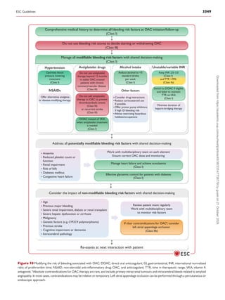
![uncontrolled bleeding.451
Dialysis can also be effective in reducing dabiga
tran concentration. Andexanet alfa rapidly reverses the activity of factor
Xa inhibitors (apixaban, edoxaban, rivaroxaban) (see Supplementary data
online, Additional evidence Table S14). An open-label RCT comparing
andexanet alfa to usual care in patients presenting with acute ICH within
6 h of symptom onset was stopped early due to improved control of
bleeding after 450 patients had been randomized.452
As DOAC-specific
antidotes are not yet available in all institutions, prothrombin complex
concentrates are often used in cases of serious bleeding on factor Xa in
hibitors, with evidence limited to observational studies.453
Due to the complexities of managing bleeding in patients taking
OAC, it is advisable that each institution develop specific policies involv
ing a multidisciplinary team that includes cardiologists, haematologists,
emergency physicians/intensive care specialists, surgeons, and others.
It is also important to educate patients taking anticoagulants on the
signs and symptoms of bleeding events and to alert their healthcare
provider when such events occur.335
The decision to reinstate OAC will be determined by the severity,
cause, and subsequent management of bleeding, preferably by a multidis
ciplinary team and with close monitoring. Failure to reinstitute OAC after
a bleed significantly increases the risk of MI, stroke, and death.454
However, if the cause of severe or life-threatening bleeds cannot be trea
ted or reversed, the risk of ongoing bleeding may outweigh the benefit of
thromboembolic protection.335
7. [R] Reduce symptoms by rate
and rhythm control
Most patients diagnosed with AF will need therapies and/or interven
tions to control heart rate, revert to sinus rhythm, or maintain sinus
rhythm to limit symptoms or improve outcomes. While the concept
of choosing between rate and rhythm control is often discussed, in real
ity most patients require a combination approach which should be con
sciously re-evaluated during follow-up. Within a patient-centred and
shared-management approach, rhythm control should be a consider
ation in all suitable AF patients, with explicit discussion of benefits
and risks.
7.1. Management of heart rate in patients
with AF
Limiting tachycardia is an integral part of AF management and is often
sufficient to improve AF-related symptoms. Rate control is indicated as
initial therapy in the acute setting, in combination with rhythm control
therapies, or as the sole treatment strategy to control heart rate and
reduce symptoms. Limited evidence exists to inform the best type
and intensity of rate control treatment.457
The approach to heart
rate control presented in Figure 7 can be used for all types of AF, includ
ing paroxysmal, persistent, and permanent AF.
Recommendation Table 14 — Recommendations for
heart rate control in patients with AF (see also
Evidence Table 14)
Recommendations Classa
Levelb
Rate control therapy is recommended in patients
with AF, as initial therapy in the acute setting, an
adjunct to rhythm control therapies, or as a sole
treatment strategy to control heart rate and reduce
symptoms.458–460
I B
Beta-blockers, diltiazem, verapamil, or digoxin are
recommended as first-choice drugs in patients with
AF and LVEF >40% to control heart rate and reduce
symptoms.48,461,462
I B
Beta-blockers and/or digoxin are recommended in
patients with AF and LVEF ≤40% to control heart
rate and reduce symptoms.40,185,463–465
I B
Combination rate control therapy should be
considered if a single drug does not control
symptoms or heart rate in patients with AF,
providing that bradycardia can be avoided, to control
heart rate and reduce symptoms.
IIa C
Lenient rate control with a resting heart rate of
< 110 b.p.m. should be considered as the initial
target for patients with AF, with stricter control
reserved for those with continuing AF-related
symptoms.459,460,466
IIa B
Atrioventricular node ablation in combination with
pacemaker implantation should be considered in
patients unresponsive to, or ineligible for, intensive
rate and rhythm control therapy to control heart
rate and reduce symptoms.467–469
IIa B
Atrioventricular node ablation combined with
cardiac resynchronization therapy should be
considered in severely symptomatic patients with
permanent AF and at least one hospitalization for HF
to reduce symptoms, physical limitations, recurrent
HF hospitalization, and mortality.470,471
IIa B
Intravenous amiodarone, digoxin, esmolol, or
landiolol may be considered in patients with AF who
have haemodynamic instability or severely depressed
LVEF to achieve acute control of heart rate.472,473
IIb B
©
ESC
2024
AF, atrial fibrillation; b.p.m., beats per minute; HF, heart failure; LVEF, left ventricular
ejection fraction.
a
Class of recommendation.
b
Level of evidence.
Recommendation Table 13 — Recommendations for
management of bleeding in anticoagulated patients
(see also Evidence Table 13)
Recommendations Classa
Levelb
Interrupting anticoagulation and performing
diagnostic or treatment interventions is
recommended in AF patients with active bleeding
until the cause of bleeding is identified and resolved.
I C
Prothrombin complex concentrates should be
considered in AF patients on VKAs who develop a
life-threatening bleed, or bleed into a critical site, to
reverse the antithrombotic effect.450
IIa C
Specific antidotes should be considered in AF
patients on a DOAC who develop a life-threatening
bleed, or bleed into a critical site, to reverse the
antithrombotic effect.451,455,456
IIa B
©
ESC
2024
AF, atrial fibrillation; DOAC, direct oral anticoagulant; VKA, vitamin K antagonist.
a
Class of recommendation.
b
Level of evidence.
ESC Guidelines 3351
Downloaded
from
https://academic.oup.com/eurheartj/article/45/36/3314/7738779
by
guest
on
21
October
2024](https://image.slidesharecdn.com/atriafibr24-241118112337-7c4f5251/85/ATRIAL-FIBRILLATION-2024-Guidelines-ESC-38-320.jpg)
![7.1.1. Indications and target heart rate
The optimal heart rate target in AF patients depends on the setting,
symptom burden, presence of heart failure, and whether rate control
is combined with a rhythm control strategy. In the RACE II (Rate
Control Efficacy in Permanent Atrial Fibrillation) RCT of patients
with permanent AF, lenient rate control (target heart rate <110 [beats
per minute] b.p.m.) was non-inferior to a strict approach (<80 b.p.m. at
rest; <110 b.p.m. during exercise; Holter for safety) for a composite of
clinical events, NYHA class, or hospitalization.186,459
Similar results
were found in a post-hoc combined analysis from the AFFIRM (Atrial
Fibrillation Follow-up Investigation of Rhythm Management) and the
RACE (Rate Control versus Electrical cardioversion) studies.474
Therefore, lenient rate control is an acceptable initial approach, unless
there are ongoing symptoms or suspicion of tachycardia-induced car
diomyopathy, where stricter targets may be indicated.
7.1.2. Heart rate control in the acute setting
In acute settings, physicians should always evaluate and manage under
lying causes for the initiation of AF prior to, or in parallel to, instituting
acute rate and/or rhythm control. These include treating sepsis, addres
sing fluid overload, or managing cardiogenic shock. The choice of drug
(Table 12) will depend on the patient’s characteristics, presence of heart
failure and LVEF, and haemodynamic profile (Figure 7). In general for
acute rate control, beta-blockers (for all LVEF) and diltiazem/verapamil
(for LVEF >40%) are preferred over digoxin because of their more
rapid onset of action and dose-dependent effects.462,475,476
More se
lective beta-1 receptor blockers have a better efficacy and safety profile
than unselective beta-blockers.477
Combination therapy with digoxin
may be required in acute settings (combination of beta-blockers with
diltiazem/verapamil should be avoided except in closely monitored
situations).177,478
In selected patients who are haemodynamically
unstable or with severely impaired LVEF, intravenous amiodarone,
landiolol, or digoxin can be used.472,473,479
7.1.3. Long-term heart rate control
Pharmacological rate control can be achieved with beta-blockers,
diltiazem, verapamil, digoxin, or combination therapy (Table 12) (see
Supplementary data online, Additional Evidence Table S15).480
The choice of rate control drugs depends on symptoms, comorbid
ities, and the potential for side effects and interactions. Combination
therapy of different rate-controlling drugs should be considered only
when needed to achieve the target heart rate, and careful follow-up
to avoid bradycardia is advised. Combining beta-blockers with verap
amil or diltiazem should only be performed in secondary care
with regular monitoring of heart rate by 24 h ECG to check for
bradycardia.459
Some antiarrhythmic drugs (AADs) also have rate-
limiting properties (e.g. amiodarone, sotalol), but they should generally
be used only for rhythm control. Dronedarone should not be instituted
for rate control since it increases rates of heart failure, stroke, and
cardiovascular death in permanent AF.481
Beta-blockers, specifically beta-1 selective adrenoreceptor an
tagonists, are often first-line rate-controlling agents largely based on
their acute effect on heart rate and the beneficial effects demonstrated
in patients with chronic HFrEF. However, the prognostic benefit of
beta-blockers seen in HFrEF patients with sinus rhythm may not be pre
sent in patients with AF.133,482
Verapamil and diltiazem are non-dihydropyridine calcium channel
blockers. They provide rate control461
and have a different adverse effect
profile, making verapamil or diltiazem useful for those experiencing side
effects from beta-blockers.483
In a 60 patient crossover RCT, verapamil
and diltiazem did not lead to the same reduction in exercise capacity as
seen with beta-blockers, and had a beneficial impact on BNP.480
Digoxin and digitoxin are cardiac glycosides that inhibit the
sodium–potassium adenosine triphosphatase and augment parasympa
thetic tone. In RCTs, there is no association between the use of digoxin
and any increase in all-cause mortality.185,484
Lower doses of digoxin
may be associated with better prognosis.185
Serum digoxin concentra
tions can be monitored to avoid toxicity,485
especially in patients at
higher risk due to older age, renal dysfunction, or use of interacting
medications. In RATE-AF (RAte control Therapy Evaluation in perman
ent Atrial Fibrillation), a trial in patients with symptomatic permanent
AF, there was no difference between low-dose digoxin and bisoprolol
for patient-reported quality of life outcomes at 6 months. However,
those randomized to digoxin demonstrated fewer adverse effects, a
greater improvement in mEHRA and NYHA scores, and a reduction
in BNP.48
Two ongoing RCTs are addressing digoxin and digitoxin
use in patients with HFrEF with and without AF (EudraCT-
2013-005326-38, NCT03783429).486
Table 12 Drugs for rate control in AF
Agenta
Intravenous administration Usual range for oral maintenance
dose
Contraindicated
Beta-blockersb
Metoprolol
tartrate
2.5–5 mg bolus over 2 mins; up to 15 mg
maximal cumulative dose
25–100 mg twice daily In case of asthma, non-selective
beta-blockers should be avoided.
Contraindicated in acute HF and history of
severe bronchospasm.
Metoprolol XL
(succinate)
N/A 50–200 mg once daily
Bisoprolol N/A 1.25–20 mg once daily
Atenololc
N/A 25–100 mg once daily
Esmolol 500 µg/kg i.v. bolus over 1 min; followed by
50–300 µg/kg/min
N/A
Landiolol 100 µg/kg i.v. bolus over 1 min; followed by
10–40 µg/kg/min
N/A
Nebivolol N/A 2.5–10 mg once daily
Carvedilol N/A 3.125–50 mg twice daily
Continued
3352 ESC Guidelines
Downloaded
from
https://academic.oup.com/eurheartj/article/45/36/3314/7738779
by
guest
on
21
October
2024](https://image.slidesharecdn.com/atriafibr24-241118112337-7c4f5251/85/ATRIAL-FIBRILLATION-2024-Guidelines-ESC-39-320.jpg)
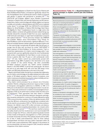
![7.2.4. Antiarrhythmic drugs
The aims of long-term rhythm control are to maintain sinus rhythm, im
prove quality of life, slow the progression of AF, and potentially reduce
morbidity related to AF episodes (see Supplementary data online,
Additional Evidence Table S19).17,445,574,575
Antiarrhythmic drugs do
not eliminate recurrences of AF, but in patients with paroxysmal
or persistent AF, a recurrence is not equivalent to treatment
failure if episodes are less frequent, briefer, or less symptomatic.
Antiarrhythmic drugs also have a role for long-term rhythm control in
AF patients that are considered ineligible or unwilling to undergo cath
eter or surgical ablation.
Before starting AAD treatment, reversible triggers should be identified
and underlying comorbidities treated to reduce the arrhythmogenic sub
strate, prevent progression of AF, and facilitate maintenance of sinus
Table 13 Antiarrhythmic drugs for sinus rhythm restoration
Drug Administration
route
Initial dosing Subsequent
dosing [long-
term approach]
Acute success
rate and time
to sinus
rhythm
Contraindications and precautions
Flecainide Oral 200–300 mg [long-term
50−150 mg twice
daily]
50%–60% at 3 h
and 75%–85% at
6–8 h (3–8 h)
• Should not be used in patients with severe
structural or coronary artery disease, Brugada
syndrome, or severe renal failure (CrCl
<35 mL/min/1.73 m2
).
• Prior documentation of safety and efficacy in
an inpatient setting is recommended prior to
pill-in-the-pocket use.
• An AVN-blocking agent should be
administered to avoid 1:1 conduction if
transformation to AFL.
• Drug infusion should be discontinued in case
of QRS widening >25% or bundle branch
block occurrence.
• Caution is needed in patients with sinus node
disease and AVN dysfunction.
• Do NOT use for conversion of atrial flutter.
Intravenous 1–2 mg/kg over
10 min
52%–95%
(Up to 6 h)
Propafenone Oral 450–600 mg [long-term
150-300 mg three
times daily]
45%–55% at 3 h,
69%–78% at 8 h
(3–8 h)
Intravenous 1.5–2 mg/kg over
10 min
43%–89%
(Up to 6 h)
Amiodarone Intravenous (/oral) 300 mg intravenous
over 30–60 min
900-1200 mg
intravenous over 24
hours (or 200 mg
oral three times
daily for 4 weeks).
[long-term 200 mg
oral daily]
44% (8–12 h to
several days)
• May cause phlebitis (use a large peripheral
vein, avoid i.v. administration >24 h and use
preferably volumetric pump).
• May cause hypotension, bradycardia/
atrioventricular block, QT prolongation.
• Only if no other option in patients with
hyperthyroidism (risk of thyrotoxicosis).
• Consider the broad range of drug
interactions.
Ibutilide Intravenous 1 mg over 10 min
(0.01 mg/kg if body
weight <60 kg)
1 mg over 10 min
(10–20 min after
the initial dose)
31%–51%
(30–90 min)
in AF
60–75% in AFL
(60 min)
• Should be used in the setting of a cardiac care
unit as it may cause QT prolongation and
torsades de pointes.
• ECG monitoring for at least 4 h after
administration to detect any proarrhythmic
effects.
• Should not be used in patients with prolonged
QT, severe LVH, or low LVEF.
Vernakalant Intravenous 3 mg/kg over 10 min
(maximum 339 mg)
2 mg/kg over 10 min
(10–15 min after
the initial dose)
(maximum 226 mg)
50% within
10 min
• Should not be used in patients with arterial
hypotension (SBP <100 mmHg), recent ACS
(within 1 month), NYHA III or IV HF, QT
prolongation or severe aortic stenosis.
• May cause arterial hypotension, QT
prolongation, QRS widening, or
non-sustained ventricular tachycardia.
©
ESC
2024
ACS, acute coronary syndromes; AF, atrial fibrillation; AFL, atrial flutter; AVN, atrioventricular node; CrCl, creatinine clearance; ECG, electrocardiogram; HF, heart failure; LVEF, left
ventricular ejection fraction; LVH, left ventricular hypertrophy; NYHA, New York Heart Association; QT, QT interval; SBP, systolic blood pressure. Long-term dosage for maintenance
of sinus rhythm is indicated in [square brackets]. Long-term oral dosing for dronedarone is 400 mg twice daily, and for sotalol 80-160 mg twice daily.
ESC Guidelines 3357
Downloaded
from
https://academic.oup.com/eurheartj/article/45/36/3314/7738779
by
guest
on
21
October
2024](https://image.slidesharecdn.com/atriafibr24-241118112337-7c4f5251/85/ATRIAL-FIBRILLATION-2024-Guidelines-ESC-44-320.jpg)
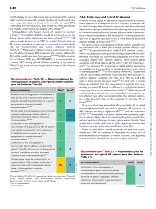
![7.2.8. AF ablation during cardiac surgery
Atrial fibrillation is a significant risk factor for early mortality, late mor
tality, and stroke in patients referred for cardiac surgery.675–677
The
best validated method of surgical ablation is the Maze procedure,
consisting of a pattern of transmural lesions including PVI, with
subsequent modifications using bipolar radiofrequency and/or
cryothermy ablation with LAA amputation (see Supplementary data
online, Additional Evidence Table S25).678–681
Education and training,
close co-operation within a multidisciplinary team, and shared
decision-making can improve the quality and outcomes of surgical
ablation.682
A number of RCTs have shown that surgical AF ablation during
cardiac surgery increases freedom from arrhythmia recur
rence.683–688
Performing surgical AF ablation, mainly targeting
those patients needing mitral valve surgery, is not associated with
increased morbidity or mortality.678,683–685
Observational data,
including large registries, have supported the potential value of
surgical AF ablation,689–700
but further RCTs are needed to evaluate
which patients should be selected, and whether this approach
contributes to the prevention of stroke, thromboembolism, and
death.
Data on pacemaker implantation rates after surgical AF ablation
are variable and are likely influenced by centre experience and the
patients treated (e.g. underlying sinus node disease). In a systematic
review of 22 RCTs (1726 patients), permanent pacemaker implant
ation rates were higher with surgical AF ablation than without con
comitant AF surgery (6.0% vs. 4.1%; RR, 1.69; 95% CI, 1.12–2.54).701
Observational registry data from contemporary cohorts (2011–
2020) suggest an overall pacemaker rate post-operatively of 2.1%
in patients selected for surgical AF ablation, with no discernible
impact of surgical ablation on the need for a pacemaker, but
higher rates in those needing multivalve surgery.702
With a safety-
first approach in mind, imaging is advised during surgical AF ablation
to exclude thrombus and help to plan the surgical approach
(e.g. with TOE), regardless of effective pre-procedural anticoagulant
use.
7.2.9. Atrial tachycardia after pulmonary vein
isolation
After any ablation for AF, recurrent arrhythmias may manifest as AF,
but also as atrial tachycardia (AT). Although AT may be perceived as
a step in the right direction by the treating physician, this view is often
not shared by the patient because AT can be equally or more symptom
atic than the original AF. Conventionally, an early arrhythmia recur
rence post-PVI (whether AT, AF, or flutter) is considered potentially
transitory.708
Recent trials using continuous implantable loop recorders
for peri-procedural monitoring have provided insight into the incidence
and significance of early arrhythmia recurrences, and have confirmed a
link between early and later recurrence.709
Discussion of management
options for AT post-ablation should ideally involve a multidisciplinary
team with experience in interventional management of complex ar
rhythmias, considering technical challenges, procedural efficacy, and
safety, in the context of patient preferences.
8. [E] Evaluation and dynamic
reassessment
The development and progression of AF results from continuous
interactions between underlying mechanisms (electrical, cellular,
neurohormonal, and haemodynamic), coupled with a broad range of
clinical factors and associated comorbidities. Each individual factor
Endoscopic and hybrid ablation procedures should
be considered in patients with symptomatic
persistent AF refractory to AAD therapy to
prevent symptoms, recurrence, and progression of
AF, within a shared decision-making rhythm
control team of electrophysiologists and
surgeons.667–671,674
IIa A
Endoscopic and hybrid ablation procedures may
be considered in patients with symptomatic
paroxysmal AF refractory to AAD therapy and
failed percutaneous catheter ablation strategy to
prevent symptoms, recurrence, and progression of
AF, within a shared decision-making rhythm
control team of electrophysiologists and
surgeons.668,669
IIb B
©
ESC
2024
AAD, antiarrhythmic drugs; AF, atrial fibrillation; LAA, left atrial appendage.
a
Class of recommendation.
b
Level of evidence.
Recommendation Table 22 — Recommendations for
AF ablation during cardiac surgery (see also Evidence
Table 22)
Recommendations Classa
Levelb
Concomitant surgical ablation is recommended in
patients undergoing mitral valve surgery and AF
suitable for a rhythm control strategy to prevent
symptoms and recurrence of AF, with shared
decision-making supported by an experienced team
of electrophysiologists and arrhythmia surgeons.683–
685,701
I A
Intraprocedural imaging for detection of left atrial
thrombus in patients undergoing surgical ablation is
recommended to guide surgical strategy,
independent of oral anticoagulant use, to prevent
peri-procedural ischaemic stroke and
thromboembolism.
I C
Concomitant surgical ablation should be considered
in patients undergoing non-mitral valve cardiac
surgery and AF suitable for a rhythm control strategy
to prevent symptoms and recurrence of AF, with
shared decision-making supported by an
experienced team of electrophysiologists and
arrhythmia surgeons.701,703–707
IIa B
©
ESC
2024
AF, atrial fibrillation.
a
Class of recommendation.
b
Level of evidence.
ESC Guidelines 3361
Downloaded
from
https://academic.oup.com/eurheartj/article/45/36/3314/7738779
by
guest
on
21
October
2024](https://image.slidesharecdn.com/atriafibr24-241118112337-7c4f5251/85/ATRIAL-FIBRILLATION-2024-Guidelines-ESC-48-320.jpg)
![has considerable variability over time, affecting its contribution to the
AF-promoting substrate. The risk profile of each patient is also far
from static, and requires a dynamic mode of care to ensure optimal
AF management.710,711
Patients with AF require periodic reassessment
of therapy based on this changing risk status if we are to improve the
overall quality of care. Timely attention to modifiable factors and
underpinning comorbidities has the potential to slow or reverse the
progression of AF, increase quality of life, and prevent adverse out
comes such as heart failure, thromboembolism, and major bleeding.
The [E] in AF-CARE encompasses the range of activity needed by
healthcare professionals and patients to: (i) thoroughly evaluate asso
ciated comorbidities and risk factors that can guide treatment; and
(ii) provide the dynamic assessment needed to ensure that treatment
plans remain suited to that particular patient. This task force recom
mends an adaptive strategy that not only reacts to changes notified
by a patient, but also proactively seeks out issues where altering man
agement could impact on patient wellbeing. Avoidance of unnecessary
and costly follow-up is also inherent in this framework, with educated
and empowered patients contributing to identifying the need for access
to specialist care or an escalation of management. The patient-centred,
shared decision philosophy is embedded to improve efficiency in mod
els of care and to address the needs of patients with AF.
Medical history and the results of any tests should be regularly re-
evaluated to address the dynamic nature of comorbidities and risk fac
tors.712
This may have impact on therapeutic decisions; e.g. resumption
of full-dose DOAC therapy after improvement in the patient’s renal
function. The timing of review of the AF-CARE pathway is patient spe
cific and should respond to changes in clinical status. In most cases, this
task force advises re-evaluation 6 months after initial presentation, and
then at least annually by a healthcare professional in primary or second
ary care (see Figure 3).
8.1. Implementation of dynamic care
A multidisciplinary-based approach is advocated to improve implemen
tation of dynamic AF-CARE (see Figure 2); although potentially re
source intensive, this is preferred to more simplistic or opportunistic
methods. For example, in a pragmatic trial of 47 333 AF patients iden
tified through health insurance claims, there was no difference in OAC
initiation at 1 year in those randomized to a single mailout of patient and
clinician education, compared with those in the usual care group.713
For
co-ordination of care there is a core role for cardiologists, general prac
titioners, specialized nurses, and pharmacists.714
If needed, and depend
ing on local resources, others may also be involved (cardiac surgeons,
physiotherapists, neurologists, psychologists, and other allied health
professionals). It is strongly advocated that one core team member co-
ordinates care, and that additional team members become involved ac
cording to the needs of the individual patient throughout their AF
trajectory.
Several organizational models of integrated care for AF have been
evaluated, but which components are most useful remains unclear.
Some models include a multidisciplinary team,715,716
while others are
nurse-led79,122,124,717
or cardiologist-led.79,122,124,717
Several published
models used computerized decision support systems or electronic
health applications.79,122,715,718
Evaluation within RCTs has demon
strated mixed results due to the variety of methods tested and differ
ences in regional care. Several studies report significant improvements
with respect to adherence to anticoagulation, cardiovascular mortality,
and hospitalization relative to standard of care.121–123
However, the
RACE 4 (IntegRAted Chronic Care Program at Specialized AF Clinic
Versus Usual CarE in Patients with Atrial Fibrillation) trial, which in
cluded 1375 patients, failed to demonstrate superiority of nurse-led
over usual care.79
New studies of the components and optimal models
for delivery for integrated care approaches in routine practice are on
going (ACTRN12616001109493, NCT03924739).
8.2. Improving treatment adherence
Advances in the care of patients with AF can only be effective if appro
priate tools are available to support the implementation of the treat
ment regimen.719
A number of factors are related to the optimal
implementation of care at the level of: (i) the individual patient (culture,
cognitive impairment, and psychological status); (ii) the treatment
(complexity, side effects, polypharmacy, impact on daily life, and
cost); (iii) the healthcare system (access to treatment and multidiscip
linary approach); and (iv) the healthcare professional (knowledge,
awareness of guidelines, expertise, and communication skills). A collab
orative approach to patient care, based upon shared decision-making
and goals tailored to individual patient needs, is crucial in promoting on
going patient adherence to the agreed treatment regimen.720
Even
when treatment seems feasible for the individual, patients often lack ac
cess to reliable and up-to-date information about risks and benefits of
various treatment options, and consequently are not empowered to
engage in their own management. A sense of ownership that promotes
the achievement of joint goals can be encouraged through the use of
educational programmes, websites (such as https://afibmatters.org),
app-based tools, and individually tailored treatment protocols which
take into account gender, ethnic, socioeconomic, environmental, and
work factors. In addition, practical tools (e.g. schedules, apps, bro
chures, reminders, pillboxes) can help to implement treatment in daily
life.721,722
Regular review by members of the multidisciplinary team en
ables the evolution of a flexible and responsive management regimen
that the patient will find easier to follow.
8.3. Cardiac imaging
A TTE is a valuable asset across all four AF-CARE domains when
there are changes in the clinical status of an individual patient
(Figure 13).723–725
The key findings to consider from an echocardiogram
are any structural heart disease (e.g. valvular disease or left ventricular
hypertrophy), impairment of left ventricular function (systolic and/or
diastolic to classify heart failure subtype), left atrial dilatation, and right
heart dysfunction.59,67,726
To counter irregularity when in AF, obtaining
measurements in cardiac cycles that follow two similar RR intervals can
improve the value of parameters compared with sequential averaging of
cardiac cycles.723,727
Contrast TTE or alternative imaging modalities
may be required where image quality is poor, and quantification of
left ventricular systolic function is needed for decisions on rate or
rhythm control. Other cardiac imaging techniques, such as cardiac mag
netic resonance (CMR), CT, TOE, and nuclear imaging can be valuable
when: (i) TTE quality is suboptimal for diagnostic purposes; (ii) addition
al information is needed on structure, substrate, or function; and (iii) to
support decisions on interventional procedures (see Supplementary
data online, Figure S1).59,724,725,728
As with TTE, other types of cardiac
imaging can be challenging in the context of AF irregularity or with rapid
heart rate, requiring technique-specific modifications when acquiring
ECG-gated sequences.729–731
3362 ESC Guidelines
Downloaded
from
https://academic.oup.com/eurheartj/article/45/36/3314/7738779
by
guest
on
21
October
2024](https://image.slidesharecdn.com/atriafibr24-241118112337-7c4f5251/85/ATRIAL-FIBRILLATION-2024-Guidelines-ESC-49-320.jpg)
![8.4. Patient-reported outcome measures
Patients with AF have a lower quality of life compared with the general
population.732
Improvement in quality of life and functional status should
play a key role in assessing and reassessing treatment decisions (see
Supplementary data online, Additional Evidence Table S26).36
Patient-reported outcome measures are valuable to measure quality of
life, functional status, symptoms, and treatment burden for patients
with AF over time.55,733–735
Patient-reported outcome measures are
playing an increasing role in clinical trials to assess the success of treat
ment; however, they remain under-utilized.736,737
They can be divided
into generic or disease-specific tools, with the latter helping to provide
insight into AF-related impacts.738
However, multimorbidity can still con
found the sensitivity of all PROMs, impacting on association with other
established metrics of treatment performance such as mEHRA symptom
AF-CARE
pathway
Objective for
imaging
To identify
comorbidities which
are associated with
recurrence and
progression of AF
To determine
stroke risk, choice
of anticoagulant
drug and ensure
safety for
cardioversion
To determine optimal
choice of rate and
rhythm control
strategy and likely
success of ablation
To detect changes in
the patient's heart
structure and function
which would affect
their management plan
C
Comorbidity and risk
factor management
R
Reduce symptoms
by rate and
rhythm control
Avoid stroke and
thromboembolism
A
Evaluation and
dynamic
reassessment
E
Assessment
Left ventricular ejection fraction,
wall motion abnormalities, diastolic
indices, right ventricular function and
left ventricular hypertrophy to
determine subtype and aetiology of
heart failure
Detection of valvular disease
Detection of pericardial fluid or
pericardial disease
Detection of heart failure for
CHA2
DS2
-VA score
Transoesophageal echocardiogram
for left atrial appendage assessment
to exclude thrombus prior to
cardioversion
Detection of moderate-severe
mitral stenosis to determine choice
of anticoagulation
Reassess known valve disease for
increase in severity
Reassess left ventricular size and
function if there is a change in the
patient’s clinical status or symptoms
Left ventricular ejection fraction to
determine choice of rate control
Left ventricular size and function to
determine choice of rhythm control
Severity of valvular disease to
determine choice of rhythm control
Left atrial size and function to
determine risk of arrhythmia
recurrence following ablation
Example of
pathology
Cardiac amyloid
Clot in LAA
Severe LV
impairment
Mixed mitral
valve disease
Figure 13 Relevance of echocardiography in the AF-CARE pathway. AF, atrial fibrillation; AF-CARE, atrial fibrillation—[C] Comorbidity and risk fac
tor management, [A] Avoid stroke and thromboembolism, [R] Reduce symptoms by rate and rhythm control, [E] Evaluation and dynamic reassessment;
CHA2DS2-VA, congestive heart failure, hypertension, age ≥75 years (2 points), diabetes mellitus, prior stroke/transient ischaemic attack/arterial
thromboembolism (2 points), vascular disease, age 65–74 years; LAA, left atrial appendage; LV, left ventricle.
ESC Guidelines 3363
Downloaded
from
https://academic.oup.com/eurheartj/article/45/36/3314/7738779
by
guest
on
21
October
2024](https://image.slidesharecdn.com/atriafibr24-241118112337-7c4f5251/85/ATRIAL-FIBRILLATION-2024-Guidelines-ESC-50-320.jpg)
![class and natriuretic peptides.48
Intervention studies have demonstrated
an association between improvement in PROM scores and reduction in
AF burden and symptoms.48,738
Atrial fibrillation-specific questionnaires include the AF 6 (AF6),739
Atrial Fibrillation Effect on QualiTy-of-Life (AFEQT),740
the Atrial
Fibrillation Quality of Life Questionnaire (AFQLQ),741
the Atrial
Fibrillation Quality of Life (AF-QoL),742
and the Quality of Life in
Atrial Fibrillation (QLAF).743
The measurement properties of most of
these tools lack sufficient validation.49
The International Consortium
for Health Outcomes Measurement (ICHOM) working group recom
mends the use of the AFEQT PROM or a symptom questionnaire called
the Atrial Fibrillation Severity Scale (AFSS) for measuring exercise tol
erance and the impact of symptoms in AF.744
Through wider use of pa
tient experience measures, there is an opportunity at the institutional
level to improve the quality of care delivered to patients with AF.49–55
9. The AF-CARE pathway in specific
clinical settings
The following sections detail specific clinical settings where approaches
to AF-CARE may vary. Unless specially discussed, measures for [C] co
morbidity and risk factor management, [A] avoidance of stroke and
thromboembolism, [R] rate and rhythm control, and [E] evaluation
and dynamic reassessment should follow the standard pathways intro
duced in Section 4.
9.1. AF-CARE in unstable patients
Unstable patients with AF include those with haemodynamic instability
caused by the arrhythmia or acute cardiac conditions, and severely ill
patients who develop AF (sepsis, trauma, surgery, and particularly
cancer-related surgery). Conditions such as sepsis, adrenergic over
stimulation, and electrolyte disturbances contribute to onset and recur
rence of AF in these patients. Spontaneous restoration of sinus rhythm
has been reported in up to 83% during the first 48 h after appropriate
treatment of the underlying cause.551,745
Emergency electrical cardioversion is still considered the first-choice
treatment if sinus rhythm is thought to be beneficial, despite the limita
tion of having a high rate of immediate relapse.746
Amiodarone is a
second-line option because of its delayed activity; however, it may be
an appropriate alternative in the acute setting.747,748
In a multicentre co
hort study carried out in the United Kingdom and the United States of
America, amiodarone and beta-blockers were similarly effective for
rate control in intensive care patients, and superior to digoxin and cal
cium channel blockers.749
The ultra-short acting and highly selective beta-
blocker landiolol can safely control rapid AF in patients with low ejection
fraction and acutely decompensated heart failure, with a limited impact
on myocardial contractility or blood pressure.477,750,751
9.2. AF-CARE in acute and chronic
coronary syndromes
The incidence of AF in acute coronary syndromes (ACS) ranges from
2% to 23%.752
The risk of new-onset AF is increased by 60%–77% in
patients suffering an MI,753
and AF may be associated with an increased
risk of ST-segment elevation myocardial infarction (STEMI) or
non-STEMI ACS.754
Overall, 10%–15% of AF patients undergo percu
taneous intervention (PCI) for CAD.755
In addition, AF is a common
precipitant for type 2 MI.756
Observational studies show that patients
with both ACS and AF are less likely to receive appropriate antithrom
botic therapy757
and more likely to experience adverse outcomes.758
Peri-procedural management of patients with ACS or chronic coronary
syndromes (CCS) are detailed in the 2023 ESC Guidelines for the man
agement of acute coronary syndromes and 2024 ESC Guidelines for the
management of chronic coronary syndromes.759,760
The combination of AF with ACS is the area where use of multiple
antithrombotic drugs is most frequently indicated, consisting of antiplate
let agents plus OAC (Figure 14) (see Supplementary data online,
Additional Evidence Table S27). There is a general trend to decrease the
duration of DAPT to reduce bleeding; however, this may increase ischae
mic events and stent thrombosis.761,762
In ACS there is a high risk of pre
dominantly platelet-driven atherothrombosis and thus of coronary
ischaemic events. Acute coronary syndromes treated by PCI require
DAPT for improved short- and long-term prognosis. Therefore, a peri-
procedural triple antithrombotic regimen including an OAC, aspirin, and
a P2Y12 inhibitor should be the default strategy for most patients. Major
thrombotic events vs. major bleeding risk need to be balanced when pre
scribing antiplatelet therapy and OAC after the acute phase and/or after
PCI. The combination of OAC (preferably a DOAC) and a P2Y12 inhibi
tor results in less major bleeding than triple therapy that includes aspirin.
Clopidogrel is the preferred P2Y12 inhibitor, as the evidence for ticagre
lor and prasugrel is less clear with higher bleeding risk.763–769
Ongoing
trials will add to our knowledge about safely combining DOACs with
antiplatelet agents (NCT04981041, NCT04436978). When using
VKAs with antiplatelet agents, there is consensus opinion to use an
INR range of 2.0–2.5 to mitigate excess bleeding risk.
Short–term triple therapy (≤1 week) is recommended for all pa
tients without diabetes after ACS or PCI. In pooled analyses of
RCTs, omitting aspirin in patients with ACS undergoing PCI has
the potential for higher rates of ischaemic/stent thrombosis, without
impact on incident stroke.761,762,770–772
None of the trials were
powered for ischaemic events. All patients in AUGUSTUS (an open–
label, 2 × 2 factorial, randomized controlled clinical trial to evaluate
the safety of apixaban vs. vitamin k antagonist and aspirin vs. aspirin pla
cebo in patients with AF and ACS or PCI) received aspirin plus a P2Y12
inhibitor for a median time of 6 days.773
At the end of the trial, apixaban
and a P2Y12 inhibitor without aspirin was the optimal treatment regi
men for most patients with AF and ACS and/or PCI, irrespective of
the patient’s baseline bleeding and stroke risk.774,775
Recommendation Table 23 — Recommendations to
improve patient experience (see also Evidence Table 23)
Recommendations Classa
Levelb
Evaluating quality of care and identifying
opportunities for improved treatment of AF should
be considered by practitioners and institutions to
improve patient experiences.49–55
IIa B
©
ESC
2024
AF, atrial fibrillation.
a
Class of recommendation.
b
Level of evidence.
3364 ESC Guidelines
Downloaded
from
https://academic.oup.com/eurheartj/article/45/36/3314/7738779
by
guest
on
21
October
2024](https://image.slidesharecdn.com/atriafibr24-241118112337-7c4f5251/85/ATRIAL-FIBRILLATION-2024-Guidelines-ESC-51-320.jpg)
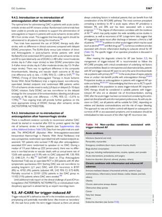
![9.6. AF-CARE in post-operative patients
Peri-operative AF describes the onset of the arrhythmia during an
ongoing intervention. Post-operative AF (POAF), defined as new-
onset AF in the immediate post-operative period, is a common com
plication with clinical impact that occurs in 30%–50% of patients
undergoing cardiac surgery,816–818
and in 5%–30% of patients under
going non-cardiac surgery. Intra- and post-operative changes and
specific AF triggers (including peri-operative complications) and
pre-existing AF-related risk factors and comorbidities increase the
susceptibility to POAF.819
Although POAF episodes may be self-
terminating, POAF is associated with 4–5 times increase in recurrent
AF during the next 5 years,820,821
and is a risk factor for stroke, MI,
heart failure, and death.822–827
Other adverse events associated
with POAF include haemodynamic instability, prolonged hospital
stay, infections, renal complications, bleeding, increased in-hospital
death, and greater healthcare cost.828–830
While multiple strategies to prevent POAF with pre-treatment or acute
drug treatment have been described, there is a lack of evidence from large
RCTs. Pre-operative use of propranolol or carvedilol plus N-acetyl
cysteine in cardiac and non-cardiac surgery is associated with a reduced
incidence of POAF,831–834
but not major adverse events.835
An umbrella
review of 89 RCTs from 23 meta-analyses (19 211 patients, but not neces
sarily in AF) showed no benefit from beta-blockers in cardiac surgery for
mortality, MI, or stroke. In non-cardiac surgery, beta-blockers were asso
ciated with reduced rates of MI after surgery (RR range, 0.08–0.92), but
higher mortality (RR range, 1.03–1.31), and increased risk of stroke (RR
range, 1.33–7.72).836
Prevention of peri-operative AF can also be achieved
with amiodarone. In a meta-analyses, amiodarone (oral or intravenous
[i.v.]) and beta-blockers were equally effective in reducing post-operative
AF,837
but their combination was better than beta-blockers alone.838
Lower cumulative doses of amiodarone (<3000 mg during the
loading phase) could be effective, with fewer adverse events.837,839,840
Withdrawal of beta-blockers should be avoided due to increased risk
of POAF.841
Other treatment strategies (steroids, magnesium, sotalol,
(bi)atrial pacing, and botulium injection into the epicardial fat pad) lack
scientific evidence for the prevention of peri-operative AF.842,843
Peri-operative posterior pericardiotomy, due to the reduction of post-
operative pericardial effusion, showed a significant decrease in POAF
in patients undergoing cardiac surgery (OR, 0.44; 95% CI, 0.27–0.70;
P = .0005).844–846
In 3209 patients undergoing non-cardiac thoracic sur
gery, colchicine did not lead to any significant reduction in AF compared
with placebo (HR, 0.85; 95% CI, 0.65–1.10; P = .22).847
The evidence for prevention of ischaemic stroke in POAF by OAC
is limited.822,827
Oral anticoagulant therapy is associated with a high
bleeding risk soon after cardiac surgery or major non-cardiac inter
ventions.827
Conversely, meta-analyses of observational cohort stud
ies suggest a possible protective impact of OAC in POAF for all-cause
mortality848
and a lower risk of thromboembolic events following car
diac surgery, accompanied by higher rates of bleeding.849
This task
force recommends to treat post-operative AF according to the
AF-CARE pathway as discussed for trigger-induced AF (with the [R]
pathway the same as for first-diagnosed AF). Ongoing RCTs in cardiac
surgery (NCT04045665) and non-cardiac surgery (NCT03968393)
will inform optimal long-term OAC use among patients with POAF.
While awaiting the results of these trials, this task force recommends
that after acute bleeding risk has settled, long-term OAC should be
considered in patients with POAF according to their thromboembolic
risk factors.
9.7. AF-CARE in embolic stroke of
unknown source
Theterm‘embolicstrokeofundeterminedsource’(ESUS)wasintroduced
to identify non-lacunar strokes whose mechanism is likely to be embolic,
but the source remains unidentified.856
Of note, these patients have a re
current risk of stroke of 4%–5% per year.856
The main embolic sources as
sociated with ESUS are concealed AF, atrial cardiomyopathy, left
ventricular disease, atherosclerotic plaques, patent foramen ovale (PFO),
valvulardiseases,andcancer.Atrialcardiomyopathyandleftventriculardis
ease are the most prevalent causes.856
AF is reported to be the underlying
mechanism in 30% of ESUS patients.857–859
The detection of AF among
ESUS patients increases the longer cardiac monitoring is provided (see
Supplementary data online, Additional Evidence Table S29).857,860–864
This
also holds for the duration of implantable cardiac monitoring, with prob
ability of AF detection ranging from 2% with 1 week to over 20% by 3
years.865
In patients with ESUS, factors associated with an increased detec
tion of AF are increasing age,866,867
left atrial enlargement,866
cortical loca
tion of stroke,868
large or small vessel disease,863
an increased number of
Recommendation Table 26 — Recommendations for
management of post-operative AF (see also Evidence
Table 26)
Recommendations Classa
Levelb
Peri-operative amiodarone therapy is recommended
where drug therapy is desired to prevent
post-operative AF after cardiac surgery.838,839,850,851
I A
Concomitant posterior peri-cardiotomy should be
considered in patients undergoing cardiac surgery to
prevent post-operative AF.845,846
IIa B
Long-term oral anticoagulation should be considered
in patients with post-operative AF after cardiac and
non-cardiac surgery at elevated thromboembolic
risk, to prevent ischaemic stroke and
thromboembolism.811,852–854
IIa B
Routine use of beta-blockers is not recommended in
patients undergoing non-cardiac surgery for the
prevention of post-operative AF.836,855
III B
©
ESC
2024
AF, atrial fibrillation.
a
Class of recommendation.
b
Level of evidence.
Recommendation Table 25 — Recommendations for
trigger-induced AF (see also Evidence Table 25)
Recommendation Classa
Levelb
Long-term oral anticoagulation should be considered
in suitable patients with trigger-induced AF at
elevated thromboembolic risk to prevent ischaemic
stroke and systemic thromboembolism.13,800,806,807,815
IIa C
©
ESC
2024
AF, atrial fibrillation.
a
Class of recommendation.
b
Level of evidence.
3368 ESC Guidelines
Downloaded
from
https://academic.oup.com/eurheartj/article/45/36/3314/7738779
by
guest
on
21
October
2024](https://image.slidesharecdn.com/atriafibr24-241118112337-7c4f5251/85/ATRIAL-FIBRILLATION-2024-Guidelines-ESC-55-320.jpg)
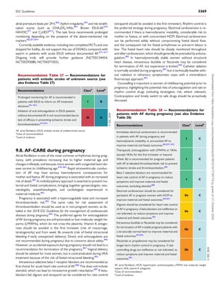

![10.3.1. Single timepoint screening ‘snapshot’
Several cluster RCTs in primary care settings have explored whether
screening performed as a snapshot of the heart rhythm at one time
point can detect more AF compared with usual care in individuals
aged ≥65 years.1138–1140
No increased detection of AF was seen in
groups randomized to single timepoint screening.1138–1140
These find
ings were confirmed in a meta-analysis of RCTs showing that screening
as a one-time event did not increase detection of AF compared with
usual care.1135
Notably, these studies were performed in healthcare
settings where the detection of AF in the population might be high,
hence the results might not be generalizable to healthcare settings
with a lower spontaneous AF detection. There are no RCTs addressing
clinical outcomes in patients with AF detected by single timepoint
screening.1123,1135
10.3.2. Prolonged screening
Studies using prolonged screening have shown an increased detection
of AF leading to initiation of OAC.1129,1135,1141
Two RCTs have inves
tigated the effect on clinical outcomes in prolonged screening for AF.5,6
In the STROKESTOP trial (Systematic ECG Screening for Atrial
Fibrillation Among 75 Year Old Subjects in the Region of Stockholm
Recommendation Table 31 — Recommendations for
screening for AF (see also Evidence Table 31)
Recommendations Classa
Levelb
Review of an ECG (12-lead, single, or multiple leads)
by a physician is recommended to provide a definite
diagnosis of AF and commence appropriate
management.1091,1121–1123,1125
I B
Routine heart rhythm assessment during healthcare
contact is recommended in all individuals aged ≥65
years for earlier detection of AF.
I C
Population-based screening for AF using a prolonged
non-invasive ECG-based approach should be
considered in individuals aged ≥75 years, or ≥65
years with additional CHA2DS2-VA risk factors to
ensure earlier detection of AF.6,1135–1137
IIa B
©
ESC
2024
AF, atrial fibrillation; CHA2DS2-VA, congestive heart failure, hypertension, age ≥75 years
(2 points), diabetes mellitus, prior stroke/transient ischaemic attack/arterial
thromboembolism (2 points), vascular disease, age 65–74 years; ECG, electrocardiogram.
a
Class of recommendation.
b
Level of evidence.
Screening
approach
Population
example
Setting for
AF detection
Type of
screening
Follow
AF-CARE
After
thromboembolic
event
Patients with
embolic stroke of
unknown source
Age ≥65 years,
or risk of
thromboembolism
Age ≥75 years,
or ≥65 years plus
other
CHA2
DS2
-VA
factors
Initiated after
index event
At the time of
routine healthcare
contact
Structured national
or regional
screening
programmes
Ad-hoc to
catch AF
In patients with
risk factors
Patient
informed
about
implications
of AF
detection
C
Comorbidity and risk
factor management
A
Avoid stroke and
thromboembolism
R
Reduce symptoms by
rate and rhythm control
E
Evaluation and
dynamic reassessment
Any rhythm check,
confirmed by ECG
Routine heart
rhythm assessment
(Class I)
Invasive or
non-invasive ECG
Prolonged
monitoring
(Class I)
Population-based
Non-invasive ECG
(Class IIa)
Figure 16 Approaches to screening for AF. AF, atrial fibrillation; AF-CARE, atrial fibrillation—[C] Comorbidity and risk factor management, [A] Avoid
stroke and thromboembolism, [R] Reduce symptoms by rate and rhythm control, [E] Evaluation and dynamic reassessment; CHA2DS2-VA, congestive
heart failure, hypertension, age ≥75 years (2 points), diabetes mellitus, prior stroke/TIA/arterial thromboembolism (2 points), vascular disease, age 65–
74 years; ECG, electrocardiogram. See Figure 15 for non-invasive ECG methods.
3374 ESC Guidelines
Downloaded
from
https://academic.oup.com/eurheartj/article/45/36/3314/7738779
by
guest
on
21
October
2024](https://image.slidesharecdn.com/atriafibr24-241118112337-7c4f5251/85/ATRIAL-FIBRILLATION-2024-Guidelines-ESC-61-320.jpg)
![and Halland, Sweden), 75- and 76-year-olds were randomized to be in
vited to prolonged screening for AF using single-lead ECGs twice daily
for 2 weeks, or to standard of care. After a median of 6.9 years there
was a small reduction in the primary combined endpoint of all-cause
mortality, stroke, systematic embolism, and severe bleeding in favour
of prolonged screening (HR, 0.96; 95% CI, 0.92–1.00; P = .045).6
In
the LOOP (Atrial Fibrillation Detected by Continuous ECG
Monitoring) trial, individuals at increased risk of stroke were rando
mized to receive an implantable loop recorder that monitored heart
rhythm for an average of 3.3 years, or to a control group receiving
standard of care. Although there was a higher detection of AF
(31.8%) and subsequent initiation of OAC in the loop recorder
group compared with standard of care (12.2%), this was not accom
panied by a difference in the primary outcome of stroke or systemic
embolism.5
In a meta-analysis of recent RCTs on the outcome of
stroke, a small but significant benefit was seen in favour of prolonged
screening (RR, 0.91; 95% CI, 0.84–0.99).1136
This was not repeated
in a second meta-analysis including older RCTs, where no risk reduc
tion was seen with regard to mortality or stroke.1135
Notably, both
these meta-analyses are likely underpowered to assess clinical
outcomes.
10.4. Factors associated with incident AF
The most common risk predictors for incident (new-onset) AF are
shown in Table 16. While the factors listed are robustly associated
with incident AF in observational studies, it is not known whether
the relationships are causal. Studies using Mendelian randomization
(genetic proxies for risk factors to estimate causal effects) robustly im
plicate systolic BP and higher BMI as causal risk factors for incident
AF.1142
A high degree of interaction occurs between all factors related to AF
development (see Supplementary data online, Additional Evidence
Table S33).1038,1039,1143–1145
For ease of clinical application, risk predic
tion tools have combined various factors, and have recently employed
machine learning algorithms for prediction.1146,1147
Classical risk scores
are also available with variable predictive ability and model performance
(see Supplementary data online, Table S7).1148
Improved outcomes
when using these risk scores have yet to be demonstrated. Although
knowledge is rapidly increasing about the genetic basis for AF in
some patients, the value of genetic screening is limited at the present
time (see Supplementary data online).
10.5. Primary prevention of AF
Preventing the onset of AF before clinical manifestation has clear
potential to improve the lives of the general population and reduce
the considerable health and social care costs associated with
development of AF. Whereas the [C] in AF-CARE is focused on the
effective management of risk factors and comorbidities to limit AF
recurrence and progression, there is also evidence they can be targeted
to prevent AF. Available data are presented below for hypertension,
heart failure, type 2 diabetes mellitus, obesity, sleep apnoea syndrome,
Table 16 Factors associated with incident AF
Demographic factors Age1149–1151
Male sex1149–1152
European ancestry1149,1150
Lower socioeconomic status1150
Lifestyle behaviours Smoking/tobacco use1149–1151
Alcohol intake1149,1150
Physical inactivity1149,1150
Vigorous exercise1153–1156
Competitive or athlete-level endurance
sports1151,1157
Caffeine1158–1160
Continued
Comorbidities and risk
factors
Hypertension1149–1151
Heart failure178,1149–1151,1161
Valvular disease1149,1151,1162–1164
Coronary artery disease1149,1151,1161,1165
Peripheral arterial disease785
Congenital heart disease1149,1166
Heart rate, heart rate variability1167,1168
Total cholesterol1149,1150
Low-density lipoprotein cholesterol1150
High-density lipoprotein cholesterol1150
Triglycerides1150
Impaired glucose tolerance,1169–1172
diabetes
mellitus1149–1151,1169
Renal dysfunction/CKD1149–1151,1173,1174
Obesity1149–1151,1175,1176
Body mass index, weight1149–1151
Height1150
Sleep apnoea1149,1151,1177,1178
Chronic obstructive pulmonary disease1179
Subclinical
atherosclerosis
Coronary artery calcification1149,1151,1180
Carotid IMT and carotid plaque1149,1151,1181,1182
ECG abnormalities PR interval prolongation1149,1151,1183
Sick sinus syndrome1149,1184,1185
Wolff–Parkinson–White1149,1186
Genetic factors Family history of AF1149,1151,1187–1190
AF-susceptible loci identified by
GWAS1149,1151,1191,1192
Short QT syndrome1149
Genetic cardiomyopathies990,1193
Biomarkers C-reactive protein1150,1151
Fibrinogen1150
Growth differentiation factor-151194
Natriuretic peptides (atrial and B-type)1195–1200
Cardiac troponins1199
Inflammatory biomarkers 1149,1151
Others Thyroid dysfunction912,1149–1151
Autoimmune diseases1150
Air pollution1149,1201
Sepsis1149,1202
Psychological factors 1203,1204
©
ESC
2024
AF, atrial fibrillation; CKD, chronic kidney disease; GWAS, genome-wide association
studies; HF, heart failure; IMT, intima-media thickness.
ESC Guidelines 3375
Downloaded
from
https://academic.oup.com/eurheartj/article/45/36/3314/7738779
by
guest
on
21
October
2024](https://image.slidesharecdn.com/atriafibr24-241118112337-7c4f5251/85/ATRIAL-FIBRILLATION-2024-Guidelines-ESC-62-320.jpg)
![10.5.7. Alcohol intake
The premise that reducing alcohol intake can prevent AF is based on
observational studies linking alcohol to an excess risk of incident AF
in a dose-dependent manner (see Supplementary data online).1220–1222
In addition, a population cohort study of those with high alcohol con
sumption (>60 g/day for men and >40 g/day for women) found that ab
stinence from alcohol was associated with a lower incidence of AF
compared with patients who continued heavy drinking.1223
11. Key messages
(1) General management: optimal treatment according to the
AF-CARE pathway, which includes: [C] Comorbidity and risk fac
tor management; [A] Avoid stroke and thromboembolism; [R]
Reduce symptoms by rate and rhythm control; and [E]
Evaluation and dynamic reassessment.
(2) Shared care: patient-centred AF management with joint decision-
making and a multidisciplinary team.
(3) Equal care: avoid health inequalities based on gender, ethnicity,
disability, and socioeconomic factors.
(4) Education: for patients, family members, caregivers, and health
care professionals to aid shared decision-making.
(5) Diagnosis: clinical AF requires confirmation on an ECG device to
initiate risk stratification and AF management.
(6) Initial evaluation: medical history, assessment of symptoms and
their impact, blood tests, echocardiography/other imaging,
patient-reported outcome measures, and risk factors for
thromboembolism and bleeding.
(7) Comorbidities and risk factors: thorough evaluation and manage
ment critical to all aspects of care for patients with AF to avoid
recurrence and progression of AF, improve success of AF treat
ments, and prevent AF-related adverse outcomes.
(8) Focus on conditions associated with AF: including hypertension,
heart failure, diabetes mellitus, obesity, obstructive sleep apnoea,
physical inactivity, and high alcohol intake.
(9) Assessing the risk of thromboembolism: use locally validated risk
tools or the CHA2DS2-VA score and assessment of other risk fac
tors, with reassessment at periodic intervals to assist in decisions
on anticoagulant prescription.
(10) Oral anticoagulants: recommended for all eligible patients, except
those at low risk of incident stroke or thromboembolism
(CHA2DS2-VA = 1 anticoagulation should be considered;
CHA2DS2-VA ≥2 anticoagulation recommended).
(11) Choice of anticoagulant: DOACs (apixaban, dabigatran, edoxa
ban, and rivaroxaban) are preferred over VKAs (warfarin and
others), except in patients with mechanical heart valves and mitral
stenosis.
(12) Dose/range of anticoagulant: use full standard doses for DOACs
unless the patient meets specific dose-reduction criteria; for
VKAs, keep INR generally 2.0–3.0, and in range for >70% of the
time.
(13) Switching anticoagulants: switch from a VKA to DOAC if risk of
intracranial haemorrhage or poor control of INR levels.
(14) Bleeding risk: modifiable bleeding risk factors should be managed
to improve safety; bleeding risk scores should not be used to de
cide on starting or withdrawing anticoagulants.
(15) Antiplatelet therapy: avoid combining anticoagulants and antipla
telet agents, unless the patient has an acute vascular event or
needs interim treatment for procedures.
(16) Rate control therapy: use beta-blockers (any ejection fraction), di
goxin (any ejection fraction), or diltiazem/verapamil (LVEF >40%)
as initial therapy in the acute setting, an adjunct to rhythm control
therapies, or as a sole treatment strategy to control heart rate and
symptoms.
(17) Rhythm control: consider in all suitable AF patients, explicitly dis
cussing with patients all potential benefits and risks of cardiover
sion, antiarrhythmic drugs, and catheter or surgical ablation to
reduce symptoms and morbidity.
(18) Safety first: keep safety and anticoagulation in mind when consid
ering rhythm control; e.g. delay cardioversion and provide at least
3 weeks of anticoagulation beforehand if AF duration >24 h, and
consider toxicity and drug interactions for antiarrhythmic
therapy.
(19) Cardioversion: use electrical cardioversion in cases of haemo
dynamic instability; otherwise choose electrical or pharmacologic
al cardioversion based on patient characteristics and preferences.
(20) Indication for long-term rhythm control: the primary indication
should be reduction in AF-related symptoms and improvement
in quality of life; for selected patient groups, sinus rhythm main
tenance can be pursued to reduce morbidity and mortality.
(21) Success or failure of rhythm control: continue anticoagulation ac
cording to the patient’s individual risk of thromboembolism, irre
spective of whether they are in AF or sinus rhythm.
(22) Catheter ablation: consider as second-line option if antiarrhyth
mic drugs fail to control AF, or first-line option in patients with
paroxysmal AF.
(23) Endoscopic or hybrid ablation: consider if catheter ablation fails,
or an alternative to catheter ablation in persistent AF despite anti
arrhythmic drugs.
(24) Atrial fibrillation ablation during cardiac surgery: perform in cen
tres with experienced teams, especially for patients undergoing
mitral valve surgery.
(25) Dynamic evaluation: periodically reassess therapy and give atten
tion to new modifiable risk factors that could slow/reverse the
progression of AF, increase quality of life, and prevent adverse
outcomes.
12. Gaps in evidence
The following bullet list gives the most important gaps in evidence
where new clinical trials could substantially aid the patient pathway:
Definition and clinical impact of AF
• Paroxysmal AF is not one entity, and patterns of AF progression and
regression are highly variable. It is uncertain what the relevance is for
treatment strategies and management decisions.
• Thirty seconds as definition for clinical AF needs validation and evalu
ation whether it is related to AF-related outcomes.
• Definition, clinical features, diagnosis, and implementation for treat
ment choices of atrial cardiomyopathy in patients with AF is
unsettled.
• Diversity in AF presentation, underlying pathophysiological mechan
isms, and associated comorbidities is incompletely understood with
regard to differences in sex, gender, race/ethnicity, socioeconomic
state, education, and differences between low-, moderate-, and high-
income countries.
• Personalized risk prediction for AF incidence, AF progression, and as
sociated outcomes remains challenging.
ESC Guidelines 3377
Downloaded
from
https://academic.oup.com/eurheartj/article/45/36/3314/7738779
by
guest
on
21
October
2024](https://image.slidesharecdn.com/atriafibr24-241118112337-7c4f5251/85/ATRIAL-FIBRILLATION-2024-Guidelines-ESC-64-320.jpg)
![• Insights into psychosocial and environmental factors and risk of AF
and adverse outcomes in AF are understudied.
Patient-centred, multidisciplinary AF management
• The benefit of additional education directed to patients, to family
members, and to healthcare professionals in order to optimize
shared decision-making still needs to be proved.
• Access to patient-centred management according to the AF-CARE
principles to ensure equality in healthcare provision and improve out
comes warrants evidence.
• The place of remote monitoring and telemedicine for identification
and follow-up of patients with AF, or its subgroups is non-established,
though widely applied.
[C] Comorbidity and risk factor management
• Methods to achieve consistent and reproducible weight loss in pa
tients with AF requires substantial improvement. Despite some evi
dence demonstrating the benefits of weight loss, widespread
adoption has been limited by the need for reproducible strategies.
• The importance of sleep apnoea syndrome and its treatment on
AF-related outcomes remains to be elucidated.
[A] Avoid stroke and thromboembolism
• Data are lacking on how to treat patients with low risk of stroke (with
a CHA2DS2-VA score of 0 or 1), as these patients were excluded
from large RCTs.
• Not enough evidence is available for OAC in elderly patients, frail
polypharmacy patients, those with cognitive impairment/dementia,
recent bleeding, previous ICH, severe end-stage renal failure, liver im
pairment, cancer, or severe obesity.
• In elderly patients, routinely switching VKAs to DOACs is associated
with increased bleeding risk; however, the reasons why this happens
are unclear.
• The selection of which patients with asymptomatic device-detected
subclinical AF benefit from OAC therapy needs to be defined.
• There is a lack of evidence whether and when to (re)start anticoagu
lation after intracranial haemorrhage.
• There is lack of evidence about optimal anticoagulation in patients
with ischaemic stroke or left atrial thrombus while being treated
with OAC.
• There is uncertainty about the place of LAA closure and how to man
age antithrombotic post-procedural management when LAAO is
performed.
• Balance of thromboembolism and bleeding is unclear in patients with
AF and incidental cerebral artery aneurysms identified on brain MRI.
[R] Reduce symptoms by rate and rhythm control
• In some patients, AF can be benign in terms of symptoms and out
comes. In which patients rhythm control is not needed warrants
investigation.
• Application of antiarrhythmic drugs has been hampered by poor ef
fectiveness and side effects; however, new antiarrhythmic drugs are
needed to increase the therapeutic arsenal for AF patients.
• The amount of AF reduction obtained by rhythm control to improve
outcomes is unknown.
• Large catheter ablation studies showed no improved outcome of
patients with AF. Some small studies in specific subpopulations
have observed an improved outcome. This warrants further inves
tigation to provide each patient with AF with personalized treat
ment goals.
• Uncertainty exists on the time of duration of AF and risk of stroke
when performing a cardioversion.
• The value of diagnostic cardioversion for persistent AF in steering
management of AF is unknown.
• Decisions on continuation of OAC are completely based on stroke
risk scores and irrespective of having (episodes) of AF; whether
this holds for patients undergoing successful catheter ablation is
uncertain.
• Large variability in ablation strategies and techniques exist for pa
tients with persistent AF, or after first failed catheter ablation for par
oxysmal AF. The optimal catheter ablation strategy and techniques,
however, are unknown.
• Sham-controlled intervention studies are lacking to determine the ef
fects on AF symptoms, quality of life, and PROMS, accounting for the
placebo effect that is associated with interventions.
The AF-CARE pathway in specific clinical settings
• The optimal duration of triple therapy in patients with AF at high risk
of recurrent coronary events after acute coronary syndrome is
unclear.
• The role of the coronary vessel involved and whether this should im
pact on the duration of combined OAC and antiplatelet treatment
needs further study.
• The role of antiplatelet therapy in patients with AF and peripheral ar
tery disease on OAC is uncertain.
• The use of DOACs in patients with congenital heart disease, particu
larly in patients with complex corrected congenital defects, is poorly
studied.
• Improved risk stratification for stroke in patients with AF and cancer,
or with post-operative or trigger-induced AF is needed to inform on
OAC treatment decisions.
Screening and prevention of AF
• There are a lack of adequately powered randomized controlled stud
ies on ischaemic stroke rate in patients screened for AF, both in the
primary prevention setting and in secondary prevention (post-
stroke), and its cost-effectiveness.
• Population selection that might benefit the most from screening, the
optimal duration of screening, and the burden of AF that might in
crease the risk for patients with screening-detected AF are uncertain.
• Evaluation of strategies to support longer-term use of technologies
for AF detection are awaited.
• The role of photoplethysmography technology for AF screening in an
effort to assess AF burden and reduce stroke is still unclear.
• How new consumer devices and wearable technology can be used
for diagnostic and monitoring purposes in routine clinical practice
needs to be clarified.
3378 ESC Guidelines
Downloaded
from
https://academic.oup.com/eurheartj/article/45/36/3314/7738779
by
guest
on
21
October
2024](https://image.slidesharecdn.com/atriafibr24-241118112337-7c4f5251/85/ATRIAL-FIBRILLATION-2024-Guidelines-ESC-65-320.jpg)
![14. Evidence tables
Evidence tables are available at European Heart Journal online.
15. Data availability statement
No new data were generated or analysed in support of this research.
16. Author information
Author/Task Force Member Affiliations: Michiel Rienstra,
Department of Cardiology, University of Groningen, University
Medical Center Groningen, Groningen, Netherlands; Karina
V. Bunting, Institute of Cardiovascular Sciences, University of
Birmingham, Birmingham, United Kingdom, Cardiology Department,
Queen Elizabeth Hospital, University Hospitals Birmingham NHS
Foundation Trust, Birmingham, United Kingdom; Ruben
Casado-Arroyo, Department of Cardiology, H.U.B.-Hôpital
Erasme, Université Libre de Bruxelles, Brussels, Belgium; Valeria
Caso, Stroke Unit, Santa della Misericordia Hospital, Perugia, Italy;
Harry J.G.M. Crijns, Cardiology Maastricht University Medical
Centre, Maastricht, Netherlands, Cardiology Cardiovascular Research
Institute Maastricht, Maastricht University, Maastricht, Netherlands;
Tom J. R. De Potter, Department of Cardiology, OLV Hospital,
Recommendations for AF patients with chronic coronary or vascular disease
Antiplatelet therapy beyond 12 months is not recommended in stable patients with chronic coronary or vascular disease treated with oral
anticoagulation, due to lack of efficacy and to avoid major bleeding.
III B
Recommendations for management of post-operative AF
Peri-operative amiodarone therapy is recommended where drug therapy is desired to prevent post-operative AF after cardiac surgery. I A
Routine use of beta-blockers is not recommended in patients undergoing non-cardiac surgery for the prevention of post-operative AF. III B
Recommendations for patients with embolic stroke of unknown source
Prolonged monitoring for AF is recommended in patients with ESUS to inform on AF treatment decisions. I B
Initiation of oral anticoagulation in ESUS patients without documented AF is not recommended due to lack of efficacy in preventing
ischaemic stroke and thromboembolism.
III A
Recommendations for patients with AF during pregnancy
Immediate electrical cardioversion is recommended in patients with AF during pregnancy and haemodynamic instability or pre-excited AF
to improve maternal and foetal outcomes.
I C
Therapeutic anticoagulation with LMWHs or VKAs (except VKAs for the first trimester or beyond Week 36) is recommended for pregnant
patients with AF at elevated thromboembolic risk to prevent ischaemic stroke and thromboembolism.
I C
Beta-1 selective blockers are recommended for heart rate control of AF in pregnancy to reduce symptoms and improve maternal and foetal
outcomes, excluding atenolol.
I C
Recommendations for prevention of thromboembolism in atrial flutter
Oral anticoagulation is recommended in patients with atrial flutter at elevated thromboembolic risk to prevent ischaemic stroke and
thromboembolism.
I B
Recommendations for screening for AF
Review of an ECG (12-lead, single, or multiple leads) by a physician is recommended to provide a definite diagnosis of AF and commence
appropriate management.
I B
Routine heart rhythm assessment during healthcare contact is recommended in all individuals aged ≥65 years for earlier detection of AF. I C
Recommendations for primary prevention of AF
Maintaining optimal blood pressure is recommended in the general population to prevent AF, with ACE inhibitors or ARBs as first-line
therapy.
I B
Appropriate medical HF therapy is recommended in individuals with HFrEF to prevent AF. I B
Maintaining normal weight (BMI 20–25 kg/m2
) is recommended for the general population to prevent AF. I B
Maintaining an active lifestyle is recommended to prevent AF, with the equivalent of 150–300 min per week of moderate intensity or 75–
150 min per week of vigorous intensity aerobic physical activity.
I B
Avoidance of binge drinking and alcohol excess is recommended in the general population to prevent AF. I B
©
ESC
2024
AAD, antiarrhythmic drugs; ACEi, angiotensin-converting enzyme inhibitor; ACS, acute coronary syndromes; AF, atrial fibrillation; AF-CARE, atrial fibrillation—[C] Comorbidity and risk
factor management, [A] Avoid stroke and thromboembolism, [R] Reduce symptoms by rate and rhythm control, [E] Evaluation and dynamic reassessment; AFL, atrial flutter; ARB,
angiotensin receptor blocker; BMI, body mass index; CHA2DS2-VA, congestive heart failure, hypertension, age ≥75 years (2 points), diabetes mellitus, prior stroke/transient ischaemic
attack/arterial thromboembolism (2 points), vascular disease, age 65–74 years; DOAC, direct oral anticoagulant; ECG, electrocardiogram; ESUS, embolic stroke of undetermined source;
HF, heart failure; HFmrEF, heart failure with mildly reduced ejection fraction; HFpEF, heart failure with preserved ejection fraction; HFrEF, heart failure with reduced ejection fraction;
INR, international normalized ratio of prothrombin time; LAA, left atrial appendage; LMWH, low molecular weight heparin; LVEF, left ventricular ejection fraction; PCI, percutaneous
intervention; SGLT2, sodium-glucose cotransporter-2; TTR, time in therapeutic range; VKA, vitamin K antagonist.
a
Class of recommendation.
b
Level of evidence.
3382 ESC Guidelines
Downloaded
from
https://academic.oup.com/eurheartj/article/45/36/3314/7738779
by
guest
on
21
October
2024](https://image.slidesharecdn.com/atriafibr24-241118112337-7c4f5251/85/ATRIAL-FIBRILLATION-2024-Guidelines-ESC-69-320.jpg)

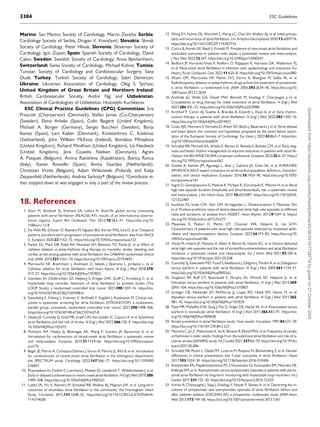
![116. Biersteker TE, Schalij MJ, Treskes RW. Impact of mobile health devices for the detec
tion of atrial fibrillation: systematic review. JMIR Mhealth Uhealth 2021;9:e26161.
https://doi.org/10.2196/26161
117. Romiti GF, Pastori D, Rivera-Caravaca JM, Ding WY, Gue YX, Menichelli D, et al.
Adherence to the ‘atrial fibrillation better care’ pathway in patients with atrial fibrilla
tion: impact on clinical outcomes—a systematic review and meta-analysis of 285,000
patients. Thromb Haemost 2022;122:406–14. https://doi.org/10.1055/a-1515-9630
118. Gallagher C, Elliott AD, Wong CX, Rangnekar G, Middeldorp ME, Mahajan R, et al.
Integrated care in atrial fibrillation: a systematic review and meta-analysis. Heart
2017;103:1947–53. https://doi.org/10.1136/heartjnl-2016-310952
119. Kirchhof P, Benussi S, Kotecha D, Ahlsson A, Atar D, Casadei B, et al. 2016 ESC
Guidelines for the management of atrial fibrillation developed in collaboration with
EACTS. Eur Heart J 2016;37:2893–962. https://doi.org/10.1093/eurheartj/ehw210
120. Hindricks G, Potpara T, Dagres N, Arbelo E, Bax JJ, Blomström-Lundqvist C, et al. 2020
ESC Guidelines for the diagnosis and management of atrial fibrillation developed in col
laboration with the European Association for Cardio-Thoracic Surgery (EACTS): the
task force for the diagnosis and management of atrial fibrillation of the European
Society of Cardiology (ESC) developed with the special contribution of the
European Heart Rhythm Association (EHRA) of the ESC. Eur Heart J 2021;42:
373–498. https://doi.org/10.1093/eurheartj/ehaa612
121. Qvist I, Hendriks JM, Møller DS, Albertsen AE, Mogensen HM, Oddershede GD, et al.
Effectiveness of structured, hospital-based, nurse-led atrial fibrillation clinics: a com
parison between a real-world population and a clinical trial population. Open Heart
2016;3:e000335. https://doi.org/10.1136/openhrt-2015-000335
122. Hendriks JM, de Wit R, Crijns HJ, Vrijhoef HJ, Prins MH, Pisters R, et al. Nurse-led care
vs. usual care for patients with atrial fibrillation: results of a randomized trial of inte
grated chronic care vs. routine clinical care in ambulatory patients with atrial fibrilla
tion. Eur Heart J 2012;33:2692–9. https://doi.org/10.1093/eurheartj/ehs071
123. Carter L, Gardner M, Magee K, Fearon A, Morgulis I, Doucette S, et al. An integrated
management approach to atrial fibrillation. J Am Heart Assoc 2016;5:e002950. https://
doi.org/10.1161/JAHA.115.002950
124. van den Dries CJ, van Doorn S, Rutten FH, Oudega R, van de Leur S, Elvan A, et al.
Integrated management of atrial fibrillation in primary care: results of the ALL-IN clus
ter randomized trial. Eur Heart J 2020;41:2836–44. https://doi.org/10.1093/eurheartj/
ehaa055
125. Abed HS, Wittert GA, Leong DP, Shirazi MG, Bahrami B, Middeldorp ME, et al. Effect
of weight reduction and cardiometabolic risk factor management on symptom burden
and severity in patients with atrial fibrillation: a randomized clinical trial. JAMA 2013;
310:2050–60. https://doi.org/10.1001/jama.2013.280521
126. Pathak RK, Middeldorp ME, Meredith M, Mehta AB, Mahajan R, Wong CX, et al.
Long-term effect of goal-directed weight management in an atrial fibrillation cohort:
a long-term follow-up study (LEGACY). J Am Coll Cardiol 2015;65:2159–69. https://
doi.org/10.1016/j.jacc.2015.03.002
127. Middeldorp ME, Pathak RK, Meredith M, Mehta AB, Elliott AD, Mahajan R, et al.
PREVEntion and regReSsive effect of weight-loss and risk factor modification on atrial
fibrillation: the REVERSE-AF study. Europace 2018;20:1929–35. https://doi.org/10.
1093/europace/euy117
128. Pathak RK, Middeldorp ME, Lau DH, Mehta AB, Mahajan R, Twomey D, et al.
Aggressive risk factor reduction study for atrial fibrillation and implications for the out
come of ablation: the ARREST-AF cohort study. J Am Coll Cardiol 2014;64:2222–31.
https://doi.org/10.1016/j.jacc.2014.09.028
129. Pinho-Gomes AC, Azevedo L, Copland E, Canoy D, Nazarzadeh M, Ramakrishnan R,
et al. Blood pressure-lowering treatment for the prevention of cardiovascular events in
patients with atrial fibrillation: an individual participant data meta-analysis. PLoS Med
2021;18:e1003599. https://doi.org/10.1371/journal.pmed.1003599
130. Parkash R, Wells GA, Sapp JL, Healey JS, Tardif J-C, Greiss I, et al. Effect of aggressive
blood pressure control on the recurrence of atrial fibrillation after catheter ablation: a
randomized, open-label clinical trial (SMAC-AF [Substrate Modification with
Aggressive Blood Pressure Control]). Circulation 2017;135:1788–98. https://doi.org/
10.1161/CIRCULATIONAHA.116.026230
131. McMurray JJ, Adamopoulos S, Anker SD, Auricchio A, Bohm M, Dickstein K, et al. ESC
guidelines for the diagnosis and treatment of acute and chronic heart failure 2012: the
task force for the diagnosis and treatment of acute and chronic heart failure 2012 of
the European Society of Cardiology. Developed in collaboration with the Heart Failure
Association (HFA) of the ESC. Eur Heart J 2012;33:1787–847. https://doi.org/10.1093/
eurheartj/ehs104
132. Olsson LG, Swedberg K, Ducharme A, Granger CB, Michelson EL, McMurray JJ, et al.
Atrial fibrillation and risk of clinical events in chronic heart failure with and without left
ventricular systolic dysfunction: results from the Candesartan in Heart
failure-Assessment of Reduction in Mortality and morbidity (CHARM) program.
J Am Coll Cardiol 2006;47:1997–2004. https://doi.org/10.1016/j.jacc.2006.01.060
133. Kotecha D, Holmes J, Krum H, Altman DG, Manzano L, Cleland JG, et al. Efficacy of
beta blockers in patients with heart failure plus atrial fibrillation: an individual-patient
data meta-analysis. Lancet 2014;384:2235–43. https://doi.org/10.1016/S0140-
6736(14)61373-8
134. Zannad F, McMurray JJ, Krum H, van Veldhuisen DJ, Swedberg K, Shi H, et al.
Eplerenone in patients with systolic heart failure and mild symptoms. N Engl J Med
2011;364:11–21. https://doi.org/10.1056/NEJMoa1009492
135. McMurray JJ, Packer M, Desai AS, Gong J, Lefkowitz MP, Rizkala AR, et al.
Angiotensin-neprilysin inhibition versus enalapril in heart failure. N Engl J Med 2014;
371:993–1004. https://doi.org/10.1056/NEJMoa1409077
136. Pandey AK, Okaj I, Kaur H, Belley-Cote EP, Wang J, Oraii A, et al. Sodium-glucose co-
transporter inhibitors and atrial fibrillation: a systematic review and meta-analysis of
randomized controlled trials. J Am Heart Assoc 2021;10:e022222. https://doi.org/10.
1161/JAHA.121.022222
137. McDonagh TA, Metra M, Adamo M, Gardner RS, Baumbach A, Bohm M, et al. 2021
ESC Guidelines for the diagnosis and treatment of acute and chronic heart failure.
Eur Heart J 2021;42:3599–726. https://doi.org/10.1093/eurheartj/ehab368
138. Solomon SD, McMurray JJV, Claggett B, de Boer RA, DeMets D, Hernandez AF, et al.
Dapagliflozin in heart failure with mildly reduced or preserved ejection fraction. N Engl
J Med 2022;387:1089–98. https://doi.org/10.1056/NEJMoa2206286
139. Anker SD, Butler J, Filippatos G, Ferreira JP, Bocchi E, Böhm M, et al. Empagliflozin in
heart failure with a preserved ejection fraction. N Engl J Med 2021;385:1451–61.
https://doi.org/10.1056/NEJMoa2107038
140. Bhatt DL, Szarek M, Steg PG, Cannon CP, Leiter LA, McGuire DK, et al. Sotagliflozin in
patients with diabetes and recent worsening heart failure. N Engl J Med 2021;384:
117–28. https://doi.org/10.1056/NEJMoa2030183
141. Pathak RK, Elliott A, Middeldorp ME, Meredith M, Mehta AB, Mahajan R, et al. Impact of
CARDIOrespiratory FITness on arrhythmia recurrence in obese individuals with atrial
fibrillation: the CARDIO-FIT study. J Am Coll Cardiol 2015;66:985–96. https://doi.org/
10.1016/j.jacc.2015.06.488
142. Hegbom F, Stavem K, Sire S, Heldal M, Orning OM, Gjesdal K. Effects of short-term
exercise training on symptoms and quality of life in patients with chronic atrial fibrilla
tion. Int J Cardiol 2007;116:86–92. https://doi.org/10.1016/j.ijcard.2006.03.034
143. Osbak PS, Mourier M, Kjaer A, Henriksen JH, Kofoed KF, Jensen GB. A randomized
study of the effects of exercise training on patients with atrial fibrillation. Am Heart J
2011;162:1080–7. https://doi.org/10.1016/j.ahj.2011.09.013
144. Malmo V, Nes BM, Amundsen BH, Tjonna AE, Stoylen A, Rossvoll O, et al. Aerobic
interval training reduces the burden of atrial fibrillation in the short term: a randomized
trial. Circulation 2016;133:466–73. https://doi.org/10.1161/CIRCULATIONAHA.115.
018220
145. Oesterle A, Giancaterino S, Van Noord MG, Pellegrini CN, Fan D, Srivatsa UN, et al.
Effects of supervised exercise training on atrial fibrillation: a meta-analysis of rando
mized controlled trials. J Cardiopulm Rehabil Prev 2022;42:258–65. https://doi.org/10.
1097/HCR.0000000000000665
146. Elliott AD, Verdicchio CV, Mahajan R, Middeldorp ME, Gallagher C, Mishima RS, et al.
An exercise and physical activity program in patients with atrial fibrillation: the
ACTIVE-AF randomized controlled trial. JACC Clin Electrophysiol 2023;9:455–65.
https://doi.org/10.1016/j.jacep.2022.12.002
147. Voskoboinik A, Kalman JM, De Silva A, Nicholls T, Costello B, Nanayakkara S, et al.
Alcohol abstinence in drinkers with atrial fibrillation. N Engl J Med 2020;382:20–8.
https://doi.org/10.1056/NEJMoa1817591
148. Holmqvist F, Guan N, Zhu Z, Kowey PR, Allen LA, Fonarow GC, et al. Impact of ob
structive sleep apnea and continuous positive airway pressure therapy on outcomes in
patients with atrial fibrillation-results from the Outcomes Registry for Better Informed
Treatment of Atrial Fibrillation (ORBIT-AF). Am Heart J 2015;169:647–654.e2. https://
doi.org/10.1016/j.ahj.2014.12.024
149. Fein AS, Shvilkin A, Shah D, Haffajee CI, Das S, Kumar K, et al. Treatment of obstructive
sleep apnea reduces the risk of atrial fibrillation recurrence after catheter ablation. J Am
Coll Cardiol 2013;62:300–5. https://doi.org/10.1016/j.jacc.2013.03.052
150. Li L, Wang ZW, Li J, Ge X, Guo LZ, Wang Y, et al. Efficacy of catheter ablation of atrial
fibrillation in patients with obstructive sleep apnoea with and without continuous posi
tive airway pressure treatment: a meta-analysis of observational studies. Europace
2014;16:1309–14. https://doi.org/10.1093/europace/euu066
151. Naruse Y, Tada H, Satoh M, Yanagihara M, Tsuneoka H, Hirata Y, et al. Concomitant
obstructive sleep apnea increases the recurrence of atrial fibrillation following radio
frequency catheter ablation of atrial fibrillation: clinical impact of continuous positive
airway pressure therapy. Heart Rhythm 2013;10:331–7. https://doi.org/10.1016/j.
hrthm.2012.11.015
152. Qureshi WT, Nasir UB, Alqalyoobi S, O’Neal WT, Mawri S, Sabbagh S, et al.
Meta-analysis of continuous positive airway pressure as a therapy of atrial fibrillation
in obstructive sleep apnea. Am J Cardiol 2015;116:1767–73. https://doi.org/10.1016/j.
amjcard.2015.08.046
153. Shukla A, Aizer A, Holmes D, Fowler S, Park DS, Bernstein S, et al. Effect of obstructive
sleep apnea treatment on atrial fibrillation recurrence: a meta-analysis. JACC Clin
Electrophysiol 2015;1:41–51. https://doi.org/10.1016/j.jacep.2015.02.014
154. Nalliah CJ, Wong GR, Lee G, Voskoboinik A, Kee K, Goldin J, et al. Impact of CPAP on
the atrial fibrillation substrate in obstructive sleep apnea: the SLEEP-AF study. JACC Clin
Electrophysiol 2022;8:869–77. https://doi.org/10.1016/j.jacep.2022.04.015
155. Kadhim K, Middeldorp ME, Elliott AD, Jones D, Hendriks JML, Gallagher C, et al.
Self-reported daytime sleepiness and sleep-disordered breathing in patients with atrial
ESC Guidelines 3387
Downloaded
from
https://academic.oup.com/eurheartj/article/45/36/3314/7738779
by
guest
on
21
October
2024](https://image.slidesharecdn.com/atriafibr24-241118112337-7c4f5251/85/ATRIAL-FIBRILLATION-2024-Guidelines-ESC-74-320.jpg)
![239. Hart RG, Pearce LA, Aguilar MI. Meta-analysis: antithrombotic therapy to prevent
stroke in patients who have nonvalvular atrial fibrillation. Ann Intern Med 2007;146:
857–67. https://doi.org/10.7326/0003-4819-146-12-200706190-00007
240. Ruff CT, Giugliano RP, Braunwald E, Hoffman EB, Deenadayalu N, Ezekowitz MD, et al.
Comparison of the efficacy and safety of new oral anticoagulants with warfarin in pa
tients with atrial fibrillation: a meta-analysis of randomised trials. Lancet 2014;383:
955–62. https://doi.org/10.1016/S0140-6736(13)62343-0
241. Sjalander S, Sjalander A, Svensson PJ, Friberg L. Atrial fibrillation patients do not benefit
from acetylsalicylic acid. Europace 2014;16:631–8. https://doi.org/10.1093/europace/
eut333
242. Connolly SJ, Eikelboom J, Joyner C, Diener HC, Hart R, Golitsyn S, et al. Apixaban in
patients with atrial fibrillation. N Engl J Med 2011;364:806–17. https://doi.org/10.1056/
NEJMoa1007432
243. van Doorn S, Rutten FH, O’Flynn CM, Oudega R, Hoes AW, Moons KGM, et al.
Effectiveness of CHA2DS2-VASc based decision support on stroke prevention in atrial
fibrillation: a cluster randomised trial in general practice. Int J Cardiol 2018;273:123–9.
https://doi.org/10.1016/j.ijcard.2018.08.096
244. Borre ED, Goode A, Raitz G, Shah B, Lowenstern A, Chatterjee R, et al. Predicting
thromboembolic and bleeding event risk in patients with non-valvular atrial fibrillation:
a systematic review. Thromb Haemost 2018;118:2171–87. https://doi.org/10.1055/s-
0038-1675400
245. van der Endt VHW, Milders J, de Vries BBLP, Trines SA, Groenwold RHH, Dekkers
OM, et al. Comprehensive comparison of stroke risk score performance: a systematic
review and meta-analysis among 6 267 728 patients with atrial fibrillation. Europace
2022;24:1739–53. https://doi.org/10.1093/europace/euac096
246. Quinn GR, Severdija ON, Chang Y, Singer DE. Wide variation in reported rates of
stroke across cohorts of patients with atrial fibrillation. Circulation 2017;135:
208–19. https://doi.org/10.1161/CIRCULATIONAHA.116.024057
247. Pisters R, Lane DA, Marin F, Camm AJ, Lip GY. Stroke and thromboembolism in atrial
fibrillation. Circ J 2012;76:2289–304. https://doi.org/10.1253/circj.CJ-12-1036
248. Hohnloser SH, Hijazi Z, Thomas L, Alexander JH, Amerena J, Hanna M, et al. Efficacy of
apixaban when compared with warfarin in relation to renal function in patients with
atrial fibrillation: insights from the ARISTOTLE trial. Eur Heart J 2012;33:2821–30.
https://doi.org/10.1093/eurheartj/ehs274
249. Fox KA, Piccini JP, Wojdyla D, Becker RC, Halperin JL, Nessel CC, et al. Prevention of
stroke and systemic embolism with rivaroxaban compared with warfarin in patients
with non-valvular atrial fibrillation and moderate renal impairment. Eur Heart J 2011;
32:2387–94. https://doi.org/10.1093/eurheartj/ehr342
250. Yaghi S, Henninger N, Giles JA, Leon Guerrero C, Mistry E, Liberman AL, et al.
Ischaemic stroke on anticoagulation therapy and early recurrence in acute cardioem
bolic stroke: the IAC study. J Neurol Neurosurg Psychiatry 2021;92:1062–7. https://doi.
org/10.1136/jnnp-2021-326166
251. Ocak G, Khairoun M, Khairoun O, Bos WJW, Fu EL, Cramer MJ, et al. Chronic kidney
disease and atrial fibrillation: a dangerous combination. PLoS One 2022;17:e0266046.
https://doi.org/10.1371/journal.pone.0266046
252. Seiffge DJ, De Marchis GM, Koga M, Paciaroni M, Wilson D, Cappellari M, et al.
Ischemic stroke despite oral anticoagulant therapy in patients with atrial fibrillation.
Ann Neurol 2020;87:677–87. https://doi.org/10.1002/ana.25700
253. Paciaroni M, Agnelli G, Falocci N, Caso V, Becattini C, Marcheselli S, et al. Prognostic
value of trans-thoracic echocardiography in patients with acute stroke and atrial fibril
lation: findings from the RAF study. J Neurol 2016;263:231–7. https://doi.org/10.1007/
s00415-015-7957-3
254. Hijazi Z, Oldgren J, Siegbahn A, Wallentin L. Application of biomarkers for risk strati
fication in patients with atrial fibrillation. Clin Chem 2017;63:152–64. https://doi.org/10.
1373/clinchem.2016.255182
255. Singleton MJ, Yuan Y, Dawood FZ, Howard G, Judd SE, Zakai NA, et al. Multiple blood
biomarkers and stroke risk in atrial fibrillation: the REGARDS study. J Am Heart Assoc
2021;10:e020157. https://doi.org/10.1161/JAHA.120.020157
256. Wu VC, Wu M, Aboyans V, Chang SH, Chen SW, Chen MC, et al. Female sex as a risk
factor for ischaemic stroke varies with age in patients with atrial fibrillation. Heart 2020;
106:534–40. https://doi.org/10.1136/heartjnl-2019-315065
257. Mikkelsen AP, Lindhardsen J, Lip GY, Gislason GH, Torp-Pedersen C, Olesen JB.
Female sex as a risk factor for stroke in atrial fibrillation: a nationwide cohort study.
J Thromb Haemost 2012;10:1745–51. https://doi.org/10.1111/j.1538-7836.2012.
04853.x
258. Antonenko K, Paciaroni M, Agnelli G, Falocci N, Becattini C, Marcheselli S, et al.
Sex-related differences in risk factors, type of treatment received and outcomes in pa
tients with atrial fibrillation and acute stroke: results from the RAF study (early recur
rence and cerebral bleeding in patients with acute ischemic stroke and atrial
fibrillation). Eur Stroke J 2017;2:46–53. https://doi.org/10.1177/2396987316679577
259. Wang X, Mobley AR, Tica O, Okoth K, Ghosh RE, Myles P, et al. Systematic approach
to outcome assessment from coded electronic healthcare records in the
DaRe2THINK NHS-embedded randomized trial. Eur Heart J - Dig Health 2022;3:
426–36. https://doi.org/10.1093/ehjdh/ztac046
260. Rivard L, Khairy P, Talajic M, Tardif JC, Nattel S, Bherer L, et al. Blinded randomized trial
of anticoagulation to prevent ischemic stroke and neurocognitive impairment in atrial
fibrillation (BRAIN-AF): methods and design. Can J Cardiol 2019;35:1069–77. https://
doi.org/10.1016/j.cjca.2019.04.022
261. Chung S, Kim TH, Uhm JS, Cha MJ, Lee JM, Park J, et al. Stroke and systemic embolism
and other adverse outcomes of heart failure with preserved and reduced ejection frac
tion in patients with atrial fibrillation (from the COmparison study of Drugs for symp
tom control and complication prEvention of Atrial Fibrillation [CODE-AF]). Am J
Cardiol 2020;125:68–75. https://doi.org/10.1016/j.amjcard.2019.09.035
262. Uhm JS, Kim J, Yu HT, Kim TH, Lee SR, Cha MJ, et al. Stroke and systemic embolism in
patients with atrial fibrillation and heart failure according to heart failure type. ESC
Heart Fail 2021;8:1582–9. https://doi.org/10.1002/ehf2.13264
263. McMurray JJ, Ezekowitz JA, Lewis BS, Gersh BJ, van Diepen S, Amerena J, et al. Left ven
tricular systolic dysfunction, heart failure, and the risk of stroke and systemic embolism
in patients with atrial fibrillation: insights from the ARISTOTLE trial. Circ Heart Fail
2013;6:451–60. https://doi.org/10.1161/CIRCHEARTFAILURE.112.000143
264. Kim D, Yang PS, Kim TH, Jang E, Shin H, Kim HY, et al. Ideal blood pressure in patients
with atrial fibrillation. J Am Coll Cardiol 2018;72:1233–45. https://doi.org/10.1016/j.jacc.
2018.05.076
265. Lip GY, Clementy N, Pericart L, Banerjee A, Fauchier L. Stroke and major bleeding risk
in elderly patients aged ≥75 years with atrial fibrillation: the Loire valley atrial fibrilla
tion project. Stroke 2015;46:143–50. https://doi.org/10.1161/STROKEAHA.114.
007199
266. American Diabetes Association Professional Practice Committee. 2. Classification and
diagnosis of diabetes: standards of medical care in diabetes—2022. Diabetes Care 2022;
45:S17–38. https://doi.org/10.2337/dc22-S002
267. Steensig K, Olesen KKW, Thim T, Nielsen JC, Jensen SE, Jensen LO, et al. Should the
presence or extent of coronary artery disease be quantified in the CHA2DS2-VASc
score in atrial fibrillation? A report from the western Denmark heart registry.
Thromb Haemost 2018;118:2162–70. https://doi.org/10.1055/s-0038-1675401
268. Zabalgoitia M, Halperin JL, Pearce LA, Blackshear JL, Asinger RW, Hart RG.
Transesophageal echocardiographic correlates of clinical risk of thromboembolism
in nonvalvular atrial fibrillation. Stroke prevention in atrial fibrillation III investigators.
J Am Coll Cardiol 1998;31:1622–6. https://doi.org/10.1016/S0735-1097(98)00146-6
269. Stroke Prevention in Atrial Fibrillation Investigators Committee on Echocardiography.
Transesophageal echocardiography in atrial fibrillation: standards for acquisition and
interpretation and assessment of interobserver variability. Stroke prevention in atrial
fibrillation investigators committee on echocardiography. J Am Soc Echocardiogr 1996;
9:556–66. https://doi.org/10.1016/S0894-7317(96)90127-3
270. Lozier MR, Sanchez AM, Lee JJ, Donath EM, Font VE, Escolar E. Thromboembolic out
comes of different anticoagulation strategies for patients with atrial fibrillation in the
setting of hypertrophic cardiomyopathy: a systematic review. J Atr Fibrillation 2019;
12:2207. https://doi.org/10.4022/jafib.2207
271. Guttmann OP, Rahman MS, O’Mahony C, Anastasakis A, Elliott PM. Atrial fibrillation
and thromboembolism in patients with hypertrophic cardiomyopathy: systematic re
view. Heart 2014;100:465–72. https://doi.org/10.1136/heartjnl-2013-304276
272. Guttmann OP, Pavlou M, O’Mahony C, Monserrat L, Anastasakis A, Rapezzi C, et al.
Prediction of thromboembolic risk in patients with hypertrophic cardiomyopathy
(HCM risk-CVA). Eur J Heart Fail 2015;17:837–45. https://doi.org/10.1002/ejhf.316
273. Vilches S, Fontana M, Gonzalez-Lopez E, Mitrani L, Saturi G, Renju M, et al. Systemic
embolism in amyloid transthyretin cardiomyopathy. Eur J Heart Fail 2022;24:
1387–96. https://doi.org/10.1002/ejhf.2566
274. Lee SE, Park JK, Uhm JS, Kim JY, Pak HN, Lee MH, et al. Impact of atrial fibrillation on
the clinical course of apical hypertrophic cardiomyopathy. Heart 2017;103:1496–501.
https://doi.org/10.1136/heartjnl-2016-310720
275. Hirota T, Kubo T, Baba Y, Ochi Y, Takahashi A, Yamasaki N, et al. Clinical profile of
thromboembolic events in patients with hypertrophic cardiomyopathy in a regional
Japanese cohort–results from Kochi RYOMA study. Circ J 2019;83:1747–54. https://
doi.org/10.1253/circj.CJ-19-0186
276. Hsu JC, Huang YT, Lin LY. Stroke risk in hypertrophic cardiomyopathy patients with
atrial fibrillation: a nationwide database study. Aging (Albany NY) 2020;12:24219–27.
https://doi.org/10.18632/aging.104133
277. Chao TF, Lip GYH, Liu CJ, Lin YJ, Chang SL, Lo LW, et al. Relationship of aging and
incident comorbidities to stroke risk in patients with atrial fibrillation. J Am Coll
Cardiol 2018;71:122–32. https://doi.org/10.1016/j.jacc.2017.10.085
278. Weijs B, Dudink E, de Vos CB, Limantoro I, Tieleman RG, Pisters R, et al. Idiopathic
atrial fibrillation patients rapidly outgrow their low thromboembolic risk: a 10-year
follow-up study. Neth Heart J 2019;27:487–97. https://doi.org/10.1007/s12471-019-
1272-z
279. Bezabhe WM, Bereznicki LR, Radford J, Wimmer BC, Salahudeen MS, Garrahy E, et al.
Stroke risk reassessment and oral anticoagulant initiation in primary care patients with
atrial fibrillation: A ten-year follow-up. Eur J Clin Invest 2021;51:e13489. https://doi.org/
10.1111/eci.13489
280. Fauchier L, Bodin A, Bisson A, Herbert J, Spiesser P, Clementy N, et al. Incident co
morbidities, aging and the risk of stroke in 608,108 patients with atrial fibrillation: a na
tionwide analysis. J Clin Med 2020;9:1234. https://doi.org/10.3390/jcm9041234
3390 ESC Guidelines
Downloaded
from
https://academic.oup.com/eurheartj/article/45/36/3314/7738779
by
guest
on
21
October
2024](https://image.slidesharecdn.com/atriafibr24-241118112337-7c4f5251/85/ATRIAL-FIBRILLATION-2024-Guidelines-ESC-77-320.jpg)