Download to read offline
![Hindawi Publishing Corporation
Evidence-Based Complementary and Alternative Medicine
Volume 2013, Article ID 587827, 12 pages
http://dx.doi.org/10.1155/2013/587827
Review Article
50 Years of Bong-Han Theory and 10 Years of
Primo Vascular System
Kwang-Sup Soh,1
Kyung A. Kang,2
and Yeon Hee Ryu3
1
Nano Primo Research Center, Advanced Institute of Convergence Technology, Seoul National University,
Suwon 443-270, Republic of Korea
2
Departments of Chemical Engineering, University of Louisville, Louisville, KY 40292, USA
3
Korea Institute of Oriental Medicine, Daejon 305-811, Republic of Korea
Correspondence should be addressed to Kwang-Sup Soh; kssoh1@gmail.com and Yeon Hee Ryu; yhryu@kiom.re.kr
Received 24 December 2012; Accepted 16 March 2013
Academic Editor: Xianghong Jing
Copyright © 2013 Kwang-Sup Soh et al. This is an open access article distributed under the Creative Commons Attribution License,
which permits unrestricted use, distribution, and reproduction in any medium, provided the original work is properly cited.
The primo vascular system (PVS) was first introduced by Bong-Han Kim via his five research reports. Among these the third report
was most extensive and conclusive in terms of the PVS anatomy and physiology relating to the acupuncture meridians. His study
results, unfortunately, were not reproduced by other scientists because he did not describe the materials and methods in detail. In
2002, a research team in Seoul National University reinitiated the PVS research, confirmed the existence of PVS in various organs,
and discovered new characteristics of PVS. Two important examples are as follows: PVS was found in the adipose tissue and around
cancer tissues. In parallel to these new findings, new methods for observing and identifying PVS were developed. Studies on the
cell and material content inside the PVS, including the immune function cells and stem cells, are being progressed. In this review,
Bong-Han Kim’s study results in his third report are summarized, and the new results after him are briefly reviewed. In the last
section, the obstacles in finding the PVS in the skin as an anatomical structure of acupuncture meridian are discussed.
1. Introduction: A Brief Historical Review
It was in 1962 when Bong-Han Kim (hereafter BH Kim or
Kim) reported his first study results on the anatomical entity
of acupuncture meridians (AM) [1]. He then established
and became the director of National Acupuncture Meridian
Research Institute in 1963 in Pyungyang, North Korea. The
Institute produced four additional reports on this new system
[2–5] until October 1965, when the institute abruptly closed
for some unknown reasons. His fate after the event is still
unknown.
The first report was very brief and mostly about the
electric response of acupoints, which was probably not con-
sidered very exciting. The second report, however, contains
the discovery of a completely new system, constituting node-
like anatomical structures at the acupoints and the tube-
like structure (Kim claimed it to be the AM) connected to
the nodes in the skin [2]. The research team named the
nodes the Bonghan corpuscles (currently renamed as primo
nodes) and the tubes the Bonghan ducts (primo vessels).
They also found that this new system existed not only in
the skin but also throughout the body, including on the
surfaces of body organs and inside the blood and lymph
vessels. This new discovery became the foundation of the
Bong-Han theory. To publicize its scientific achievements,
the North Korean government translated Kim’s second report
in various languages including English and disseminated it
to most major libraries in the world [6]. The third report
was an extension of the second one, observing the entire
network of the Bong-Han system (renamed as primo vascular
system (PVS)) in the mammalian body [3]. The fourth one
was about the “Sanal,” (renamed to be Primo-microcell (P-
microcell)), whose functions, he claimed, were regeneration
and/or repair, as totipotent stem cells [4]. Sanal is a Korean
word and its direct translation in English is “live egg.” The
last (fifth) one was a brief report about the hematopoietic
function of the Sanal [5]. These five publications were reports
rather than journal articles, and they described mainly results
with insufficient information on methods and materials. The
introduction and discussion sections were very short.](https://image.slidesharecdn.com/587827-160912034746/75/Bong-Han-Theory-1-2048.jpg)
![2 Evidence-Based Complementary and Alternative Medicine
Shortly after Kim’s fifth report, neither he nor his pub-
lications reappeared in public for some unknown reason,
and his work was completely neglected by the North Korean
government. Outside the North Korea (Democratic People’s
Republic of Korea; DPRK), there have been several attempts
to reproduce Kim’s results but without success, probably
because details of his methods to identify this new organ
were described either in any of his reports or elsewhere.
Nevertheless, until now, there has been no known seri-
ous attempts to either confirm or negate his claims, with
exceptions of the following two cases: one was by Kellner,
who thoroughly investigated the acupoints in a histological
manner but failed to find the structure that Kim had claimed
[7]. In our scientific opinion, Kellner’s conventional method
for histological study was not appropriate for detecting the
anticipated structure in the skin, because in the cross section
of the tissue containing an acupoint this new organ may
not be differentiated from its surrounding tissues. Optically
and histologically, both look very similar if they are seen
in their cross sections. Its presence can be revealed most
likely in the longitudinal view, with application of appropriate
dyes, according to our own experiences for the past ten
years. Another case was by Fujiwara and Yu, who partially
confirmed Kim’s discovery but incompletely [8]. Fujiwara,
who was an assistant professor in anatomy, then, later recalled
that it took about a half year of hard work to get some positive
results [9]. He was able to reproduce Kim’s results inside
blood vessels and on the surfaces of organs, which eventually
helped the research of Soh, one of the coauthors for this paper.
Soh was a professor in the Department of Physics at
the Seoul National University (SNU) during 1976–2011. He
formed Biomedical Physics Laboratory in 2000, initially to
investigate the biological phenomena related to the acupunc-
ture therapy using physical means, such as electricity, mag-
netism, acoustics, and optics. He realized that his inves-
tigation without anatomical bases would not lead to the
fundamental mechanism behind the acupuncture therapy.
Therefore he formed a scientific team to investigate the
Bong-Han theory with Dr. BC Lee as the main experimental
partner. The SNU team soon successfully confirmed the PVS
presence inside the blood vessel of rabbits. However, this
initial success soon faced difficulties in reproducibility. They
realized that the research requires highly skilful researcher
in microsurgery and optical imaging due to the extremely
small size and the semitransparent nature of the organ, which
frequently impeded the progress in their study.
The team, therefore, decided to seek Professor Fujiwara’s
help and Soh visited Dr. Fujiwara in Osaka. Fujiwara him-
self had experiences with failures in finding the PVS on
the surfaces of internal organs for more than six months.
Fujiwara was so kind to provide a movie containing the
experimental procedures that he had developed in 1960s.
With Dr. Fujiwara’s method, the SNU team then was able
to reproduce his results. Since then, the team identified the
PVS floating on the surfaces of intestines, liver, stomach, and
bladder of rabbits. Soon, more thorough, histological and
morphological studies on PVS were performed to confirm
Kim’s claims. With techniques and experiences obtained for
the organ-surface PVS, the team moved forward to find
the PVS floating inside lymph vessels. More importantly,
a technique of using Trypan blue for PVS identification
was developed by Dr. BC Lee, which enabled the team to
identify the PVS in other organs, such as in the bovine heart,
abdominal adipose tissues, brain ventricles, and the central
canal of spinal cords. The Trypan blue technique also led to
the discovery of the unique characteristics of the PVS in/on
cancerous tumors (cancer PVS). This may probably be one of
the most significant findings in the medical science because
cancer is one of the most serious, life-threatening diseases.
Until 2008, the SNU-team was the only one performing
the PVS research and, therefore, the research progress was
rather slow. In 2009, a review article, written by Soh, cov-
ering the PVS research progress during 2002 and 2008 was
published in the Journal of Acupuncture Meridian Studies
[10]. Since then several research teams in Korea participated
in the PVS research, and the number of teams and their
research subjects have been steadily growing. Outside Korea,
the PVS gained interest as a research topic, mainly in China
and USA. In September 2010, an international symposium
on the PVS, The Primo Vascular System, Its Role in Cancer
and Regeneration, was held in Korea, and its proceedings was
published by the Springer Publishing Company in 2011 [11].
Recently, a research team led by BS Kwon at the National
Cancer Center of Korea [12] confirmed that the PVS was
abundant with immune cells such as macrophages and mast
cells, which had been previously noticed by the SNU team
[13]. In addition, primo nodes were also found to be packed
with very small, embryonic stem cell- like cells. These data are
consistent with the claims by BH Kim’s on the properties of
the PVS on regeneration and wound healing [4].
In the remaining part of the paper, we summarize the
content of Kim’s third report, which relates the AM system
with the PVS, with our comments. His report contents
are compared with the recent works reported by the SNU
group and other PVS scientists. Kim’s study results that
were scientifically verified by the PVS researchers were
described first, and then new discoveries on the PVS after
Kim were introduced. The desired directions for the future
PVS research were also discussed.
2. Acupuncture Meridian System
The title of Kim’s third report is “The Kyungrak (經 絡)
System” [3], and it is officially submitted by the “Kyungrak
Institute (經絡 研究院) of Democratic People’s Republic of
Korea,” where Kim was the director. The English translation
of the title is “Acupuncture Meridian System.” The report
covers research results on the PVS network, and the scientific
standard of its content is more advanced and comprehensive
than that of his previous two reports. This was, in fact, his last
report relating the acupuncture meridians with the PVS. The
last two of his reports were about the “Sanals” or P-microcells
[4, 5], and they were published shortly before the institute was
abruptly closed in 1965.
The English translated table of content of the third report
was presented in Box 1 for the readers to easily capture the
breadth of Kim’s work. As can be seen in the box, the third](https://image.slidesharecdn.com/587827-160912034746/75/Bong-Han-Theory-2-2048.jpg)
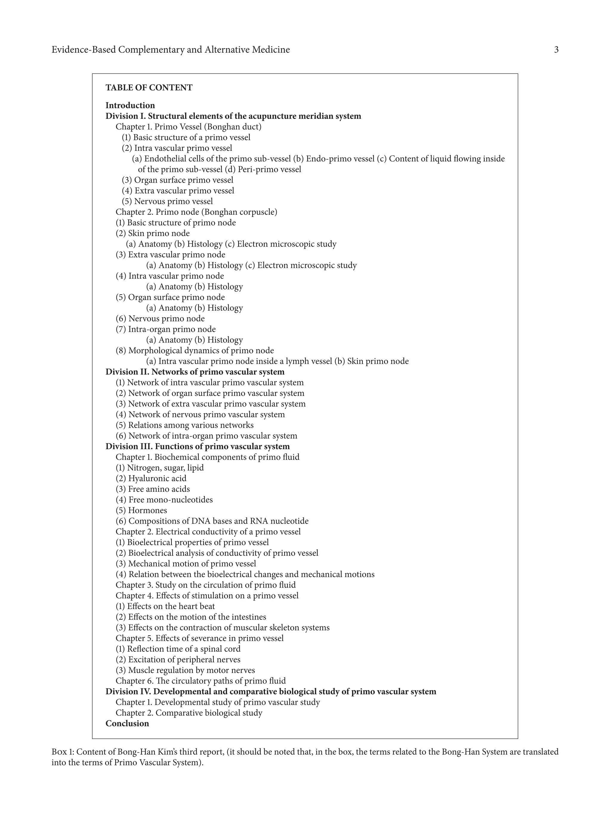
![4 Evidence-Based Complementary and Alternative Medicine
report covers not only anatomical and histological aspects of
PVS but also its basic physiological aspects. The conclusion
section of the report is in fact a good summary of the
report, which was highly beneficial to future PVS researchers.
We, therefore, translated this section (pages 36–38 of Kim’s
third report) with our own scientific comments, although
the section by itself was already published in the book The
Primo Vascular System [14]. We also added some of the
important scientific progresses in the field of PVS, since Kim’s
publications, in subsequent sections.
2.1. English Translation of Conclusion Section of Bong-Han
Kim’s Third Report with Authors’ Comments
2.1.1. The Bong-Han System (BHS) Is Composed of
Several Subsystems
(A) These subsystems have common properties of
possessing Bonghan ducts (BHDs) and Bonghan
corpuscles (BHCs). All BHCs are interconnected via
BHDs. BHDs connect BHCs. A BHD is composed of
one to tens of Bonghan ductules.
(1) A ductule has a thin layer, composed of
endothelial cells with rod-shaped nuclei. It is
surrounded by an external membrane (endo-
BHD), which is made of smooth muscle-like
cells and fine argentaffin fibers. (Comments:
Kwon et al. observed epithelial cells rather than
smooth muscle-like cells [12].) The interluminal
space in a BHD is filled with fibrous and amor-
phous materials. These ductules are wrapped
together with a membrane (peri-BHD) to form
a single BHD. This outer membrane is made of
membranous cells. In the lumen of a ductule,
basophilic the granules and nucleus-like bodies
are present.
(2) The BHC is essentially formed by the
enlargement, branching, or merging of the
ductule. Also, the basic compositions of the
BHC are the outer membrane of the ductules
and the reticular fibers, extracellular matrices
(ECM) between ductules. Inside the lumen of
the BHD, which is extended from the BHC,
basophilic granules, cell components, and chro-
maffin granules are present.
(B) The BHS is classified as described in the following.
(1) Intravascular BHS. This BHS class consists
of the intravascular (IV) BHDs and BHCs. It
is systematically distributed inside blood and
lymphatic vessels along the vessel and inside
the heart. (Comments: Up to now, the authors
observed that the BHS is only in large caliber
blood or lymphatic vessels. In the BH Kim’s
report, the size of the BHS-containing vessels is
not clearly described.) The BHDs in this class
are very fragile, and the ECM and their outer
membranes (epi-BHD) are not well developed.
The IV-BHC has a structure, particularly similar
to hematopoietic organs. In the reticular BHS,
lymphocyte series and myelocyte-like cells (cells
in bone marrow) are present. Sometimes cells
similar to organ parenchyma cells are gathered
around.
(2) Organ surface BHS. This class of the BHS
consists of the organ surface (OS) BHDs and
BHCs. They freely float on the surfaces of inter-
nal organs and are not associated with blood
or lymphatic vessels. For this class BHDs, the
interluminal materials and the outer membrane
are developed better than those for the IV-BHS.
In the lumens of the BHDs and inside BHCs,
there are cells possessing bright cytoplasm as
well as the basophilic granules.
(3) Extravascular BHS. This BHS class made of
the extravascular (EV) BHDs and BHCs. This
runs along the blood and lymphatic vessels, and
nerves. It is located just outside of them. It is
covered with thick connective tissues. In the
lumens of the BHDs and inside the BHC, many
chromaffin granules are present.
(4) Nervous BHS. This class BHS is composed
of the nervous (N) BHDs and BHCs, and it
floats in the cerebrospinal fluid. Its branches
are distributed in the parenchyma of the central
nervous system and in the peripheral nervous
system.
(5) Intraorgan BHS. Inside the parenchyma of
internal organs, there are intraorgan (IO) BHDs
and BHCs, terminal BHDs, and terminal BHCs.
(Comment: the terminal BHD has only a single
lumen, and it is a type of the ductule.) These
are extension of the BHDs originated from IV-,
EV-, or N-BHDs and present inside of the organ.
Individual BHDs merge together in an IO-BHC
and eventually form terminal sub-BHDs. These
individual terminal sub-BHDs are directly con-
nected to each nucleus of the organ cells. Again,
these fine ductules come out from these cells.
(Comment: In summary, there are five subsystems,
namely, IV-, OS-, EV-, N-, and IO-BHS). The BHS
subclasses are well connected to each other. The IV-
BHS is connected to the OS-BHS after coming out of
the vessel wall. It is also connected to the EV-BHS via
the EV-BHC. The OS-BHS is connected to the EV-
BHS and the N-BHS. The communication among
BHS subclasses is well established.
2.1.2. The BHS Is Made of Multiple Systems Circulating Bong-
han Liquor
(A) Biochemical compositions of Bonghan liquor
(i.e., liquid flowing inside the PVS) are](https://image.slidesharecdn.com/587827-160912034746/75/Bong-Han-Theory-4-2048.jpg)

![6 Evidence-Based Complementary and Alternative Medicine
3. Confirmation of BH Kim’s Study Results
The nature and the scale of BH Kim’s accomplishments on
the PVS are enormous. Since 2002, selected parts of BH
Kim’s studies were repeated, and his results were confirmed
to be accurate. This section summarizes BH Kim’s findings
that were verified fifty years later, by various scientists. Here,
we briefly list the original terms and the new ones of the
system, for your convenience. Bong-Han system (BHS) =
primo vascular system (PVS), Bonghan duct (BHD) = primo
vessel (PV), Bonghan corpuscle (BHC) = primo node (PN),
Bonghan ductule = P-subvessel, Bonghan liquor = primo
fluid (P-fluid), and Sanal = p-microcell.
3.1. Animal Species and Organs Studied. Animal species
studied for identifying the PVS were mainly rabbits, rats, and
mice. For a few cases, pigs, dogs, cows, and human placentas
were also studied. Among the PVS subclasses, defined by Kim
(Box 1), the IV-, OS-, and N-PVS were confirmed partially, if
not completely.
The IV-PVS floating inside blood vessels was first iden-
tified in the abdominal artery and the caudal vena cava of
rabbits [14], rats [15], and mice [16]. More importantly, the
PVS in the atrium of a bovine heart was found to form a
floating network [17]. The PVS in the sagittal sinus of a rat
brain was recently identified [18].
The IV-PVS floating inside lymph vessels, was visualized
with help of Janus Green B [19], fluorescent nanoparticles
[20], Alcian blue [21] and with no contrast agent [22]. The
morphological information on PVS [23] and protocols used
for PVS related experiments [24] were recently published.
The fact that the PNs possess large amount of cells and
granules related the immune system may imply that the PVS
is involved in protecting function of the body [12].
The OS-PVS floating on the surfaces of internal organs
was observed in rabbits [25] rats [26], mice [27], dogs
[28], and pigs [29]. The structure of this PVS subclass was
characterized by the optical [25], and electron microscopy
[13, 30].
The N-PVS floating in the cerebrospinal fluid (CSF) in the
brain of a rabbit [31] and a rat [32] was optically observed, by
use of Trypan blue. The PVS in the subarachnoidal space of
the rabbit [33] and rat [34] brains, and in the spine of the rat
[35] and pig [36] were also identified.
All PVS observed and listed previously were floating in
the body fluid, such as blood, lymph, abdominal fluid, and
CSF.
3.2. Brief Summary of Important PVS Characteristics Verified
3.2.1. Confirmation of PV Features. One of the most dis-
tinguished anatomical features of the PV is its bundle-like
structure made of multiple subvessels. Figure 1(a) shows a
schematic diagram of a PV prepared by Kim [3]. Throughout
BH Kim’s five reports, he, however, did not provide any actual
image or the method to observe the PVS, corresponding
to the diagram. It has been, therefore, difficult to obtain
the PV image with its cross section. The image published
in 2009, with hematoxylin and eosine staining, is shown
in Figure 1(b) [37]. The existence of P-subvessel lumens is
indeed shown in the figure, but the shape of lumens is still
not clearly shown. Thanks to better techniques in the sample
preparation and microscopy, imaging of a PV cross section,
without severe deformation, is now possible. In one of most
recent study results with an OS-PVS, a PV cross section
imaged utilizing transmission electron microscopy (TEM;
Figure 1(c)) [37] shows even the endothelial cell layer of a PV.
The surrounding ECM is made of collagen fibers, as BH Kim
claimed 3. Figure 1(d) shows confocal microscopy images of
the longitudinal and cross sections of a PV sample harvested
from the superior sagittal sinus of a rabbit brain [33], which
clearly shows multiple lumens in a PV. We, therefore, suspect
that the difficulties in appropriately preparing PV samples
and in having good microscopy techniques might be the
reason why BH Kim did not provide images of PV cross
sections. Figure 1(e) is a schematic illustration of the PV
compared with blood and lymphatic vessels displayed by
Ogay et al., which summarizes most distinctive anatomical
features of the PVS [37].
3.2.2. Cells inside the Primo Node. The PN is surrounded
by a thin membrane, usually connected to two or more
PVs (Figure 2(a)), and contains many cells (Figure 2(b)) and
embryonic stem cell-like bodies (Figure 2(c)). Recent studies
on PN have been mainly on the P-microcells (or Sanals) and
other types of cells in it. Presence of many cells involved
in immune functions was first noticed by TEM in the PN
sample obtained from the rabbit organ surfaces [13]. The cell
types and the ratio of the cells in the PNs harvested from the
internal organ surfaces and inside the lymphatic vessels of rats
are [12] mast cells (20%), eosinophils (16%), neutrophils (5%),
histiocytes (53%), lymphocytes (1%), and round immature
stem cells (3%). Although the presence of immune cells in
PNs was not specifically mentioned in BH Kim’s report, he
did emphasize the abundance of chromaffin cells [3], which
was confirmed by Kwon et al. [12].
The presence of BH Kim’s Sanals (P-microcells) in the
PVS was studied by the SNU team from its inception [25]. The
cells were found to show a peculiar motion to the light in the
UV-A range (360 nm) [40, 41]. Later, the budding process for
cell proliferation was identified by atomic force microscopy
[42–44]. An implication for these cells to be embryonic-like
stem cells were made after confirming the expression of the
stem cell biomarkers oct4, nanog, and CD133 [12, 38]. More
studies on the Sanal are being in progress.
3.2.3. The Primo Fluid and Its Flow Direction. BH Kim’s study
results on the primo fluid circulation were confirmed only in
a very limited level. The flow of the primo fluid in a certain
path was demonstrated in the study using Alcian blue, from
the rat acupoint BL-23 in the dorsal skin to the PVS on the
surface of internal organs [45]. The experiment was, however,
not always repeatable, and the reason for this inconsistency is
still unknown.
When Chrome-hematoxylin and fluorescent nanoparti-
cles were injected into testis they were found in the PVs on](https://image.slidesharecdn.com/587827-160912034746/75/Bong-Han-Theory-6-2048.jpg)
![Evidence-Based Complementary and Alternative Medicine 7
Bonghan duct
L
L L
L L
L
E EEAC CBL IBL
AF
AF
Bonghan ductule
(a)
(b)
(A) (B)
(c)
(A) (B)
(D)(C)
(d)
(e)
Blood capillary Lymphatic capillary
20 𝜇m
Porous outer
membrane
Rod-shaped
nucleus
Multiple
ductules (sub-PVs)
inside a PV
Figure 1: PV (Bonghan duct) illustrated in various publications. (a) Schematic diagram of a primo vessel, described by Bong-Han Kim with
Russian terminologies. English translation of the terminology is added in bold [3]. (b) Optical images showing histological characteristics of
a PV. (A) Phase-contrast image of a PV with DAPI. Several sub-PVs (arrow) with rod-shaped nuclei (light blue, arrowhead) are seen. (B) The
cross section of a PV: several lumens (arrows) are seen [37]. (c) Electron microscopy of a partial cross section of a PV. (A) A lumen (asterisk)
of a sub-PV can be seen. (B) Magnified image of the lumen (rectangular area in (A)). The wall (W) of the sub-PV consists of a single layer of
endothelial cells surrounded by fibrin-like fibers. ((C), (D)) Magnified image of (B). L: lumen; M: mitochondria; GER: granular endoplasmic
reticulum; P: cytoplasmic protrusion; PV: pinocytotic vesicles; V: vacuole; and F: fibrin-like fiber [37]. (d) Confocal laser scanning microscope
image of a PV. The main panel is optical microscopy of the longitudinal section of a PV (the middle section, cells with rod-shaped nuclei
in blue), accompanied by a venule and an arteriole on each side (cells with circular nuclei). The lower panel is a cross section of the PV,
showing multiple lumens (open arrowheads), and the venule (asterisk) and the arteriole (two asterisks) on its each side. The PV diameter was
approximately 30 𝜇m [33]. (e) Anatomical comparisons of characteristics of the PV, blood, and lymphatic capillaries. The PV has multiple
sub-PVs within, and the PV’s outermost layer has large pores. The endothelial cells of the sub-PV possess rod-shaped nuclei. E: endothelium;
L: lumen; AC: accessory cell; CBL: complete basal lamina; IBL: incomplete basal lamina; and AF: anchoring filaments [37].
the organ surfaces between the abdominal cavity and the
abdominal wall, although farther tracing was not technically
possible with optical microscopy [46]. Tracing primo fluid
in the PV on rabbit organ surfaces was possible using Alcian
blue, and the flow speed was measured to be 0.3±0.1 mm/sec
[30]. This value was consistent with the values in BH Kim’s
reports [3].
One of the important biochemicals in the primo fluid,
which was mentioned in BH Kim’s report, was catecholamine
(adrenalin and noradrenalin) [3], and its presence was later](https://image.slidesharecdn.com/587827-160912034746/75/Bong-Han-Theory-7-2048.jpg)
![8 Evidence-Based Complementary and Alternative Medicine
(a) (b)
(c)
(d)
5 𝜇m
5 𝜇m
Figure 2: Images of PVS. (a) An image of a PN (Bonghan corpuscle; arrowhead) found on the rabbit small intestine, with PVs (arrows) at
both ends, using methylene blue as the contrast agent [37]. (b) An image of a PN (arrows), which was identified lower part of the superior
sagittal sinus of a rabbit brain [33]. DAPI staining of the nuclei of the cells inside the PN. Very small cells are packed in the PN. ((c), (d))
Immunostaining of the small cells isolated from PNs on the surfaces of rat intestine, for the embryonic stem cell markers Oct4 (red) and
Nanog (green). The scale bar indicates 5 𝜇m [38].
(a) (b)
Figure 3: Images of PNs with different dyes. (a) A PN (BHC) and a PV (BHD) connected to the PN, on the adipose tissue around the rat
small intestine. Alcian blue flow through the PVS left it pale blue. Notice that Alcian blue did not remain in the PVS and, therefore, in situ
tracking of the PVS using this dye was difficult. (b) A PN and PVs, observed near the rat small intestine, stained with Trypan blue. Notice
that the blood vessel and adipose tissue were not stained [39].
confirmed in the PNs on the organ surface of rabbits [47] and
rats [12], using ELISA.
4. New Discoveries
Since the initiation of the PVS research, the SNU team made
significant discoveries on PVS as well as developing new
techniques for identifying PVS in the mammalian body. A few
important and medically relevant findings are presented here.
4.1. The PVS in Adipose Tissues. The presence of the PVS
entering the adipose tissues around the rat small intestine was
first noticed via optical imaging using Alcian blue, which was
injected intravenously at the femoral vein. The Alcian blue
entering a PVS floating inside a blood vessel reached to a PN
in an adipose tissue as shown in Figure 3(a). Unfortunately,
the dye flowed away without showing its previous paths.
Therefore, in situ tracking of the PVS using this dye was found
to be inappropriate [39].
Trypan blue, an alternative, was, therefore, tested, and it
remained inside the PVS, allowing us to visualize the system
in the adipose tissue. Trypan blue was found to stain the
PVS preferentially to the adipose tissues or blood vessels [39].
Figure 3(b) illustrates images of the PVS, using Trypan blue,
in the adipose tissues around the small intestine of a rat. The
presence of PVS in adipose tissues raises conjectures on its
possible roles in connection with regeneration, obesity, and
obesity-related diseases.
4.2. Cancer PVS. Existence of PVS on the surface of the
tumor membranes in a xenografted mouse was first observed
by Yoo et al. [48] of the SNU team. It was soon realized
that PVS was more densely populated in proximity of tumors
(xenografts) in various type of cancerous, human origin](https://image.slidesharecdn.com/587827-160912034746/75/Bong-Han-Theory-8-2048.jpg)
![Evidence-Based Complementary and Alternative Medicine 9
tumors, and, therefore, they were easily identified [27]. More
importantly, significantly large number of cancer cells was
found in PVs connecting the primary to the secondary
tumors [49].
Since these initial findings by the SNU team, research
teams of Akers [50], Hong [51], Heo [52], Islam [53], and Kang
[54] confirmed the presence of high density PVS (cancer
PVS) in close proximity of cancer xenografts in mice. A PV
floating inside a lymph vessel originated from the tumor
xenografted in the abdominal skin of a mouse was also
reported [55].
The Miller team also reported that the cells obtained from
the cancer-PVS of murine xenograft of human originated
lymphoma U937 expressed CD68, CD45, and lysozome. They
also revealed that the immunophenotype of cells inside the
cancer PVS is of U937 cell. The cells also showed hundreds to
thousandsfold, upregulated KLF4, one of the human cancer
stem cell specific transcription factors and an upstream
regulator of NANOG, which maintain the pluripotent and
undifferentiated state of stem cells [53].
4.3. Other New Findings. For the PVS studies, the SNU team
has been using traditional histochemistry techniques with
dyes such as Alcian blue, Trypan blue, chrome-hematoxylin.
Among these the Trypan blue spraying technique invented
by Dr. BC Lee was most effective for many applications.
The team also adopted modern imaging techniques utilizing
fluorescent nanoparticles, quantum dots, immune-affinity
technique, electron microscopy [56], X-ray microscopy, and
GFP expressing cells and animals, which were not available
at the time of BH Kim. DAPI staining to check the shape of
cell nuclei is one of the important and frequently used new
techniques for the PV identification.
In Kim’s reports, only white blood cells among cells
and biochemicals are involved in the immune system. The
concept of the stem cell, especially of the adult stem cell,
was not very well known in his time, but he claimed that P-
microcell (Sanal) is the main agent for wound healing and
regeneration, which are the two fundamental roles of stem
cells [4]. Considering that the Sanal size is very small (1–
5 𝜇m), an important question is the relation between the
Sanal and the very small embryonic-like stem cells (VSEL)
described by Ratajczak et al. [57], because both seem to
have very similar characteristics. The budding of Sanals was
previously observed by atomic force microscopy [43], and
the expressions of several stem cell biomarkers on/in Sanals
are confirmed. Detailed and various aspects on the Sanal
should be investigated because the important nature of this
particular cell. Kim also claimed that the proliferation of
Sanals was affected by the light, which has not been confirmed
yet, although the average movement activities of the Sanal in
liquid was found to increase when the light at 360 nm (UV-A)
was illuminated [40].
PVS does not express CD31 (blood vessel specific marker)
and LYVE-1 (lymphatic vessel biomarker), confirming that
the PVS is different from the blood or lymphatic systems.
Proteomics study results on the PV and P-fluid harvested
from the rabbit organ surface [58] revealed that keratin 10
was present in the PVS. Results of western blot [59] and
immunohistochemistry [12] revealed that the keratin 10 was
found to be from the epithelial cells of the PVS outer surface.
Epithelial marker protein 3 (EMP-3) [12] was also found in the
outer membrane of PVS, and von Willebrand Factor (vWF)
was present in the PVS endothelial cells.
5. Concluding Remarks
Fifty years ago, BH Kim showed the relationship between
the acupuncture meridian and the PVS by injecting a blue
dye into an acupuncture point and by observing the dye
flowing via the meridian, and concluded that the meridian
system belongs to the Bong-Han system (PVS). Until now,
the blue dye that BH Kim used is not known, and most of
his claims on the meridian and the Bong-Han system are still
to be verified, although many of his study results that could
be repeated until now are found to be very important for
the modern biomedical sciences. Approximately fifty years
after BH Kim, the presence of the PVS inside the blood and
lymphatic vessels, cerebrospinal fluids of the central nervous
system, and on the surface of the various internal organs
was indeed confirmed by various techniques developed by
the SNU team [60]. The most significant unconfirmed part
is ironically the PVS in skin, which is supposed to be the
acupoint. BH Kim claimed that the acupuncture meridians
extended into the PVS inside the mammalian body, which
still needs to be verified because the techniques used for the
PVS inside the body do not appear to work for the PVS in the
skin.
A proposed detection procedure for identifying the PVS
in the skin is as follows. (1) First, perform proteomics and
genomics of the PVS with the PVS specimens that can be now
harvested, and identify PVS specific biomarkers. (2) Then,
develop these biomarker-specific, targeting biomolecules,
such as antibodies or aptamers. (3) Apply the appropriate
image contrast agents conjugated-targeting molecules into
PNs. (4) Trace the contrast agents in appropriate image
modalities to map the PVS in the entire body [61]. In this
way, the entire PVS network, including the ones in the skin
(acupoints), is expected to be visualized.
Currently, the main obstacles to this proposed approach
are in the difficulties in obtaining sufficient quantity of pure
PVS samples using the current techniques, due to the very
small size of the PVS, and in identifying proper imaging
modalities that can provide both sufficient sensitivity and
resolution. Nevertheless once the PVS-specific biomarkers
are identified, the rest is expected to be resolved with less
difficulties.
We strongly believe that the thousand-year-old acupunc-
ture therapy and traditional eastern medicine will become
a true sense of the scientific medicine when the entire
network of PVS and its roles in mammalian body are fully
uncovered. This will then shift the level of the oriental
medicine from the traditional wisdom and art with a long
history to the biomedical sciences in true sense. Furthermore,
it will also bring a paradigm change in the regenerative
medicine, cancer, immune deficiency or hyperactivity, pain](https://image.slidesharecdn.com/587827-160912034746/75/Bong-Han-Theory-9-2048.jpg)
![10 Evidence-Based Complementary and Alternative Medicine
control, stem cell therapy, and other important issues in the
human health care in general.
Remaining Challenges
(1) Up to now, three of five PVS classes of BH Kim
classified have been confirmed for their existence. All PVS
classes need to be verified as soon as possible to fully utilize
them for the medical purpose.
(2) Due to the small size and transparent nature of the
PVS, identifying the system has been extremely difficult.
Although there have been many progresses in PVS imaging
techniques during the past ten years, which are described
in this article, more user-friendly techniques, especially for
beginning researchers, need to be developed.
(3) Due to the enormity of the potential that the
PVS related knowledge to the future medicine, complete
understanding of the system needs to be expedited. This
would be feasible only by well-organized and -focused multi-
disciplinary research efforts.
(4) Now, with the confirmation on the existence of
the PVS, is the time to vigorously pursue to elucidate the
physiological roles of the PVS, both in western and eastern
biomedical terms. Some of the PVS functions related to
the meridian were already reported by Wang et al. [62].
The PVS on the surface of internal organs was involved
neither in the inhibition of the gastric motility induced
by acupuncturing at CV12 nor in the facilitation of gastric
motility induced by acupuncturing at ST36, both of which
are related to the subclass OS-PVS. However, the nature of
the communication among the five subclass PVS networks
is extremely complicated. The most important ones for the
intestinal motility may be those along blood vessels (EV-PVS)
and nerves (N-PVS) as implied in BH Kim’s work [2]. The OS-
PVS is deeply related to stem cell-like functions and immune
functions [12]. The work on cancer PVS suggested that primo
vessel could provide a path for metastasis [48, 49, 53]. In sum,
elucidating the functional relationship between the PVS and
acupuncture meridian may be one of many important ways
of connecting the eastern medicine to the western.
Conflict of Interests
The authors declare no conflict of interests.
Acknowledgments
This work was supported in part by a grant from the National
Research Foundation of Korea and Korea Institute of Oriental
Medicine (K13290).
References
[1] B. H. Kim, “Study on the reality of acupuncture meridians,”
Journal of Jo Sun Medicine, vol. 9, pp. 5–13, 1962 (Korean).
[2] B. H. Kim, “On the acupuncture meridian system,” Journal of Jo
Sun Medicine, vol. 90, pp. 6–35, 1963 (Korean).
[3] B. H. Kim, “The Kyungrak system,” Journal of Jo Sun Medicine,
vol. 108, pp. 1–38, 1965 (Korean).
[4] B. H. Kim, “Sanal theory,” Journal of Jo Sun Medicine, vol. 108,
pp. 39–62, 1965 (Korean).
[5] B. H. Kim, “Sanal and hematopoiesis,” Journal of Jo Sun
Medicine, vol. 108, pp. 1–6, 1965 (Korean).
[6] B. H. Kim, On the Kyungrak System; (Journal of the Academy of
Medical ScienceS of the Democratic People’S Republic of Korea),
vol. 90, 1963.
[7] G. Kellner, “Bau und Funktion der Haut,” Dtsch Zschr Akup, vol.
15, pp. 1–31, 1966 (German).
[8] S. Fujiwara and S. B. Yu, “Bonghan theory’morphological
studies,” Igaku noAyumi, vol. 60, pp. 567–577, 1967 (Japanese).
[9] S. Fujiwara and S. B. Yu, “A follow-up study on the morpholog-
ical characteristics in Bong-Han theory: an interim report,” in
The Primo Vascular System: Its Role in Cancer and Regeneration,
K. S. Soh, K. A. Kang, and D. Harrison, Eds., pp. 19–22, Springer,
New York, NY, USA, 2011.
[10] K.-S. Soh, “Bonghan circulatory system as an extension of
acupuncture meridians,” Journal of Acupuncture and Meridian
Studies, vol. 2, no. 2, pp. 93–106, 2009.
[11] K. S. Soh, K. A. Kang, and D. Harrison, Eds., The Primo Vascular
System: Its Role in Cancer and Regeneration, Springer, New York,
NY, USA, 2011.
[12] B. S. Kwon, M. H. Chang, S. S. Yu, B. C. Lee, J. Y. Ro, and S.
Hwang, “Microscopic nodes and ducts inside lymphatics and
on the surfaces of internal organs are rich in granulocytes and
secretory granules,” Cytokine, vol. 60, no. 2, pp. 587–592, 2012.
[13] B.-C. Lee, J. S. Yoo, V. Ogay et al., “Electron microscopic study
of novel threadlike structures on the surfaces of mammalian
organs,” Microscopy Research and Technique, vol. 70, no. 1, pp.
34–43, 2007.
[14] J. Kim, J. Jung, and M. Potroz, “Summary of Bong-Han Kim’s
publications,” in The Primo Vascular System, K. S. Soh, K. A.
Kang, and D. Harrison, Eds., pp. 7–17, Springer, New York, NY,
USA, 2011.
[15] B.-C. Lee, K. Y. Baik, H.-M. Johng et al., “Acridine orange
staining method to reveal the characteristic features of an
intravascular threadlike structure,” Anatomical Record B, vol.
278, no. 1, pp. 27–30, 2004.
[16] J. S. Yoo, M. S. Kim, V. Ogay, and K.-S. Soh, “In vivo visualization
of Bonghan ducts inside blood vessels of mice by using an
Alcian blue staining method,” Indian Journal of Experimental
Biology, vol. 46, no. 5, pp. 336–339, 2008.
[17] B.-C. Lee, H. B. Kim, B. Sung et al., “Network of endocardial
vessels,” Cardiology, vol. 118, no. 1, pp. 1–7, 2011.
[18] H. S. Lee, W. H. Park, A. Je, H. S. Kweon, and B. C. Lee,
“Evidence for novel structures (primo vessels and primo nodes)
floating in the venous sinuses of rat brains,” Neuroscience Letters,
vol. 522, no. 2, pp. 98–102, 2012.
[19] B.-C. Lee, J. S. Yoo, K. Y. Baik, K. W. Kim, and K.-S. Soh, “Novel
threadlike structures (Bonghan ducts) inside lymphatic vessels
of rabbits visualized with a Janus Green B staining method,”
Anatomical Record B, vol. 286, no. 1, pp. 1–7, 2005.
[20] H.-M. Johng, J. S. Yoo, T.-J. Yoon et al., “Use of magnetic
nanoparticles to visualize threadlike structures inside lymphatic
vessels of rats,” Evidence-based Complementary and Alternative
Medicine, vol. 4, no. 1, pp. 77–82, 2007.
[21] C. Lee, S.-K. Seol, B.-C. Lee, Y.-K. Hong, J.-H. Je, and K.-S.
Soh, “Alcian blue staining method to visualize Bonghan threads](https://image.slidesharecdn.com/587827-160912034746/75/Bong-Han-Theory-10-2048.jpg)
![Evidence-Based Complementary and Alternative Medicine 11
inside large caliber lymphatic vessels and X-ray microtomog-
raphy to reveal their microchannels,” Lymphatic Research and
Biology, vol. 4, no. 4, pp. 181–190, 2006.
[22] B.-C. Lee and K.-S. Soh, “Contrast-enhancing optical method
to observe a Bonghan duct floating inside a lymph vessel of a
rabbit,” Lymphology, vol. 41, no. 4, pp. 178–185, 2008.
[23] Y. I. Noh, M. Rho, Y. M. Yoo, S. Jung, and S. S. Lee, “Isolation and
morphological features of primo vssels in rabbit lymph vessel,”
Journal of Acupuncture and Meridian Studies, vol. 5, no. 5, pp.
201–205, 2012.
[24] S. Jung, S. Y. Cho, K. H. Bae et al., “Protocol for the observation
of the primo vascular system in the lymph vessels of rabbits,”
Journal of Acupuncture and Meridian Studies, vol. 5, no. 5, pp.
234–240, 2012.
[25] H.-S. Shin, H.-M. Johng, B.-C. Lee et al., “Feulgen reaction
study of novel threadlike structures (Bonghan ducts) on the
surfaces of mammalian organs,” Anatomical Record B, vol. 284,
no. 1, pp. 35–40, 2005.
[26] B.-C. Lee, S.-U. Jhang, J.-H. Choi, S.-Y. Lee, P.-D. Ryu, and K.-S.
Soh, “DiI staining of fine branches of Bonghan Ducts on surface
of rat abdominal organs,” Journal of Acupuncture and Meridian
Studies, vol. 2, no. 4, pp. 301–305, 2009.
[27] J. S. Yoo, M. Hossein Ayati, H. B. Kim, W.-B. Zhang, and
K.-S. Soh, “Characterization of the primo-vascular system in
the abdominal cavity of lung cancer mouse model and its
differences from the lymphatic system,” PLoS ONE, vol. 5, no.
4, Article ID e9940, 2010.
[28] Z. Jia, K. S. Soh, Q. Zhou, B. Dong, and W. Yu, “Observation of
primo vascular system on intestinal fascia of dogs,” in The Primo
Vascular System, K. S. Soh, K. A. Kang, and D. Harrison, Eds.,
pp. 71–76, Springer, New York, NY, USA, 2011.
[29] M. A. Hossein, Y. Y. Tian, T. Huang, Y. Q. Zhang, Y. Z. Che, and
W. B. Zhang, “Finding a novel threadlike structure on the intra-
abdominal organ surface of small pigs by using in vivo trypan
blue staining,” in The Primo Vascular System, K. S. Soh, K. A.
Kang, and D. Harrison, Eds., pp. 63–70, Springer, New York, NY,
USA, 2011.
[30] B. Sung, M. S. Kim, B.-C. Lee et al., “Measurement of flow speed
in the channels of novel threadlike structures on the surfaces of
mammalian organs,” Naturwissenschaften, vol. 95, no. 2, pp. 117–
124, 2008.
[31] B.-C. Lee, S. Kim, and K.-S. Soh, “Novel anatomic structures
in the brain and spinal cord of rabbit that may belong to the
Bonghan system of potential acupuncture meridians,” Journal of
Acupuncture and Meridian Studies, vol. 1, no. 1, pp. 29–35, 2008.
[32] J. X. Dai, B.-C. Lee, P. An et al., “In situ staining of the primo
vascular system in the ventricles and subarachnoid space of the
brain by trypan blue injection into the lateral ventricle,” Neural
Regeneration Research, vol. 6, no. 28, pp. 2171–2175, 2011.
[33] M. H. Nam, J. K. Lim, S. H. Choi, S. C. Kim, and K. S. Soh, “A
primo vascular system underneath the superior sagittal sinus
in the brain of a rabbit,” Journal of Acupuncture and Meridian
Studies, vol. 5, no. 5, pp. 210–217, 2012.
[34] B. C. Lee and H. S. Lee, “Visualization of the network of primo
vessels and primo nodes above the pia mater of the brain and
spine of rats by using alcian blue,” Journal of Acupuncture and
Meridian Studies, vol. 5, no. 5, pp. 218–225, 2012.
[35] J. Lim, J. H. Jung, S. W. Lee et al., “Estimating the density of
fluorescent nanoparticles in the primo vessels in the fourth
ventricle and the spinal cord of a rat,” Journal of Biomedical
Optics, vol. 16, no. 11, Article ID 116010, 2011.
[36] S. H. Moon, R. Cha, M. S. Lee, S. C. Kim, and K. S. Soh, “Primo
vascular system in the subarachnoid space of the spinal cord of
a pig,” Journal of Acupuncture and Meridian Studies, vol. 5, no.
5, pp. 226–233, 2012.
[37] V. Ogay, K. H. Bae, K. W. Kim, and K.-S. Soh, “Comparison
of the characteristic features of Bonghan Ducts, blood and
lymphatic capillaries,” Journal of Acupuncture and Meridian
Studies, vol. 2, no. 2, pp. 107–117, 2009.
[38] V. Ogay and K. S. Soh, “Identification and characterization
of small stem-like cells in the primo vascular system of adult
animals,” in The Primo Vascular System, K. S. Soh, K. A. Kang,
and D. Harrison, Eds., pp. 14–155, Springer, New York, NY, USA,
2011.
[39] B.-C. Lee, K.-H. Bae, G.-J. Jhon, and K.-S. Soh, “Bonghan
system as mesenchymal stem cell niches and pathways of
macrophages in adipose tissues,” Journal of Acupuncture and
Meridian Studies, vol. 2, no. 1, pp. 79–82, 2009.
[40] B. Sung, V. Ogay, J. S. Yoo et al., “UV-A induced activation of
Bonghan granules in motion,” Journal of International Society
of Life Information Science, vol. 23, no. 2, pp. 297–301, 2005.
[41] B. Sung, M. S. Kim, A. Corrigan, A. M. Donald, and K.-S. Soh,
“In situ microextraction method to determine the viscosity of
biofluid in threadlike structures on the surfaces of mammalian
organs,” Physical Review E, vol. 79, no. 2, Article ID 022901,
2009.
[42] J. Kwon, K. Y. Baik, B.-C. Lee, K.-S. Soh, N. J. Lee, and C. J. Kang,
“Scanning probe microscopy study of microcells from the organ
surface Bonghan corpuscle,” Applied Physics Letters, vol. 90, no.
17, Article ID 173903, 3 pages, 2007.
[43] K. Y. Baik, V. Ogay, S. C. Jeoung, and K.-S. Soh, “Visualization of
Bonghan microcells by electron and atomic force microscopy,”
Journal of Acupuncture and Meridian Studies, vol. 2, no. 2, pp.
124–129, 2009.
[44] K. Y. Baik, C. H. Kim, S. Y. Woo, S. C. Jeoung, and K. S. Soh,
“Membrane mechanical property of primo microcells,” in The
Primo Vascular System, K. S. Soh, K. A. Kang, and D. Harrison,
Eds., pp. 163–170, Springer, New York, NY, USA, 2011.
[45] H.-J. Han, B. Sung, V. Ogay, and K.-S. Soh, “The flow path of
alcian blue from the acupoint BL23 to the surface of abdominal
organs,” Journal of Acupuncture and Meridian Studies, vol. 2, no.
3, pp. 182–189, 2009.
[46] H.-J. Han, V. Ogay, S.-J. Park et al., “Primo-vessels as new
flow paths for intratesticular injected dye in rats,” Journal of
Acupuncture and Meridian Studies, vol. 3, no. 2, pp. 81–88, 2010.
[47] J. D. Kim, V. Ogay, B.-C. Lee et al., “Catecholamine-producing
novel endocrine organ: Bonghan system,” Medical Acupuncture,
vol. 20, no. 2, pp. 97–102, 2008.
[48] J. S. Yoo, H. B. Kim, V. Ogay, B.-C. Lee, S. Ahn, and K.-S. Soh,
“Bonghan Ducts as possible pathways for cancer metastasis,”
Journal of Acupuncture and Meridian Studies, vol. 2, no. 2, pp.
118–123, 2009.
[49] J. S. Yoo, H. B. Kim, N. Won et al., “Evidence for an additional
metastatic route: in vivo imaging of cancer cells in the primo-
vascular system around tumors and organs,” Molecular Imaging
and Biology, vol. 13, no. 3, pp. 471–480, 2011.
[50] W. Akers, Y. Liu, G. Sudlow et al., “Identification of primo
vascular system in murine tumors and viscera,” in The Primo
Vascular System, K. S. Soh, K. A. Kang, and D. Harrison, Eds.,
pp. 179–183, Springer, New York, NY, USA, 2011.
[51] M. Hong, S. S. Park, H. Do, G.-J. Jhon, M. Suh, and Y. Lee,
“Primo vascular system of murine melanoma and heterogeneity](https://image.slidesharecdn.com/587827-160912034746/75/Bong-Han-Theory-11-2048.jpg)
![12 5 CONCLUDING REMARKS
of tissue oxygenation of the melanoma,” Journal of Acupuncture
and Meridian Studies, vol. 4, no. 3, pp. 159–163, 2011.
[52] C. Heo, M. Y. Hong, A. Jo, Y. H. Lee, and M. Suh, “Study of
the primo vascular system utilizing a melanoma tumor model
in a green fluorescence protein expressing mouse,” Journal of
Acupuncture and Meridian Studies, vol. 4, no. 3, pp. 198–202,
2011.
[53] A. Islam, S. Thomas, K. Sedoris, and D. Miller, “Tumor-
associated primo vascular system is derived from xenograft, not
host,” Experimental and Molecular Pathology, vol. 94, no. 1, pp.
84–90, 2013.
[54] K. A. Kang, “Mapping PVS by molecular imaging with contrast
agents,” in The Primo Vascular System: Its Role in Cancer and
Regeneration, K. S. Soh, K. A. Kang, and D. Harrison, Eds., pp.
227–234, Springer, New York, NY, USA, 2012.
[55] S. W. Lee, J. K. Lim, Y. H. Ryu, J. M. Cha, J. K. Lee, and K. S. Soh,
“Primo vessel in a lymph vessel emerging from a cancer tissue,”
Journal of Acupuncture and Meridian Studies, vol. 5, no. 5, pp.
206–209, 2012.
[56] S. Hong, S. Y. Jung, Y. H. Ju et al., “Immunohistochemical and
electron microscopic study of the meridian-like system on the
surface of internal organs of rats,” Acupuncture and Electro-
Therapeutics Research, vol. 32, no. 3-4, pp. 195–210, 2007.
[57] M. Z. Ratajczak, M. Kucia, J. Ratajczak, and E. K. Zuba-Surma,
“A multi-instrumental approach to identify and purify very
small embryonic like stem cells (VSELs) from adult tissues,”
Micron, vol. 40, no. 3, pp. 386–393, 2009.
[58] J. L. Soo, B.-C. Lee, H. N. Chang et al., “Proteomic analysis
for tissues and liquid from Bonghan ducts on rabbit intestinal
surfaces,” Journal of Acupuncture and Meridian Studies, vol. 1,
no. 2, pp. 97–109, 2008.
[59] S. R. Kim, S. K. Lee, S. H. Jang et al., “Expression of keratin
10 in rat organ surface primo-vascular tissues,” Journal of
Acupuncture and Meridian Studies, vol. 4, no. 2, pp. 102–106,
2011.
[60] K. S. Soh, “Current state of research on the primo vascular
system,” in The Primo Vascular System: Its Role in Cancer and
Regeneration, K. S. Soh, K. A. Kang, and D. Harrison, Eds., pp.
25–40, Springer, New York, NY, USA, 2011.
[61] K. A. Kang, C. Maldonado, Perez-Aradia, P. An, and K. S.
Soh, “Primo vascular system and its role in cancer metastasis,”
Advances in Experimental Medicine and Biology. In print.
[62] X. Y. Wang, H. Shi, H. Y. Shang et al., “Are primo vessels
(PVs) on the surface of gastrointestine involved in regulation
of gastric motility induced by stimulating acupoints ST36 or
CV12?” Based Complementary and Alternative Medicine, vol.
2012, Article ID 787683, 8 pages, 2012.](https://image.slidesharecdn.com/587827-160912034746/75/Bong-Han-Theory-12-2048.jpg)


The document summarizes Bong-Han Kim's third report from 1962 on the primo vascular system (PVS), which he claimed to be the anatomical basis for acupuncture meridians. It discusses the key findings and structures described in Kim's report, including primo vessels (tubes connecting primo nodes), and primo nodes (node-like structures found in various tissues). It also summarizes research since 2002 that has helped confirm and expand on some of Kim's findings, such as the discovery of PVS in adipose tissue and around cancers. However, many details of Kim's methods were unclear, making replication difficult. The document reviews progress in PVS research over the last 50 years and discusses future directions.


