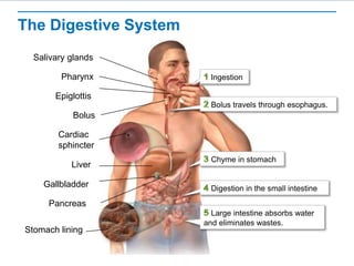
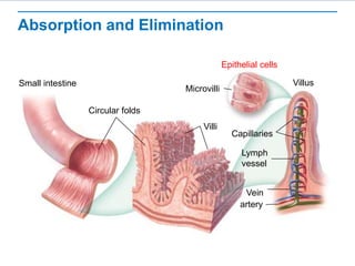
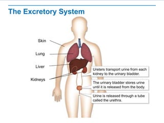
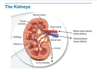
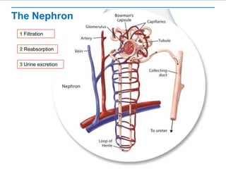
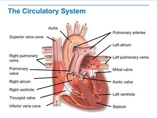
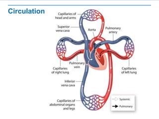
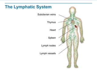
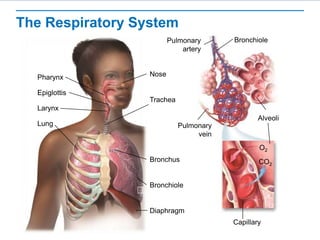
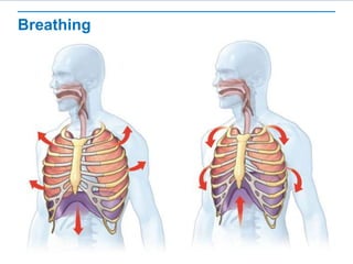
Human Body Systems I
Learning Objectives
 Describe the structures and functions of the digestive,
excretory, circulatory, lymphatic, and respiratory systems.
FC Video
1) What is the main role of the digestive system?
2) What is chemical digestion?
3)Where does the first step of digestion occur?
https://www.youtube.com/watch?v=1UvuBYUbFk0
The Digestive System
Ingestion
Salivary glands
Bolus travels through esophagus.
Pharynx
Epiglottis
Bolus
Chyme in stomach
Cardiac
sphincter
Liver
Pancreas
Gallbladder Digestion in the small intestine
Large intestine absorbs water
and eliminates wastes.
Stomach lining
Absorption and Elimination
Circular folds
Villi
Villus
Microvilli
Capillaries
Lymph
vessel
Vein
artery
Epithelial cells
Small intestine
The Excretory System
Skin
Lung
Liver
Kidneys
Ureters transport urine from each
kidney to the urinary bladder.
The urinary bladder stores urine
until it is released from the body.
Urine is released through a tube
called the urethra.
Interactivity:
https://www.savvasrealize.com/community/program/30e5a5c6-42b8-3c91-
a2c6-1ade91f020ac/20/tier/500d6ce8-6d8e-36fb-b1cc-
18ac5e7e4bca/20/lesson/e775da07-9fea-329e-84e2-
550f8577114a/20/content/f2ff353b-2bb5-3b47-8c80-b1c8802add54/23
The Kidneys
The Nephron
Filtration
Reabsorption
Urine excretion
Circulatory System Video
https://www.youtube.com/watch?v=_qmNCJxpsr0
1) What are the names of the 4 chambers of the heart?
2) What are the Valves found in the heart? And What are their
Functions?
The Circulatory System
Aorta
Superior vena cava
Right pulmonary
veins
Right atrium
Right ventricle
Tricuspid valve
Inferior vena cava
Pulmonary arteries
Left atrium
Left pulmonary veins
Pulmonary
valve
Aortic valve
Left ventricle
Septum
Mitral valve
Circulation
(Albumin, Globulin, Fibrinogen)
Blood Clotting
Thromboplastin
Blood
Capillary wall breaks.
Platelets take action.
Clot forms.
Blood Vessels
Capillary Vein
Artery
Arteriole Venule
Connective
tissue
Connective
tissue
Smooth muscle Smooth muscle
Endothelium
Valve
The Lymphatic System
Subclavian veins
Thymus
Heart
Spleen
Lymph nodes
Lymph vessels
The Respiratory System
Nose
Epiglottis
Pharynx
Larynx
Bronchiole
Lung
Bronchus
Diaphragm
Pulmonary
artery
Alveoli
Pulmonary
vein
Capillary
O2
CO2
Trachea
Bronchiole
Breathing

Body System.ppt

Editor's Notes

  • #2 Read the lesson title aloud to students.
  • #3 Click to show the learning objective.
  • #5 Tell students that the need for food presents every animal with at least two challenges—how to obtain it and how to convert that food into molecules the body can use. In humans and many other animals, this is the job of the digestive system. The digestive system converts food into small molecules that can be used by the cells of the body. Food is processed by the digestive system in four phases—ingestion, digestion, absorption, and elimination. Explain that food in the digestive system is broken down in two ways—by mechanical and chemical digestion. Mechanical digestion is the physical breakdown of large pieces of food into smaller pieces. These smaller pieces can be swallowed and accessed by digestive enzymes. During chemical digestion, enzymes break down food into the small molecules the body can use. Once food has been broken into small molecules, it can be absorbed by cells in the small intestine. Tell students that from the small intestine, the molecules enter the circulatory system, which transports them throughout the body. The digestive system cannot digest and absorb all the substances in food that enter the body. Some materials, such as cellulose, travel through the large intestine and are eliminated from the body as feces. Many students think that enzymes are made up of cells. Point out that the cell is the basic unit of life, but not all substances within the body are made up of cells. Remind students that enzymes are proteins, a kind of macromolecule. Students should review the information about the chemistry of life if they have more questions about enzymes. Ask students to identify what is happening in the first step of digestion Answer: The first step in the process is ingestion, the act of putting food into your mouth. Teeth tear and grind food into small pieces. Enzymes in saliva kill some pathogens and start to break down carbohydrates. Click to reveal the answer. Explain that this process takes about a minute and that the salivary glands release an enzyme called salivary amylase that breaks down starches into disaccharides. Ask students to describe what happens next. Answer: The bolus travels from the mouth to the stomach via the esophagus. Food is squeezed through by peristalsis. Explain that this step takes two to three seconds. Click to reveal the answer. Ask: What is the role of the epiglottis? Answer: to prevent the bolus from traveling down the wrong tube to the lungs rather than to the stomach Ask the students to describe what happens next. Answer: Muscle contractions produce a churning motion that breaks up food and forms a liquid mixture called chyme. Protein digestion begins. Click to reveal the answer. Explain that this takes two to four hours. The cardiac sphincter closes after food passes into the stomach. Glands in the stomach lining release hydrochloric acid, pepsin, and mucus. The enzyme pepsin breaks down proteins into large peptides. Ask students to describe what happens next. Answer: Chyme is slowly released into the small intestine. Bile, which is made in the liver, is released from the gallbladder into the small intestine and aids in fat digestion. Enzymes from the pancreas and duodenum complete digestion. Nutrients are absorbed through the small intestine wall. Click to reveal the answer. Explain that this takes three to five hours. There are numerous enzymes in the small intestines. Pancreatic amylase is released from the pancreas and breaks down starches. Trypsin is released from the pancreas and breaks down proteins. Lipase is released from the pancreas and breaks down fats. Maltase, sucrase, and lactase break down disaccharides into monosaccharides. Peptidase breaks down dipeptides into amino acids. Ask students to describe what happens next. Answer: The large intestine absorbs water as undigested material moves through and is eliminated from the body. Click to reveal the answer.
  • #6 Tell students that once the small intestine has completed the digestive process, it’s time to absorb nutrients. Most nutrients from food are absorbed through the walls of the small intestine. The large intestine absorbs water and several vitamins and prepares waste for elimination from the body. Explain that after leaving the duodenum, chyme moves along the rest of the small intestine. By this time, most of the chemical digestion has been completed. The chyme is now a rich mixture of small and medium-sized nutrient molecules. The small intestine’s folded surface provides an enormous surface area for absorption of nutrients. Its fingerlike projections, called villi (singular: villus), are covered with tiny projections known as microvilli, which absorb the nutrients. Help students understand the scale of the image. Ask students to arrange the following from largest to smallest: villi, microvilli, and circular folds. Answer: circular folds, villi, microvilli. Have students identify which image shows individual cells. Click to reveal the answer. Ask: How is the structure of the small intestine related to its function? Answer: The many folds and projections of the small intestine result in an enormous surface area. Because absorption requires contact between the nutrient molecules and the cells lining the small intestine, a large surface area is critical to the function of the small intestine. Tell students that nutrient molecules are then passed into the circulatory system. Sugars and amino acids pass into capillaries, while most fats and fatty acids are absorbed by lymph vessels. By the time chyme leaves the small intestine, most nutrients have been absorbed, leaving only water, cellulose, and other indigestible substances behind. When chyme leaves the small intestine, it enters the large intestine, or colon. Explain that the large intestine gets its name due to its diameter, which is greater than that of the small intestine. The primary function of the large intestine is to remove water from the material that is left. Large colonies of bacteria present in the large intestine produce compounds that the body is able to absorb and use, including vitamin K. Tell students that the concentrated waste material—feces—that remains after most of the water has been removed passes into the rectum and is eliminated from the body through the anus. When something happens that interferes with the removal of water by the large intestine, you usually become aware of it right away. If not enough water is absorbed, a condition known as diarrhea occurs. If too much water is absorbed, constipation occurs.
  • #7 Tell students that every living system produces chemical waste products, some of which are so toxic that they will cause death if they are not eliminated. Ammonia, one of the most toxic of these waste compounds, is produced when amino acids from proteins are used for energy. Ammonia is converted to a less toxic compound called urea, but it, too, must be eliminated from the body. The process by which these metabolic wastes are eliminated is called excretion. Explain that excretion is one part of the many processes that maintain homeostasis. The excretory system, which includes the skin, lungs, liver, and kidneys, excretes metabolic wastes from the body. The ureters, urinary bladder, and urethra are also involved in excretion. Click to reveal the label for the skin. Ask students how the skin might be involved in excretion. Answer: The skin excretes excess water, salts, and a small amount of urea in sweat. By releasing sweat in very small amounts, this process eliminates wastes even when you may not think you’re sweating. Click to reveal the label for the lung. Ask students how the lungs might be involved in excretion. Answer: The blood transports carbon dioxide, a waste product of cellular respiration, from the body cells to the lungs. When you exhale, your lungs excrete carbon dioxide and small amounts of water vapor. Click to reveal the label for the liver. Ask students how the liver might be involved in excretion. Answer: The liver plays many important roles in excretion. One of its principal activities is the conversion of dangerous nitrogen wastes into less toxic urea. Urea is then transported through the blood to the kidneys for elimination from the body. Click to reveal the label for the kidneys. Ask students how the kidneys might be involved in excretion. Answer: The major organs of excretion are the kidneys, a pair of fist-sized organs located on either side of the spinal column near the lower back. The kidneys remove excess water, urea, and metabolic wastes from the blood. The kidneys produce and excrete a waste product known as urine. Ureters transport urine from the kidneys to the urinary bladder, where urine is stored until it is released through the urethra.
  • #9 Point out that the function of the kidneys is to filter out wastes from the blood and reabsorb useful substances. Model how the kidney works by pouring water mixed with food coloring and clean sand through a coffee filter. Invite students to examine the contents of the coffee filter and the colored water that emerges from it. Ask: What process did this model demonstrate? Answer: filtration Ask: What substances in blood are modeled by the sand in this demonstration? Answer: blood cells and large molecules Ask: In the kidneys, what process follows filtration? Answer: reabsorption Ask students to describe what this image shows. Answer: Waste-laden blood enters the kidney through the renal artery. Filtered blood leaves the kidney through the renal vein. Waste products are released through the ureter and travel to the bladder. Explain that each kidney contains nearly a million individual processing units called nephrons. These nephrons are where most of the work of the kidney takes place. Blood purification in the kidneys involves two distinct processes: filtration and reabsorption.
  • #10 Explain that this is a zoomed-in image of the nephron. This process can be confusing for students, so encourage students to look at the image one portion at a time. Click to reveal the first step: filtration. Explain that filtration takes place in the glomerulus, a dense network of capillaries encased in Bowman’s capsule. Blood pressure forces much of the fluid from the capillaries into Bowman’s capsule. The result is a filtrate containing water, urea, glucose, salts, amino acids, and some vitamins. Click to reveal the next step: reabsorption. Explain that dissolved salts and other materials are removed from the filtrate by active transport and reabsorbed by capillaries, causing nearly all of the filtered water to follow by osmosis. In effect, the kidney first throws away nearly everything and then takes back only what the body needs. The loop of Henle conserves water and minimizes the volume of filtrate. Click to reveal the last step: urine excretion. Explain that the fluid entering collecting ducts is now known as urine. From the collecting ducts, urine flows to the ureter of each kidney. The ureters carry urine to the urinary bladder for storage until the urine leaves the body through the urethra.
  • #12 Tell students that some animals are so small that all of their cells are in direct contact with the environment. Diffusion and active transport across cell membranes supply their cells with oxygen and nutrients and remove waste products. The human body, however, contains millions of cells that are not in direct contact with the external environment. Because of this, humans need a circulatory system. The circulatory system transports oxygen, nutrients, and other substances throughout the body and removes wastes from tissues. Explain that blood is pumped through the body by the heart, a hollow organ composed almost entirely of muscle. An adult’s heart contracts on average 72 times a minute, pumping about 70 milliliters of blood with each contraction. The heart is divided into four chambers. Click to reveal the label for the septum. Tell students that a wall, called the septum, separates the right side of the heart from the left side. The septum prevents oxygen-poor and oxygen-rich blood from mixing. Students are often confused by the left and right sides because they seem revered from the viewer’s standpoint. Point out that left and right is designated from the standpoint of the organism containing the organ. Click to reveal the atria and ventricles. On each side of the septum is an upper and a lower chamber. Ask: Which chambers receive blood? Answer: Each upper chamber, or atrium (plural: atria), receives blood from the body. Explain that the right atrium accepts oxygen-poor blood from the body while the left atrium accepts oxygen-rich blood from the lungs. Ask: Where does blood travel from the ventricles? Answer: Each lower chamber, or ventricle, pumps blood out of the heart. Explain that the right ventricle pumps oxygen-poor blood to the lungs while the left ventricle pumps oxygen-rich blood to the body. Click to reveal the valves. Explain that the valves in the heart prevent blood from flowing in the wrong direction. Identify the valves between the chambers and between the ventricles and arteries. Click to reveal the superior and inferior venae cavae. Explain that these bring blood from the body to the heart. Ask: Is this blood oxygen rich or oxygen poor? Answer: oxygen poor Ask: Where does the blood go next? Answer: to the right atrium Ask: Where does the blood go next? Answer: through the tricuspid valve to the right ventricle Ask: Where does the blood go next? Answer: though the pulmonary valve to the lungs Click to reveal the pulmonary arteries. Ask: What happens to the blood in the lungs? Answer: The blood is oxygenated. Click to reveal the pulmonary veins. Explain that the blood travels back to the heart through the left and right pulmonary veins. Ask: Where does the blood go next? Answer: to the left atrium Ask: Where does the blood go next? Answer: through the mitral valve to the left ventricle Ask: Where does the blood go next? Answer: through the aortic valve into the aorta and to the body Click to reveal the aorta.
  • #13 Tell students that the heart functions as two pumps. One pump pushes blood to the lungs, while the other pushes blood to the rest of the body. The right side of the heart pumps oxygen-poor blood from the heart to the lungs through the pulmonary circulation. In the lungs, carbon dioxide diffuses from the blood, and oxygen is absorbed. Oxygen-rich blood then flows to the left side of the heart. The left side of the heart pumps oxygen-rich blood to the rest of the body through the systemic circulation. Cells absorb much of the oxygen and load the blood with carbon dioxide by the time it returns to the heart. Ask: How is pulmonary circulation different from systemic circulation? Answer: Pulmonary circulation is the blood flow between the lungs and the heart; systemic circulation is blood flow between the heart and the body. Ask: In pulmonary circulation, through what organs does blood flow? Answer: heart and lungs Ask: What happens to blood in the capillaries of the lungs? Answer: The blood absorbs oxygen and gives off carbon dioxide. Ask: What structures does blood flowing through the systemic pathway serve? Answer: the rest of the body
  • #16 Tell students that, in addition to serving as the body’s transportation system, components of blood also help regulate body temperature, fight infections, and produce clots to minimize the loss of body fluids from wounds. Explain that the human body contains 4 to 6 liters of blood. About 55 percent of total blood volume is a fluid called plasma. Plasma is about 90 percent water and 10 percent dissolved gases, salts, nutrients, enzymes, hormones, waste products, plasma proteins, cholesterol, and other compounds. Plasma proteins consist of three types—albumin, globulins, and fibrinogen. Albumin and globulins transport substances such as fatty acids, hormones, and vitamins. Albumin also plays an important role in regulating osmotic pressure. Some globulins fight viral and bacterial infections. Fibrinogen is necessary for blood to clot. Tell students that the most numerous cells in blood are red blood cells (erythrocytes). The main function of red blood cells is to transport oxygen. They get their crimson color from the iron in hemoglobin, a protein that binds oxygen. Red blood cells are produced by cells in the bone marrow. As they mature and fill with hemoglobin, nuclei and other organelles are forced out. Explain that white blood cells (leukocytes) are the “army” of the circulatory system. White blood cells guard against infection, fight parasites, and attack bacteria. A sudden increase in white blood cells is a sign that the body is fighting a serious infection. Different types of white blood cells perform different protective functions. For example, macrophages engulf pathogens. Lymphocytes are involved in the immune response. B lymphocytes produce antibodies that fight infection and provide immunity. T lymphocytes help fight tumors and viruses. In a healthy person, white blood cells are outnumbered by red blood cells by almost 1,000 to 1. Point out that a minor cut or scrape may bleed for a bit, but then the bleeding stops. Why? Because blood can clot. Blood clotting is made possible by plasma proteins and cell fragments called platelets. When platelets come in contact with the edges of a broken blood vessel, their surfaces become sticky, and they release proteins called clotting factors that start reactions to produce a solid clot. Click to reveal the first step of blood clot formation: Capillary wall breaks. Explain that a blood vessel is injured by a cut or scrape. Click to reveal the next step of blood clot formation: Platelets take action. Ask students to describe what is shown in this image. Answer: Platelets clump at the site and release the clotting factor thromboplastin, which triggers a series of reactions. Thromboplastin converts the protein prothrombin into the enzyme thrombin. Click to reveal the next step of blood clot formation: Clot forms. Ask students to describe what is shown in this image. Answer: Thrombin converts the soluble plasma protein fibrinogen into insoluble, sticky fibrin filaments, which form the clot. The clot seals the damaged area and prevents further loss of blood. Ask the following questions to help students understand the functions of each component of blood. Ask: Which two components of blood are most directly involved in blood clotting? Answer: plasma proteins and platelets Ask: Anemia is a term for a group of disorders characterized by a deficiency of red blood cells. What function of blood is impaired in individuals with anemia? Answer: delivery of oxygen to body cells Ask: If an individual has a disorder that results in a reduction of white blood cells, what is a likely result? Answer: reduced ability to fight infection
  • #17 Tell students that blood leaving the heart passes through the aorta, the first of a series of vessels that carries blood through the systemic circulation. As blood flows through the circulatory system, it moves through three types of blood vessels—arteries, capillaries, and veins. Arteries are large vessels that carry blood from the heart to the tissues of the body. Except for the pulmonary arteries, all arteries carry oxygen-rich blood. Arteries have thick, elastic walls. The smallest blood vessels are the capillaries. Most capillaries are so narrow that blood cells pass through them in single file. Their thin walls allow oxygen and nutrients to diffuse from blood into tissues and allow carbon dioxide and other waste products to move from tissues into blood. After blood passes through the capillaries, it returns to the heart through veins. Many veins are located near and between skeletal muscles. When you move, the contracting skeletal muscles squeeze the veins, pushing blood toward the heart. Many veins contain valves, which ensure blood flows in one direction through these vessels. Ask: How are the structures of veins and arteries similar? Answer: Veins and arteries have an inner layer of endothelium, a middle layer of smooth muscle, and an outer layer of connective tissue. Ask: How do the functions of arteries and veins differ? Answer: Most arteries carry oxygen-rich blood from the heart to the tissues of the body. Most veins carry oxygen-poor blood from the body back to the heart. Ask: How are capillaries similar to veins and arteries? Sample answer: Capillaries, veins, and arteries all carry blood in the body. Ask: Why does diffusion of materials between blood and body cells occur across capillary walls but not across veins or arteries? Answer: because of capillaries’ extremely thin walls
  • #18 Tell students that as blood passes through capillaries, some blood cells and components of plasma move through capillary walls and into the fluid between cells. Each day about 3 liters of fluid leaves the blood in this way. Most of this fluid, known as lymph, is reabsorbed into capillaries, but not all of it. The rest goes into the lymphatic system. The lymphatic system is a network of vessels, nodes, and organs that collects the lymph that leaves capillaries, “screens” it for microorganisms, and returns it to the circulatory system. Explain that lymph collects in a system of lymphatic capillaries that slowly conducts it into larger lymph vessels. Pressure on lymph vessels from surrounding skeletal muscles helps move lymph through the system into larger and larger ducts. Lymph vessels have valves, similar to the valves in large veins, that prevent lymph from flowing backward. These ducts return lymph to the blood through openings in veins just below the shoulders. When injury or disease blocks lymphatic vessels, lymph can accumulate in tissues, causing swelling called edema. Tell students that the lymphatic system also plays an important role in the absorption of nutrients. A system of lymph vessels runs alongside the intestines. The vessels pick up fats and fat-soluble vitamins from the digestive tract and transport these nutrients into the bloodstream. Explain that hundreds of small, bean-shaped enlargements—called lymph nodes—are scattered along lymph vessels throughout the body. Lymph nodes act as filters, trapping microorganisms, stray cancer cells, and debris. White blood cells inside lymph nodes destroy this cellular “trash.” When large numbers of microorganisms are trapped in lymph nodes, the nodes become enlarged. Tell students that the thymus and spleen also play important roles in the immune functions of the lymphatic system. T lymphocytes mature in the thymus before they can function in the immune system. The functions of the spleen are similar to those of lymph nodes. However, instead of lymph, blood flows through the spleen, where it is cleansed of microorganisms and other debris. The spleen also removes old or damaged blood cells and stores platelets. Ask: How is the location of lymph nodes and lymph vessels throughout the body related to the role of the lymphatic system in circulation? Answer: Lymph can be collected throughout the body and returned to the circulatory system. Ask: Where is lymph returned to the circulatory system? Answer: through openings in the subclavian veins Ask: In what role of the lymphatic system are the thymus and spleen directly involved? Answer: immunity
  • #19 Tell students that for multicellular organisms, respiration means the process of gas exchange with the environment. The respiratory system picks up oxygen from the air we inhale and releases carbon dioxide as we exhale. With each breath, air enters the body through the air passageways and fills the lungs, where gas exchange takes place, passing oxygen to the circulatory system. The respiratory system consists of the nose, pharynx, larynx, trachea, bronchi, and lungs. Explain that as air enters, the lining of the nose starts the filtering process by trapping large particles. Incoming air is warmed in the inner nasal cavity and sinuses. These areas produce mucus that moistens the air and catches even more dust particles. Air then moves from the nose to a cavity at the back of the mouth called the pharynx, or throat, and then into the trachea, or windpipe. A flap of tissue called the epiglottis covers the entrance to the trachea, ensuring that food or liquid goes into the esophagus instead. Tell students that between the pharynx and the trachea is the larynx, which contains two highly elastic folds of tissue known as the vocal cords. Your ability to speak, shout, and sing comes from these tissues. Mucus produced in the trachea traps inhaled particles, which cilia then sweep away from the lungs toward the pharynx. Explain that from the trachea, air moves into two large tubes called bronchi (singular: bronchus) leading to the lungs. These tubes divide into smaller bronchi, then into even smaller bronchioles. Bronchi and bronchioles are surrounded by smooth muscles that regulate the size of air passageways. The bronchioles lead to millions of tiny air sacs called alveoli (singular: alveolus). Alveoli are grouped in clusters, like bunches of grapes. A delicate network of capillaries surrounds each alveolus. Tell students that as air enters the alveoli, oxygen dissolves in their moist surfaces and diffuses across thin capillary walls into the blood. Meanwhile, carbon dioxide diffuses in the opposite direction. When you inhale, a muscle called the diaphragm contracts and flattens, creating a partial vacuum inside the tightly sealed chest cavity. Atmospheric pressure does the rest, filling the lungs as air rushes into the breathing passages. Diffusion of oxygen from alveoli into capillaries is a passive process. But hemoglobin in red blood cells actively binds oxygen, increasing the blood’s oxygen-carrying capacity more than 60 times. Most carbon dioxide enters red blood cells and combines with water, forming carbonic acid. The rest of it dissolves in plasma or binds to hemoglobin and proteins in plasma. Ask: What structures does air pass through between the nose and the lungs? Answer: pharynx, larynx, trachea Ask: Why can you breathe through your mouth if your nose is stuffed up? Answer: The mouth also opens into the pharynx. Ask: In what respiratory structures does gas exchange occur? Answer: alveoli or lungs A common misconception among students is that oxygen-poor blood is blue. Explain that in illustrations, the color blue is often used to represent oxygen-poor blood, but this does not reflect the actual color of oxygen-poor blood. Oxygen-rich blood is bright red and oxygen-poor blood is deeper red. Although veins look blue in some people, the blood flowing in them is not.
  • #20 Tell students that there are no muscles in our lungs or connected directly to them that participate in breathing. The force that drives air into the lungs comes from ordinary air pressure, the diaphragm, and muscles associated with the ribs. Movements of the diaphragm and rib cage change air pressure in the chest cavity during inhalation and exhalation. You can control your breathing almost any time you want, to blow up a balloon or to play a trumpet. But this doesn’t mean that breathing is purely voluntary. Your nervous system has final control of your breathing muscles whether you are conscious or not. This is why people who drown have water in their lungs. When they lose consciousness, they “breathe” water into their lungs. Explain that breathing is initiated by the breathing center in the part of the brain stem called the medulla oblongata. Sensory neurons in or near the medulla and in some large blood vessels gather information about carbon dioxide levels in the body and send the information to the breathing center. When stimulated, the breathing center sends nerve impulses that cause the diaphragm and chest muscles to contract, bringing air into the lungs. The higher the blood carbon dioxide level, the stronger the impulses. If the blood carbon dioxide level reaches a critical point, the impulses become so powerful that you cannot keep from breathing. Ask: How does the volume of the chest cavity change during inhalation? Answer: It increases. Ask: How does the volume of the chest cavity change during exhalation? Answer: It decreases. Ask: How can a chest wound impair breathing? Answer: The processes of inhalation and exhalation work because the chest cavity is a sealed compartment. If a chest wound opens the chest cavity to the environment outside the body, the pressure changes that drive inhalation and exhalation will not occur.