1. ChrisMiller
BMP influence on Differentiation of Embryonic Stem Cells into Cardiomyocytes
Introduction:
Heart disease continues to be problematic around the world and the leading cause of
death in the United States. The American Heart Association reports heart disease being the most
costly health problem which includes surgical operations, medications, and the diagnostic
process racking up over $ 228 billion in expenditures in 2008 (1). Many issues arise with regard
to heart disease, as this intricate organ delivers oxygen and nutrients around the body while also
playing a role in the immune system. Using stem cells coupled with growth factors to generate
cardiac cells could be the resolution the world needs for medical conditions patients suffer from
such as heart attack, heart defects, and other degenerative diseases associated with the heart.
The idea of using stem cells for regenerative medicine is a relatively recent discovery in
the medical world. Taking a pluripotent cell and manipulating it in a way to achieve a specialized
cell has raised many questions. The medical community continues to strive for a way to master
this technique as these cells are extremely sensitive to the environment they reside in. Working
with stem cells in culture creates an issue as the environment inside and outside the body are
very different. Understanding the growth of stem cells outside the body is crucial to saving future
lives.
People every day sit on transplant lists hoping to survive long enough to make it to the
top. The possibility of finding the right donor match is something else that one needs to think
about. Graft vs Host can develop if the right donor match is not found. Immune cells will attack
the new heart resulting in certain death, so it is another problem associated with finding the right
donor. Heart transplants have risen from 22 in 1975 to over 2000 in 2010 (1). The need is there
and ever growing. It can sometimes be years though before the right donor and opportunity arise.
Harvesting cells to create a usable heart or creating cells to repair the damaged one lies within
the future of this field. Stem cell differentiation to create cardiac cells can be this life saving
process for many people around the world; more importantly embryonic stem cells (ESCs) are of
interest with regard to the heart because they can easily form cardiac tissue. A research group out
of Tokyo worked with mice and a line of ESCs known as P19Cl6 obtained from the inner mass
of the blastocyst early in embryonic development (2). This is a common method used in
obtaining ESCs. Endoderm and ectoderm derived tissues were created from the use of the
P19C16 cell line that mimicked cardiac cells. Because these ESCs are pluripotent in nature, they
can produce any cell type within the body. As long as the right environmental conditions are met
along with other cell specific transcription factors, ESCs allow researchers to generate a
particular cell of interest. The stem cells ability to differentiate into muscle cells are of
importance for future tissue engineering and therapeutic medical procedures; specifically in the
area of developing treatment options for cardiac tissue damage (3).
Cardiac damage can occur anywhere in the heart making it important to understand the
heart is a heterogeneous muscle tissue. Any muscle cell can fall under the broad category of a
myocyte. They can be further categorized from there to smooth, skeletal, and cardiac. Cardiac
2. ChrisMiller
cells are a type of myocyte found within the heart; they are also known as cardiomyocytes.
Ventricular, atrial, and specialized sinoatrial and atrioventricular node regions exist throughout
the heart producing varied cell characteristics (4). The inability to generate specific cell types in
culture is another problem still needing to be addressed. Cultures today produce a variety of each
cell type found in the heart.
Embryonic stem cells have the ability, under the right conditions, to become any type of
cardiomyocyte in culture through methods such as using specific growth factors, embroid body
formation, and the use of END- 2 cells cocultured with ESCs (5). The use of the END-2 cells
treated with mitomycin C can replace mouse embryonic fibroblasts (MEFs) as feeders for human
ESCs and facilitate differentiation into cardiomyocytes. The mitomycin C acts to control
proliferation and help produce secretive factors before differentiation can take place. Embryoid
body formation relies on spontaneous formation of cardiomyocytes in a suspension culture.
However, the most successful technique to date for generating cardiomyocytes in vitro is from
the use of growth factors such as bone morphogenetic protein (BMP) along with other
transcription factors it associates with like AFT-2 (6). BMP appears in 20 different forms within
the body and has been identified as a crucial protein in developmental processes as well. Apart
from its obvious intentions of generating bone tissues, BMP regulates roles in the body such as
tissue homeostasis, cell growth, differentiation, apoptosis, and vascular remodeling (7). BMP-2
and BMP-4 play vital roles in heart development in embryogenesis and will be focused on
throughout the literature review. BMP-2 and BMP-4 are also part of the transforming growth
factor beta (TGF-β) super family because of their ability to play multiple roles in the body
influencing cell growth and transformation. This review aims to look at the BMP signaling
cascade, including its role in development, and generating cardiomyocytes in culture using the
BMP growth factor.
BMP Signaling Cascade:
The cascade of events that occur with the introduction of BMP in cellular development
and all metabolic pathways must be a process thoroughly understood so future therapies can be
provided. BMPs allow for many basic biological processes to function like that of bone
formation, cell growth, and differentiation. Without these proteins the body would not be able to
thrive and maintain homeostasis. BMP-2 and BMP-4 are two closely related members of the
TGF-beta superfamily. The TGF-beta superfamily is comprised of many different proteins that
exist within the body handling many different bodily functions. Regulation of cell growth,
proliferation, differentiation, adhesion, migration, and apoptosis are all processes controlled by
this superfamily of proteins (8). Other proteins in this family include activin/inhibin, growth
differentiation factors, and TGF-beta (9). All members of the BMPs share a distinct feature on
the C-terminus end; this is a region containing seven cysteine residues (8). The BMP family can
be further classified into subclasses. Subclass A includes BMP-2 and BMP-4 due to 80%
homology seen between the two proteins. They are also 92% identical on the carboxyl-terminal
regions in regards to their amino acid sequence (10).
3. ChrisMiller
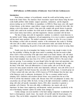
Figure 1. Chemical structure of a BMP molecule before secretion out of the cell (10)
The homology seen between BMP-2 and BMP-4 is something to note. This indicates similar
functions for each of these two proteins recognized as factors to induce differentiation into
cardiomyocytes. In the absence of BMP-4, BMP-2 can compensate to temporarily resolve the
cascade of events. Experiments performed with mice demonstrated this situation as gastrulation
of embryos does not normally take place in the absence of BMP-4. BMP-2 overlaps BMP-4
localization sites in the heart, amnion, and allantois due to the protein homology. This can help
induce further development than expected in embryos if BMP-4 is absent or dysfunctional (10).
BMP-2’s involvement would be anticipated to replace BMP-4 as the genetic sequence allows for
the system to temporarily rely on BMP-2 until an adequate supply of BMP- 4 can be used once
again. BMP-4 was removed from a cell culture in vitro in order to see the effects it had on
embryogenesis and the ability to maintain homeostasis. Most embryos did not survive past one
week, but some embryos did survive past this stage. In these cases, BMP-2 needed to be
overexpressed and up-regulated as BMP-2 would still need to perform the normal functions it
associates with such as preserving functional characteristics of the amnion, gut, and heart (6).
The developing embryos still ended in fatality in the absence of BMP-4 though, suggesting that
BMP-2 cannot sustain all of the functions of BMP-4 long term.
The actual BMP signaling cascade starts with an initial precursor molecule inside the
cytoplasm of the cell before secretion of the mature BMP molecule can take place. Here, the pre-
protein consists of a signal peptide, propeptide domain, and a mature peptide sequence on the
carboxy-terminal (8). Once a signal generates a need for the protein to be used, the signal peptide
is cleaved to start the process. The protein then undergoes glycosylation and dimerization of the
pro-domain and mature peptide molecule. The prodomain is cleaved after the glycosylation and
dimerization but before secretion. A conformational change occurs to form the newly created
protein ready to be used by cellular mechanisms throughout the body. This mature BMP is
derived from the carboxy-terminal region as a heterodimer or a homodimer (8). These
homodimers and heterodimers allow for different receptor binding capabilities. There can be
several combinations formed allowing for different conformational changes of BMP for specific
tissue and cell binding.
Now, the protein has been properly synthesized and is ready to be used upon secretion
out of the cell. Figure 2 is a representation of the BMP pathway with a cell surface receptor to
generate a cardiomyocyte dependent on environmental conditions along with other transcription
factors including NKX2-5, ATF-2, MEF2C, HAND2 and MYOCD.
4. ChrisMiller
Figure 2. Schematic of the mechanistic process of the signaling cascade for BMP and its
associated receptor. (11)
BMPs first bind to cell surface receptors where a signal complex initiates the process.
There are two different receptor types. The type II receptor is activated by a BMP. This signals a
close by type I receptor to be phosphorylated creating a high energy complex. Smad proteins
activate in the cytoplasm by another phosphorylation event. These Smad proteins act as signal
transducers. They interpret an initial signal from the phosphorylation events to activate a
transcriptional response (8). There are several different Smad proteins that exist in different
locations throughout the body forming different complexes dependent on the type of BMP bound
to the receptor of the cell. Generally, Smad-1, Smad-5, and Smad-8 form heteromeric complexes
with Smad-4 within these activated complexes. A transcriptional response can take place to
generate the desired transcriptional response needed to form a particular cell type within the
nucleus. Transcription factor ATF-2 is highly expressed in this process and a key component for
terminal cardiomyocyte differentiation (11). Smad-6 and Smad-7 proteins act as inhibitors to the
system if overexpression occurs. Other modulators such as Noggin exist outside the cell and bind
once a BMP finds a receptor of choice.
BMPs and their signaling cascade are vital during embryogenesis. They play key roles in
both mesoderm formation and heart development. Either BMP-2 or BMP-4 deficiency are the
only two of the 20 known BMPs that lead to complete embryo lethality. The absence of these
two prevents mesoderm formation along with amnion and cardiac defects. These three aspects of
5. ChrisMiller
embryogenesis are essential to forming a healthy fetus early in development. Absences of other
BMP molecules generally result in death upon birth due to the inability of the fetus to maintain
homeostasis outside of the mother. Birth defects are common when BMPs are absent or
dysfunctional from mutations presented such as abnormal lymphatic development, increased
bone length and density, bone fusion in ankles and wrist, and failure of ventral body wall closure
(11). These molecules have been proven to be essential with embryogenesis and throughout
adulthood. A lack of the BMP signaling cascade will certainly cause death in mammalian
species.
Cardiomyocyte generation from culture of ESCs:
There have been a variety of methods proposed for the process of differentiating human
ESCs into cardiomyocytes as this has yet to be a perfected process. ESC lines are acquired from
all sorts of places across the world including medical research labs, universities, and cancer
institutes. As described earlier, ESCs are found within the inner mass of a blastocyst during
embryogenesis. Once the cells are obtained, the cells can be cultured on a media that supports
their growth before differentiation to reach an ideal confluency. A medium comprised of growth
factors can help to stimulate the division of the ESCs to a desired number. DMEM is a common
media used consisting of high levels of glucose to stimulate cell proliferation along with a
percentage of fetal bovine serum (FBS). The serum consists of embryonic growth factors to aid
in the proliferation of the undifferentiated ESCs (12). 10%-20% FBS is often used in culturing
stem cells as this appears to be the ideal concentration range of embryonic factors used by many
researchers. This medium can then be treated with a mouse embryonic fibroblast (MEF) feeder
layer with the undifferentiated cells lying on top. This MEF layer allows for growth of the cells
by secreting factors such as activin-A promoting growth of the ESCs while suspending the
differentiation of the ESCs (13).
Three novel methods have provided promising results differentiating ESCs into
cardiomyocytes. By far the most widely used method is by spontaneous differentiation through
the formation of embryoid bodies. The ESCs cells are first dispersed into smaller clusters using a
collagen degrading enzyme such as collagenase IV. This enzyme disrupts the collagen network
of the ECM between adjacent cells. They are then transferred to a petri dish in suspension such
as the hanging drop method. The suspension allows the clusters of ESCs to form embryoid
bodies in the petri dish in 7-10 days. Next, the cells are transferred to a 0.1 % gelatin-coated petri
dish. Spontaneous contracting areas can be observed as early as 5 days after being plated (13).
This was the first method to characterize the ability of ESCs to differentiate into cardiomyocytes.
Coculture of endoderm like cells or END-2 cells has also been proposed as an adequate
source of differentiating ESCs into cardiomyocytes. This process involves first isolating the
END-2 cells from HepG2 cells of mice. The END-2 cells are then cultured in DMEM media
consisting of 10% FBS. After both are cultured, the two are brought together to form a coculture
with mitomycin C. The END-2 cells were treated with mitomycin C for 3 hours to suspend
proliferation while allowing secretive factors to be produced to stimulate differentiation. The
END-2 cells then provide a feeder layer for the ESCs to differentiate into cardiomyocytes. The
6. ChrisMiller
culture was grown up to five weeks and observations of beating areas were observed after 10
days of the coculture. 35 ± 10% of colonies observed within 12-well plates exhibited a beating
area out of their 30 total plates (5). This number was much higher in the results compared to that
found with the spontaneous EB in suspension method. The mechanism of this system is still
poorly understood though. The way in which these cells differentiate are not described. Secretive
factors and cell to cell contact are thought to be the origins of this END-2 cells ability to help
facilitate differentiation of the ESCs into cardiomyocytes (7).
By far the best way to differentiate an ESC into a cardiomyocyte is through growth factor
initiation. This makes sense as these same molecules such as BMP-2 and BMP-4 are active early
in embryogenesis and throughout life. Growth factors act as a molecular switch to excite a cell
into a permanent state allowing transcription of cell specific genes for cardiomyocytes. Several
growth factors have been identified including BMP4, FGF, and Wnt3a with the inclusion of
Activin A (7). One study used BMP4 with Activin A to produce over 30% of total ESCs into
cardiomyocytes (14). They were then able to purify their culture through a Percoll gradient
centrifugation to achieve an average of 86% cardiomyocytes from these cultures. This extra step
allowed for a more precise isolation of their cardiomyocytes through this recently identified
gradient protocol. Another study utilized a combination of BMP4, FGF2, VEGF, and DKK1 in a
serum free media that saw populations exceeding 50% of contracting cardiomyocytes from ESCs
(15). Another novel method was used by combining END-2 cells and BMP-2 to produce high
confluency of cardiomyocytes. The two methods, using either END-2 or BMP-2, in their own
right would be able to differentiate ESCs into cardiomyocytes, but this research group thought to
combine both factors to further induce a desired differentiation. 92% cardiomyocyte generation
of murine ES-D3 cell line were generated by their novel method. The END-2 provided secretive
factors for differentiation while BMP-2 provided further genes to be transcribed for
cardiomyocytes. This same research group also wanted to compare BMP-2 only cell cultures
without the use of END-2 cells coupled with BMP-2. The cultured ESCs were placed in a BMP-
2 only media yielding just 44% of cardiomyocyte differentiation (16). It appears by combining
END-2 and BMP-2 suits conditions like that found in the body. Increased expression of cardiac-
specific genes such as NKx2.5 and apha-MHC were observed in the combined method of END-2
and BMP-2. Results such as these can further fuel other methods to generate purified cultures of
cardiomyocytes. No one method has proven to be efficient in differentiation. However, the purity
of the cardiomyocyte culture appears to be much greater by growth factor initiation than
compared to that of the previous two methods described on their own. The use of growth factors
appear to be the best way of achieving the highest concentrations of cardiomyocytes, but not
widely used due to the added cost of reagents compared to that of other methods previously
described as well as simplicity.
Immunostaining, western blots, calcium transients, and RT-PCR can be used for
confirmation of the differentiated cells to be cardiomyocytes. The expression of key cardiac
genes and proteins are needed to test whether the cells generated are in this excited and
permanent state expressing genes needed for a cardiomyocyte. Western blots combine
7. ChrisMiller
electrophoresis with an antibody probe to identify proteins of the heart in culture such as NKX2-
5, MEF2C, HAND2 and MYOCD (11). Many proteins are expressed in cardiomyocytes, so
creating these antibodies coupled with some sort of fluorescent dye will allow for further
recognition. Immunostaining is related as receptors expressed on cardiomyocytes will essentially
light up the cells to be recognized as cardiomyocytes. Immunostaining of dispersed cells from a
beating embryoid body with anti-cardiac α/β-myosin heavy chain mAb’s allow for
characterization of cardiomyocytes (13). Calcium transients can be run looking at the cells ability
to uptake calcium in and out of the cell. This relates to the cells ability to form a contracting beat
as the influx of calcium binds to receptors of the sarcomere to establish a contraction to take
place. RT-PCR allows for specific genes to be amplified and run on an agarose gel to again be
run on electrophoreses (17). All of these contributing aspects signify key cardiomyocyte
characteristics observed under a microscope. They are unique methods in their own respect to
characterize whether the cell being observed has attributes of a cardiomyocyte.
Conclusion:
This review aimed to look at the BMP signaling cascade, including its role in
development, and generating cardiomyocytes in culture using the BMP growth factor. Other
methods of differentiating cardiomyocytes were discussed as well to compare the efficiencies of
each method. Growth factor initiation appears to be the best method to differentiate ESCs into
cardiomyocytes. BMP-2 and BMP-4 are two crucial proteins of the TGF-β super family found in
the body and expressed heavily in embryogenesis. BMP-2 and BMP-4 play important roles in
tissue homeostasis, cell growth, differentiation, apoptosis, and vascular remodeling throughout
life in humans. Mutations or removal of both proteins have resulted in fatality in embryogenesis.
BMP-2 and BMP-4 are the only two proteins of the BMP family to be crucial in the first week of
embryogenesis. BMP-2 can temporarily replace BMP-4 but long term stability cannot yet be
achieved without BMP-4. The homology between the carboxy-terminal ends allow for BMP-2 to
resolve the signaling cascade for BMP-4 if overexpressed. However, the signaling process is
complex and not fully understood why exactly BMP-2 acts in this way for only a short time.
Generating cardiomyocytes from ESCs is a process needing to be further researched as
no one process allows for an efficient method to date. ESCs are pluripotent and can produce any
cell type in the body such as a cardiomyocyte. This provides huge potential for tissue
engineering. Generating a method to efficiently produce cardiomyocytes in vitro can lead to a
better understanding of the mechanisms for in vivo use. The way cells act in the body are much
different than how they act outside the body. The environment plays a huge role in the
differentiation of these cells.
Ultimately, the goal of stem cell research is to produce a product to be used within the
human body. 2015 marked the first clinical case of ESCs being differentiated into
cardiomyocytes for in vivo use in humans. A Surgical site of fibrin gel consisting of 1 million
cardiomyocytes were loaded under the pericardial flap of an infarcted heart (18). Three and six
month evaluations have been taken with 10% increase in blood pumping efficiency. These are
8. ChrisMiller
promising results but the process is still in its infancy. Long term stability has not yet been
monitored in this patient and still being evaluated.
The need for new innovative therapies for heart disease remains at a high due to the
ongoing prevalence of increasing heart disease seen across the world. More research needs to be
done in order to grasp the full knowledge of ESCs development in vitro and in vivo. As we move
forward there is hope and promise for such new therapies for people with heart disease and for
future stem cell research. It does not just stop at ESCs being induced onto cardiomyocytes either.
The ESC has the capacity to differentiate into all cells of the body. Unlocking the answers can
have such a large impact on the future of the human race.
9. ChrisMiller
References
1. Roger VL, Goa S, Lloyd-Jones D, et al. AHA statistical update heart disease and
stroke statistics—2012 update. American Heart Association. 2012. Available at:
http://www.ncbi.nlm.nih.gov/pmc/articles/PMC4440543/
2. Huch M, Koo B. Modeling mouse and human development using organoid cultures.
Development for Advances in developmental biology and stem cells. 2015. Available
at: http://dev.biologists.org/content/142/18/3113
3. Engler A, Krieger C, Discher D, et al. Embryonic cardiomyocytes beat best on a
matrix with heart-like elasticity: scar-like rigidity inhibits beating. Journal of Cell
Science. 2008. Available at: http://www.ncbi.nlm.nih.gov/pmc/articles/PMC2740334/
4. FU J, Jjang P, Li R, et al. Na+/Ca2+ Exchanger is a Determinant of Excitation–
Contraction Coupling in Human Embryonic Stem Cell–Derived Ventricular
Cardiomyocytes. Stem Cells Dev. 2010. Available at:
http://www.ncbi.nlm.nih.gov/pubmed/19719399
5. Mummary C, Ostwaard D, Tertoolen L, et al. Differentiation of Human Embryonic
Stem Cells to Cardiomyocytes Role of Coculture With Visceral Endoderm-Like Cells.
2003. Avaiable at: http://www.ncbi.nlm.nih.gov/pubmed/12742992
6. Monzen K. Smads, Tak1, and Their Common Target Atf-2 Play a Critical Role in
Cardiomyocyte Differentiation. 2001. Available at:
http://jcb.rupress.org/content/153/4/687.full
7. Rajala K, Mattila M, Setala K. Cardiac Differentiation of Pluripotent Stem Cells. Stem
Cells International. 2011. Available at:
http://www.hindawi.com/journals/sci/2011/383709/
8. Jain, A. Bone morphogenetic proteins: The anomalous molecules. 2013. Available at:
http://www.ncbi.nlm.nih.gov/pmc/articles/pmc3808010/.
9. Kollias H, Mcdermott J. Transforming growth factor- and myostatin signaling in
skeletal muscle. JAP. 2008. Available at:
http://www.ncbi.nlm.nih.gov/pubmed/18032576
10. Winnier G, Blessing M, et al. Bone morphogenetic protein-4 is required for mesoderm
formation and patterning in the mouse. Genes and Development. 2015. Available at:
http://genesdev.cshlp.org/content/9/17/2105
11. Wang, R. Bone Morphogenetic Protein (BMP) signaling in development and human
diseases . 2014. Available at:
http://www.sciencedirect.com/science/article/pii/s2352304214000105.
12. Kehat I, Karsenti D, Gepstein A, et al. Human embryonic stem cells can differentiate
into myocytes with structural and functional properties of cardiomyocytes. J Clin
Invest. 2001. Available at: http://www.ncbi.nlm.nih.gov/pmc/articles/PMC209357/
13. Kehat I, Karsenti D, Gepstein A, et al. Human embryonic stem cells can differentiate
into myocytes with structural and functional properties of cardiomyocytes. J Clin
Invest. 2001;108(3):407–414. doi:10.1172/JCI12131.
14. Laflamme M, Chen K, Murry C, et al. Cardiomyocytes derived from human
embryonic stem cells in pro-survival factors enhance function of infarcted rat hearts.
Nature Biotechnology. 2007. Available at:
http://www.nature.com/nbt/journal/v25/n9/abs/nbt1327.html
10. ChrisMiller
15. B Moore J, Fu J, Li R, et al. Distinct cardiogenic preferences of two human embryonic
stem cell (hESC) lines are imprinted in their proteomes in the pluripotent state.
Biochem Biophys Res Commun. 2008. Available:
http://www.ncbi.nlm.nih.gov/pmc/articles/PMC4055054/
16. Bin Z, Hui S, et al. Efficient cardiomyocyte differentiation of embryonic stem cells by
bone morphogenetic protein-2 combined with visceral endoderm-like cells. Cell
Biology International. 2006. Available at:
http://www.ncbi.nlm.nih.gov/pubmed/18503758
17. Keung W, Boheler K, Li R. Developmental cues for the maturation of metabolic,
electrophysiological and calcium handling properties of human pluripotent stem cell-
derived cardiomyocytes. Stem Cell Research and Therapy. 2014. Available at:
18. Menashe P, Vanneaux V, Larghero J. Human embryonic stem cell-derived cardiac
progenitors for severe heart failure treatment: first clinical case report. 2015. Available
at: http://dx.doi.org/10.1093/eurheartj/ehv189 ehv189