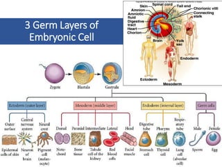
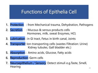
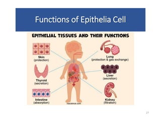
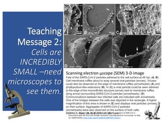
Download to read offline
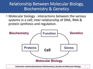
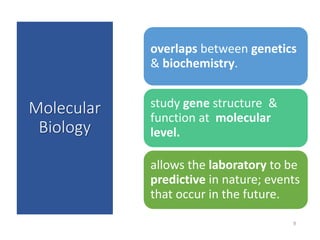
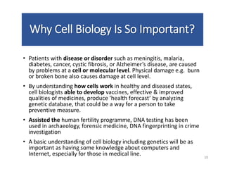
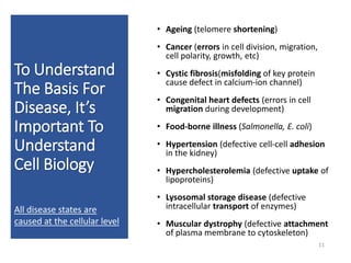
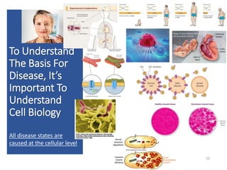
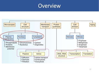
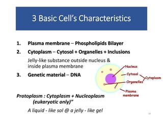
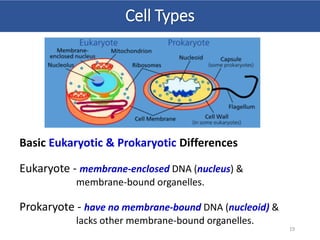
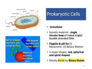

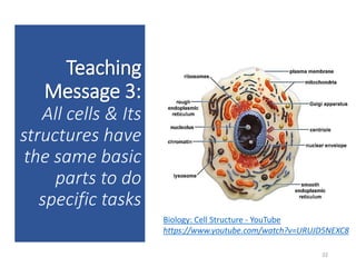
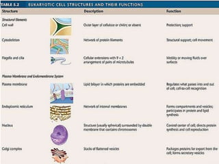
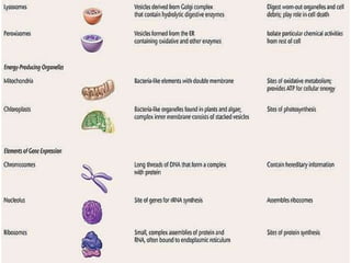

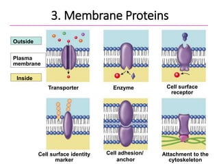
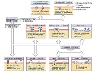
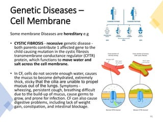

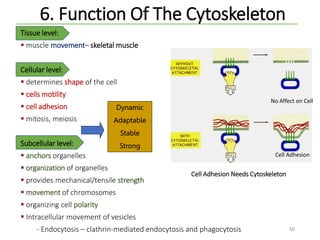
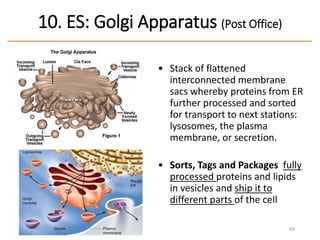
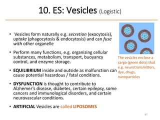


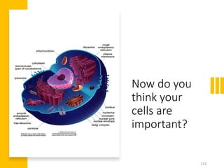


This document outlines the structure and function of cells, covering molecular cell biology, the principles of cell theory, and the significance of understanding cell biology in relation to diseases. It emphasizes that all diseases are caused at the cellular level and highlights various examples, such as cancer and cystic fibrosis, alongside the basics of cellular composition and the differences between eukaryotic and prokaryotic cells. Additionally, it explains the functions of different cell types and organelles, the importance of cell communication, and the role of the cytoskeleton in maintaining cell structure and function.


