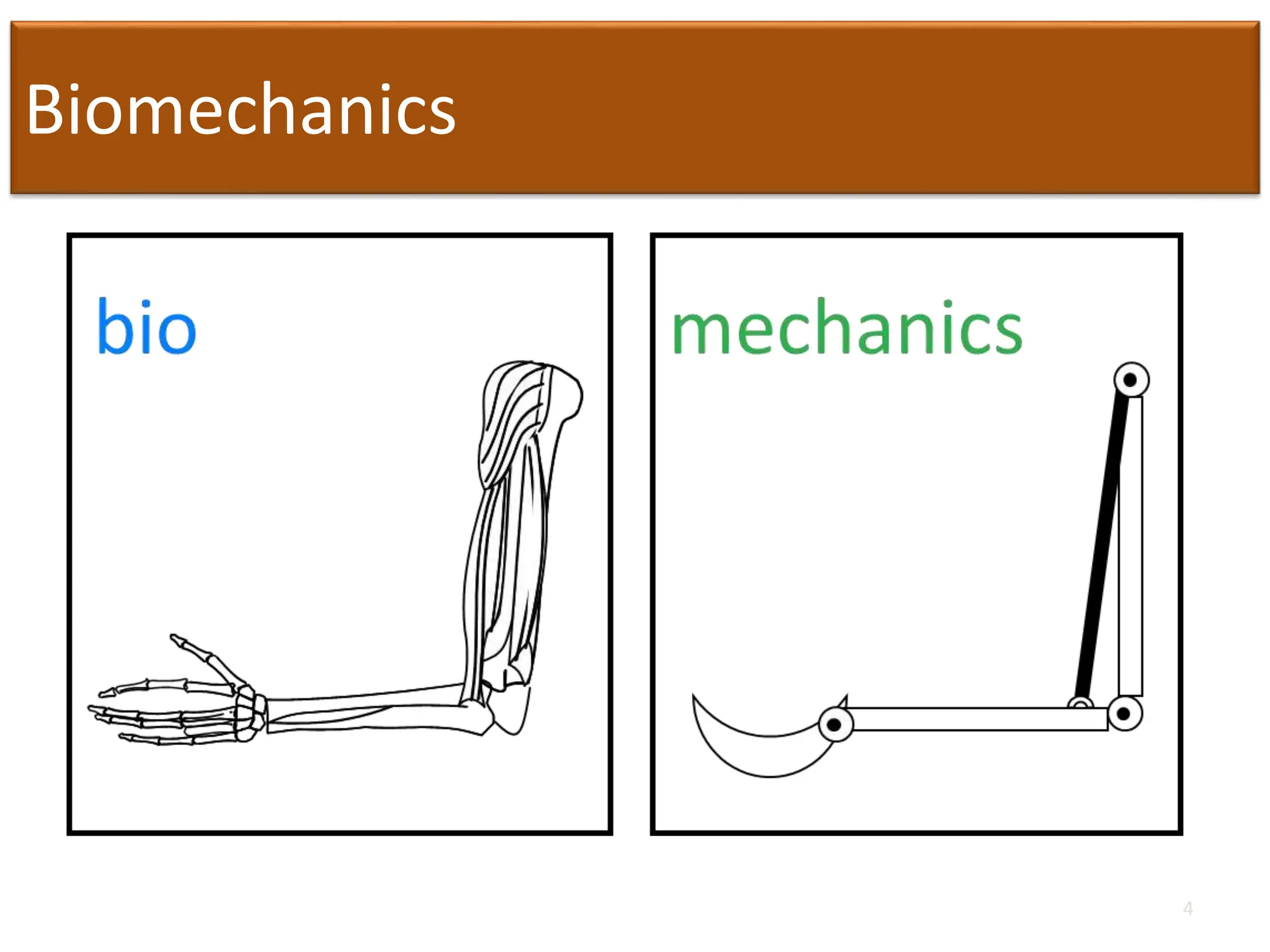
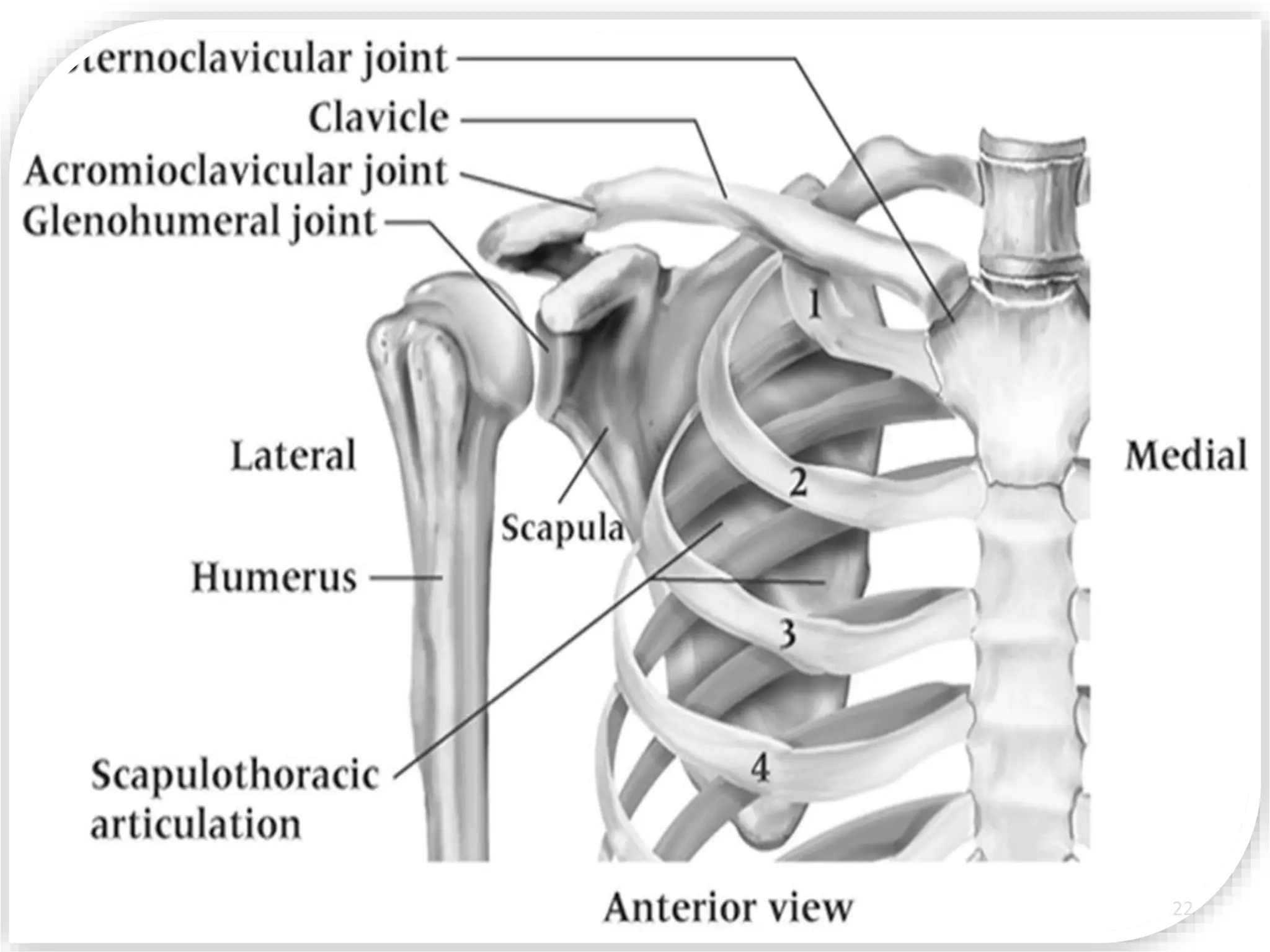
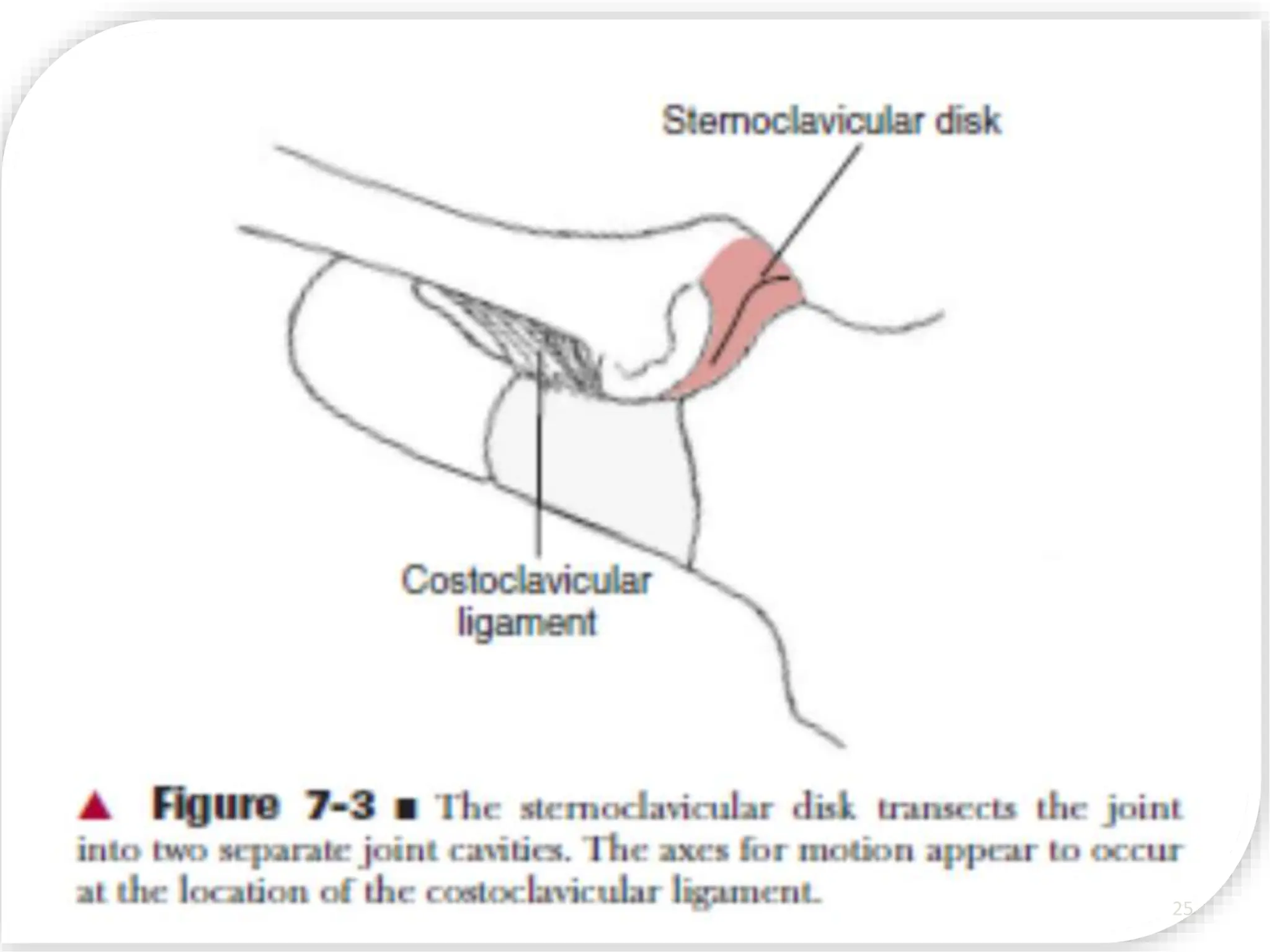
2. OBJECTIVES
At the end of this lecture students should be able to:
• Define different terms of biomechanics
• Identify different structures in shoulder complex
• Explain kinetics and kinematics of shoulder joint
• Describe different pathologies of shoulder complex
2
5. Biomechanics
• The term biomechanics combines the prefix
bio, meaning “life,” with the field of
mechanics, which is the study of the actions of
forces, (both internal muscle forces and
external forces.) In biomechanics we analyze
the mechanical aspects of living organisms.
5
8. Subdivisions
• statics: study of systems in constant motion,
(including zero motion)
• dynamics: study of systems subject to
acceleration
• kinematics: study of the appearance or
description of motion
• kinetics: study of the actions of forces (Force
can be thought of as a push or pull acting on a
body.)
8
9. kinematics
• What we visually observe of a body in motion
is called the kinematics of the movement.
Kinematics is the study of the size,
sequencing, and timing of movement, without
regard for the forces that cause or result from
the motion. The kinematics of an exercise or a
sport skill is known, more commonly, as form
or technique.
9
23. SC JOINT
Clavicle articulates with manubrium of the sternum
Weak bony structure but held by strong ligaments
Fibrocartilaginous disk between articulating
surfaces
• Shock absorber and helps prevent displacement
forward
• Clavicle permitted to move up and down, forward and
backward and in rotation
• Clavicle must elevate 40 degrees to allow upward
rotation of scapula and thus shoulder abduction
23
24. SC JOINT
• The only attachment of the upper extremity to axial
skeleton
• Plane synovial joint with degree of freedom 6, having
joint capsule, joint disk and three major ligaments
• Movement of the SC joint produces scapular
movements, if it is fused the equal amount of
movement will occur at AC joint
24
28. MOVEMENTS OF SC JOINT
Movements in horizontal plane:
• Protraction (30 degree)
limited by costoclavicular and
post. capsule
• Retraction (30 degree) limited
by costoclavicular and ant.
capsule
28
29. MOVEMENTS OF SC JOINT
• Elevation (48 degree)
– limited by costoclavicular
• Depression (less than15 degree)
–limited by first rib
Axial Rotation
Ant. Rot. (very limited – 10 degree)
Post. Rot. (50 degree)
29
32. AC JOINT
• Lateral end of clavicle with
acromion process of scapula
• Weak joint and susceptible
to sprain and separation
• Joint capsule n two major
ligaments and disk – present
or absent
32
33. AC JOINT
LIGAMENTS:
• Coracoclavicular
– Medial: Conoid
– Lateral: Trapezoid
• Acromioclavicular
– Superior
– Inferior
• Coracoacromial:
– Coracoids process to acromiom
process
• Closed packed position is
when the humerus is abducted to
90 degree. 33
34. MOVEMENTS OF AC JOINT
• Internal and external rotation
– Bringing the glenoid fossa of the scapula
anteromedially and posterolaterally, respectively
• Anterior and posterior tiping or tilting
– Ant. - acromion tipping forward and the inferior
angle tipping backward
– Post. - rotate the acromion backward and the
inferior angle forward.
• Upward and downward rotation
– Upward rotation tilts the glenoid fossa upward and
downward rotation is the opposite motion.
34
38. CORACOACROMIAL ARCH
Arch over the GH joint formed by Coracoacromial
arch,acromion and coracoid process
• Sub acromial space: area in between CA arch and
humeral head
• Supraspinatus tendon, long head biceps tendon, and
sub acromial bursa
• Subject to irritation and inflammation as a result of
excessive humeral head translation or impingement from
repeated overhead activity
38
40. Structures Within Suprahumeral Space
1. Long head of biceps
2. Superior capsule
3. Supraspinatus tendon
4. Upper margins of
subscapularis &
infraspinatus tendons
5. Subacromial bursa
6. Inferior surface of
the A-C joint
40
41. SUBACROMIAL SPACE
Clinical Relevance
Avoidance of impingement during elevation of
the arm requires
• External rotation of humerus to clear greater
tuberosity
• Upward rotation of scapula to elevate lateral end of
acromiom
41
42. SUBACROMIAL SPACE
• Primary Impingement
Structural stenosis of subacromial space
• Secondary Impingement
Functional stenosis of subacromial space due
to abnormal arthrokinematics
42
43. Glenohumeral Joint
Ball and socket, synovial joint in which round head of humerus
articulates with shallow glenoid fossa of scapula
stabilized slightly by fibrocartilaginous rim called the Glenoid Labrum
Humeral head larger than glenoid fossa
• At any point during elevation of shoulder only 25 to 30% of humeral
head is in contact with glenoid Statically
stabilized by labrum and capsular ligaments Dynamically
stabilized by deltoid and rotator cuff muscles
• Three degrees of freedom
Stability provided by
• Passive restraints
• Active restraints
43
46. Glenoid Labrum
• When the arms hang dependently at the side, the two articular
surfaces of the GH joint have little contact. The majority of the time,
the inferior surface of the humeral head rests on only a small inferior
portion of the fossa. The total available articular
• surface of the glenoid fossa is enhanced by an accessory structure,
the glenoid labrum. This structure surrounds and is attached to the
periphery of the glenoid fossa enhancing the depth or curvature of
the fossa by approximately 50%.
• the labrum was traditionally thought to be synoviumlined
fibrocartilage, more recently it has been proposed that it is actually a
redundant fold of dense fibrous connective tissue with little
fibrocartilage other than at the attachment of the labrum to the
periphery of the fossa.
• The labrum superiorly is loosely attached, whereas the inferior
portion is firmly attached and relatively immobile.The glenoid labrum
also serves as the attachment site for the glenohumeral ligaments and
the tendon of the long head of the biceps brachii. 46
47. GH CAPSULE
• The entire GH joint is surrounded by a large, loose capsule that is
taut superiorly and slack anteriorly and inferiorly in the resting
position (arm dependent at the side).The capsular surface area is
twice that of the humeral head.39 More than 2.5 cm of distraction of
the head from the glenoid fossa is allowed in the loose-packed
position.
• The relative laxity of the GH capsule is necessary for the large
excursion of joint surfaces but provides little stability without the
reinforcement of ligaments and muscles. When the humerus is
abducted and laterally rotated on the glenoid fossa, the capsule
twists on itself and tightens, making abduction and lateral rotation
the close-packed position for the GH joint
47
51. Restraints to External Rotation
• Dependent on arm position
• 0° - SGHL, C-H & subscapular
• 45° - SGHL & MGHL
• 90° - anterior band IGHLC
51
52. Restraints to Internal Rotation
• Dependent on arm position
• 0° - posterior band of IGHLC
• 45° - anterior & posterior band of IGHLC
• 90° - anterior & posterior band of IGHLC
52
59. Scapulo thoracic (ST) Joint
Not a true joint, but movement of scapula on thoracic
cage is critical to joint motion
• Scapula capable of upward/downward rotation,
external/internal rotation & anterior/posterior tipping
• In addition to rotating other motions include scapular
elevation and depression & protraction (abduction) and
retraction (adduction)
59
60. ST Joint
During humeral elevation (flexion, abduction
and scaption) scapula and humerus must move
in synchronous fashion
Often termed scapulohumeral rhythm
• Total range 180°: 120° @ GH joint, 60° of scapular
moments
• Ratio of 2:1, degrees of GH movement to scapular
movement after 30 degrees of abduction and 45 to 6
degrees of flexion
– Maintain joint congruency
– Length-tension relationship for numerous muscles
– Adequate subacromial space
60
61. Scapulo humeral rhythm
– During humeral elevation
• Scapula upwardly rotates
• Posteriorly tips
• Externally rotates
• Elevates
• & Retracts
–Alterations in these movement patterns
can cause a variety of shoulder
conditions
61
The fibers of the superior AC ligament are reinforced
by aponeurotic fibers of the trapezius and deltoid muscles,
which makes the superior joint support stronger
than the inferior.The trapezoid
ligament is quadrilateral in shape and is nearly
horizontal in orientation. The conoid ligament, medial
and slightly posterior to the trapezoid, is more triangular
and vertically oriented.