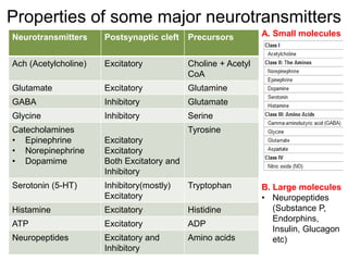
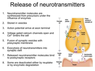
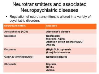
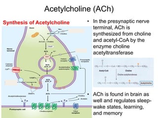
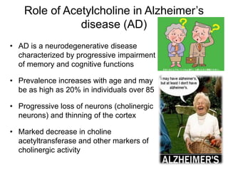
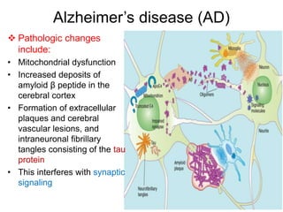
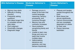
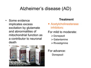
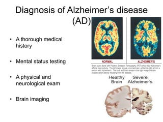
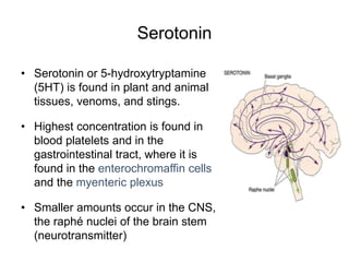
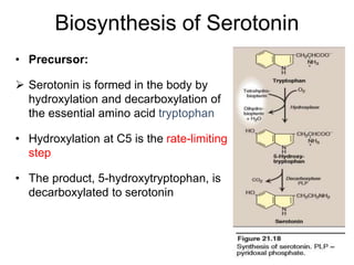
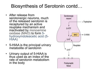
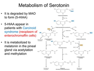
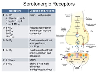
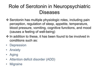
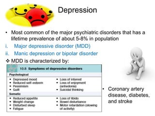
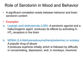
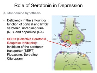
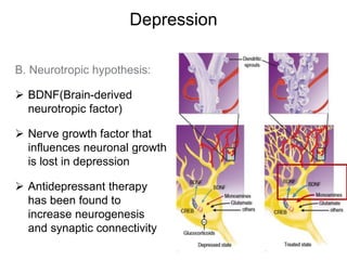
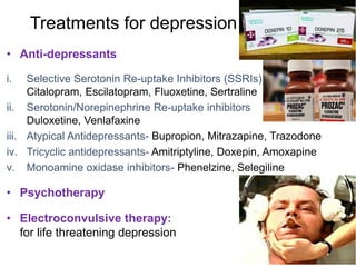
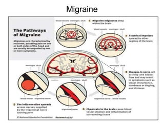
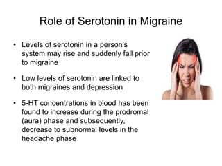
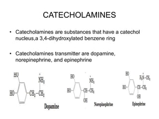
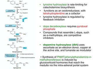
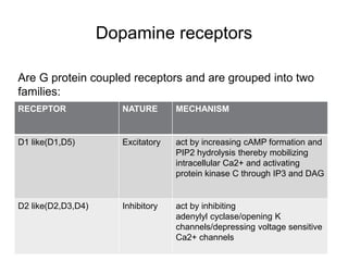
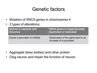
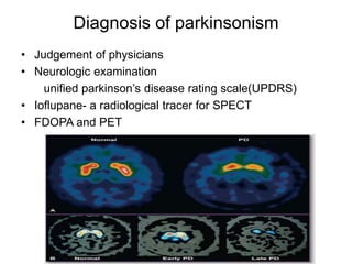
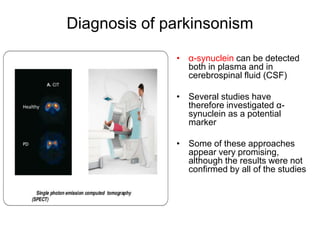
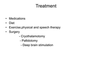
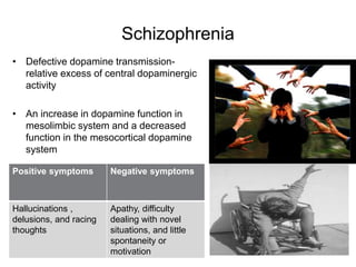
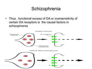
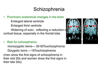
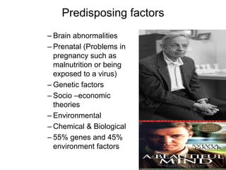
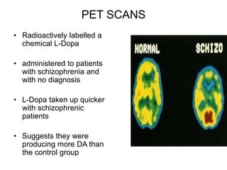
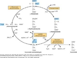
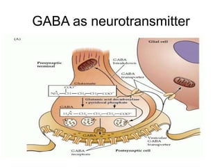
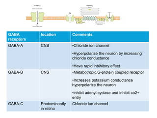
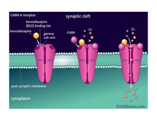
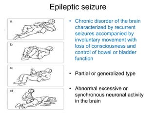
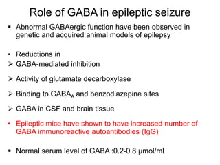
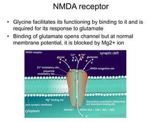
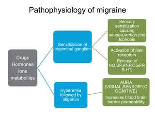
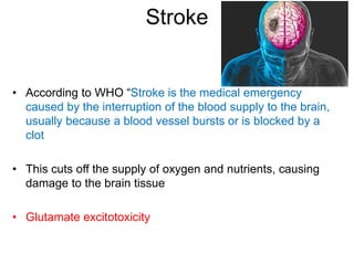
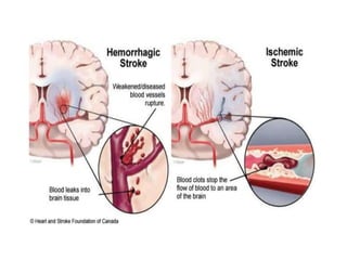
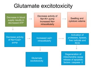
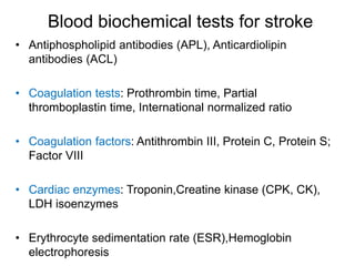
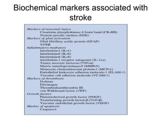
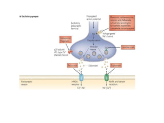
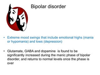
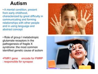
Role of Neurotransmitters in
Neuropsychiatric Disorders
Neuropsychiatric disorders
Neuropsychiatry is a field of
scientific medicine that
concerns itself with the
complex relationship between
human behavior and brain
function, and endeavors to
understand abnormal behavior
and behavioral disorders on the
basis of an interaction of
neurobiological and
psychological–social factors
Neuropsychiatric Symptoms:
Cognitive impairment , Disturbance
of consciousness
Neurotic complaints, Anxiety, Mood
changes ,
Psychotic States: Hallucination &
Delusion
Behavioral and Personality
changes
Neuropsychiatric disorders contd…
• The World Health Organization’s International
Classification of Disease (10th edition) or ICD-10
classification of neuropsychiatric disorders:
Neurotransmitters
• Chemical messengers released
from presynaptic nerve terminals
into the synaptic cleft
 Criteria:
• The substance must be present
within the presynaptic neuron
• The substance must be released in
response to presynaptic
depolarization, which must occur in
a Ca2+ dependent manner
• Specific receptors for the substance
must be present on the
postsynaptic cell
Properties of some major neurotransmitters
Neurotransmitters Postsynaptic cleft Precursors
Ach (Acetylcholine) Excitatory Choline + Acetyl
CoA
Glutamate Excitatory Glutamine
GABA Inhibitory Glutamate
Glycine Inhibitory Serine
Catecholamines
• Epinephrine
• Norepinephrine
• Dopamime
Excitatory
Excitatory
Both Excitatory and
Inhibitory
Tyrosine
Serotonin (5-HT) Inhibitory(mostly)
Excitatory
Tryptophan
Histamine Excitatory Histidine
ATP Excitatory ADP
Neuropeptides Excitatory and
Inhibitory
Amino acids
A. Small molecules
B. Large molecules
• Neuropeptides
(Substance P,
Endorphins,
Insulin, Glucagon
etc)
Release of neurotransmitters
1. Neurotransmitter molecules are
synthesized from precursors under the
influence of enzymes
2. Stored in vesicles
3. Action potential arrive at axon terminal
4. Voltage gated calcium channels open and
Ca2+ enters the cell
5. Fusion of synaptic vesicles with
presynaptic membrane
6. Exocytosis of neurotransmitters into
synaptic cleft
7. Released neurotransmitter molecules bind
to postsynaptic receptors
8. Some are deactivated either by reuptake
or by enzymatic degradation
Neurotransmitters and associated
Neuropsychiatric diseases
• Regulation of neurotransmitters is altered in a variety of
psychiatric disorders
Neurotransmitters Diseases
Acetylcholine (ACh) Alzheimer’s disease
Serotonin Depression
Migraine, Aging
Attention deficit disorder (ADD)
Anxiety
Dopamine (High) Schizophrenia
(Low) Parkinsonism
GABA (γ-Aminobutyrate) Epileptic seizures
Glutamate Migraine
Stroke
Autism
Acetylcholine (ACh)
• In the presynaptic nerve
terminal, ACh is
synthesized from choline
and acetyl-CoA by the
enzyme choline
acetyltransferase
• ACh is found in brain as
well and regulates sleep-
wake states, learning,
and memory
Synthesis of Acetylcholine
Role of Acetylcholine in Alzheimer’s
disease (AD)
• AD is a neurodegenerative disease
characterized by progressive impairment
of memory and cognitive functions
• Prevalence increases with age and may
be as high as 20% in individuals over 85
• Progressive loss of neurons (cholinergic
neurons) and thinning of the cortex
• Marked decrease in choline
acetyltransferase and other markers of
cholinergic activity
Alzheimer’s disease (AD)
 Pathologic changes
include:
• Mitochondrial dysfunction
• Increased deposits of
amyloid β peptide in the
cerebral cortex
• Formation of extracellular
plaques and cerebral
vascular lesions, and
intraneuronal fibrillary
tangles consisting of the tau
protein
• This interferes with synaptic
signaling
Mild Alzheimer’s Disease Moderate Alzheimer’s
Disease
Severe Alzheimer’s
Disease
 Memory loss starts
 Difficulty in finding
direction
 Frequently asking
questions
 One takes longer time
to complete normal
daily tasks
 Poor judgment
 Personality changes
 Diagnostic stage
 Damage occurs in
areas of the brain that
control language,
reasoning, sensory
processing, and
conscious thought
 Memory loss and
confusion increases
 Have problems
recognizing family and
friends
 Unable to learn new
things and carry out
tasks that involve
multiple steps
 Hallucinations,
delusions and
paranoia, and may
behave impulsively
 Plaques and tangles
spread throughout the
brain
 Brain tissue has
shrunk significantly
 Cannot communicate
and are completely
dependent on others
for their care
 Person becomes
bedridden
Alzheimer’s disease (AD)
• Some evidence
implicates excess
excitation by glutamate
and abnormalities of
mitochondrial function as
a contributor to neuronal
death
Treatment
 Acetylcholinesterase
inhibitors:
For mild to moderate:
Donepezil
Galantamine
Rivastigmine
For advance:
Donepezil
Diagnosis of Alzheimer’s disease
(AD)
• A thorough medical
history
• Mental status testing
• A physical and
neurological exam
• Brain imaging
Serotonin
• Serotonin or 5-hydroxytryptamine
(5HT) is found in plant and animal
tissues, venoms, and stings.
• Highest concentration is found in
blood platelets and in the
gastrointestinal tract, where it is
found in the enterochromaffin cells
and the myenteric plexus
• Smaller amounts occur in the CNS,
the raphé nuclei of the brain stem
(neurotransmitter)
Biosynthesis of Serotonin
• Precursor:
 Serotonin is formed in the body by
hydroxylation and decarboxylation of
the essential amino acid tryptophan
• Hydroxylation at C5 is the rate-limiting
step
• The product, 5-hydroxytryptophan, is
decarboxylated to serotonin
Biosynthesis of Serotonin contd…
• After release from
serotonergic neurons, much
of the released serotonin is
recaptured by an active
reuptake mechanism and
inactivated by monoamine
oxidase (MAO) to form 5-
hydroxyindoleacetic acid (5-
HIAA)
• 5-HIAA is the principal urinary
metabolite of serotonin
• Urinary output of 5-HIAA is
thus used as an index of the
rate of serotonin metabolism
in the body
Metabolism of Serotonin
• It is degraded by MAO
to form (5-HIAA)
• 5-HIAA appear in
patients with Carcinoid
syndrome (neoplasm of
enterochromaffin cells)
• It is metabolized to
melatonin in the pineal
gland via acetylation
and methylation
Serotonergic Receptors
Receptors Location and Actions
 5-HT1
5-HT1A, 5-HT1B, 5-
HT1D, 5-HT1E, 5-
HT1F, 5-HT1P
Brain, Raphe nuclei
 5-HT2
5-HT2A
5-HT2B
5-HT2C
Platelet aggregation
and smooth muscle
contraction
 5-HT3 Gastrointestinal tract,
area postrema;
vomiting
 5-HT4 Gastrointestinal tract;
brain; secretion and
peristalsis
 5-HT5A,B Brain;
 5-HT6,7 Brain; 5-HT6 high
affinity for
antidepressant drugs
Role of Serotonin in Neuropsychiatric
Diseases
 Serotonin has multiple physiologic roles, including pain
perception, regulation of sleep, appetite, temperature,
blood pressure, vomiting, cognitive functions, and mood
(causes a feeling of well-being)
 In addition to these, it has been found to be involved in
conditions such as:
• Depression
• Anxiety
• Aging
• Attention deficit disorder (ADD)
• Migraine
Depression
• Most common of the major psychiatric disorders that has a
lifetime prevalence of about 5-8% in population
i. Major depressive disorder (MDD)
ii. Manic depression or bipolar disorder
 MDD is characterized by:
• Coronary artery
disease, diabetes,
and stroke
Role of Serotonin in Mood and Behavior
• A significant correlation exists between behavior and brain
serotonin content
• Examples:
a. Lysergic acid diethylamide (LSD): A serotonin agonist and a
hallucinogenic agent produces its effects by activating 5-
HT2 receptors in the brain
b. MDMA (3,4-Methylenedioxymethamphetamine) or ecstasy:
A popular drug of abuse
It produces euphoria initially which is followed by difficulty
in concentrating, depression, and, in monkeys, insomnia
Role of Serotonin in Depression
A. Monoamine hypothesis:
• Deficiency in the amount or
function of cortical and limbic
serotonin, norepinephrine
(NE), and dopamine (DA)
• SSRIs (Selective Serotonin
Reuptake Inhibitors):
Inhibition of the serotonin
transporter (SERT)
Fluoxetine, Sertraline,
Citalopram
Depression
B. Neurotropic hypothesis:
 BDNF(Brain-derived
neurotropic factor)
 Nerve growth factor that
influences neuronal growth
is lost in depression
 Antidepressant therapy
has been found to
increase neurogenesis
and synaptic connectivity
Clinical Diagnosis of Depression
i. Structural imaging studies in
major depression is associated
with a 5–10% loss of volume in
the hippocampus of brain
ii. Biological markers of depression
are:
a. CSF level of 5-HIAA is low
b. 24hr urinary collection of 5-HIAA
c. Hypocholesterolemia
d. Low blood folate levels
e. Decreased 5-HT1A receptor
expression
f. Decreased BDNF in serum
Reference range: < or =210 mcg/24
hrs
However, depressive
disorders are heterogeneous
and diagnosed on the basis
of a patient’s symptoms, not
on the basis of a laboratory
test
Treatments for depression
• Anti-depressants
i. Selective Serotonin Re-uptake Inhibitors (SSRIs)
Citalopram, Escilatopram, Fluoxetine, Sertraline
ii. Serotonin/Norepinephrine Re-uptake inhibitors
Duloxetine, Venlafaxine
iii. Atypical Antidepressants- Bupropion, Mitrazapine, Trazodone
iv. Tricyclic antidepressants- Amitriptyline, Doxepin, Amoxapine
v. Monoamine oxidase inhibitors- Phenelzine, Selegiline
• Psychotherapy
• Electroconvulsive therapy:
for life threatening depression
Migraine
Role of Serotonin in Migraine
• Levels of serotonin in a person's
system may rise and suddenly fall prior
to migraine
• Low levels of serotonin are linked to
both migraines and depression
• 5-HT concentrations in blood has been
found to increase during the prodromal
(aura) phase and subsequently,
decrease to subnormal levels in the
headache phase
CATECHOLAMINES
• Catecholamines are substances that have a catechol
nucleus,a 3,4-dihydroxylated benzene ring
• Catecholamines transmitter are dopamine,
norepinephrine, and epinephrine
• tyrosine hydroxylase is rate-limiting for
catecholamine biosynthesis
• functions as an oxidoreductase, with
tetrahydropteridine as a cofactor
• tyrosine hydroxylase is regulated by
feedback inhibition
• dopa decarboxylase requires pyridoxal
phosphate
• Compounds that resemble L-dopa, such
as α-methyldopa, are competitive
inhibitors
• dopamine hydroxylase (dbh) uses
ascorbate as an electron donor, copper at
the active site, and fumarate as modulator
 Synthesis of PNMT(phenylethanolamine-n-
methyltransferase) is induced by
glucocorticoid hormones that reach the
medulla via the intra-adrenal portal system
Degradation of catecholamines
Catecholamines
Norepinephrine
• stimulatory processes
• elevated levels
ANXIETY,MOOD
DAMPENING
• Low levels LOW
ENERGY, DECREASED
FOCUS ability and sleep
cycle problems
epinephrine
elevated attention
deficit disorder with
hyperactivity (ADDH)
Depleted level Long
term STRESS or
INSOMNIA
Diagnostic use
Ref. Range:
Younger than 1 year: < 27 mg/g creatinine
Age 1-2 years: < 18 mg/g creatinine
Age 2-4 years: < 13 mg/g creatinine
Age 5-9 years: < 8.5 mg/g creatinine
Age 10-14 years: < 7 mg/g creatinine
The reference range in persons aged 15 years and older is
2-7 mg/24 hours
Conditions associated with elevations in urinary
VMA include the following: stress and anxiety
Mechanism of action of dopamine
Receptor subfamily location action
D1 and D2 Substantia nigra and
striatum
Motor control
D1 and D2 Limbic cortex and
associated structure
Information processing
D2 Anterior pituitary Inhibits prolactin release
• Action depend upon type of receptor with which it interacts
• Central effects of dopamine
Dopamine receptors
RECEPTOR NATURE MECHANISM
D1 like(D1,D5) Excitatory act by increasing cAMP formation and
PIP2 hydrolysis thereby mobilizing
intracellular Ca2+ and activating
protein kinase C through IP3 and DAG
D2 like(D2,D3,D4) Inhibitory act by inhibiting
adenylyl cyclase/opening K
channels/depressing voltage sensitive
Ca2+ channels
Are G protein coupled receptors and are grouped into two
families:
Manifestation of dopamine level
• Increased level of dopamine schizophrenia
• Decreased level of dopamine Parkinsonism
• Plasma reference ranges :
Supine adults - < 10 ng/ml
Ambulatory adults - < 20 ng/mL
Age 3-15 years - < 60 pg/mL
Parkinsonism
• Parkinsonism is a neurodegenerative
disorder characterized by rigidity, tremor
and hypokinesia with secondary
manifestations like defective posture
and gait, mask-like face and sialorrhoea;
dementia may accompany
• Dopaminergic neurons and dopamine
receptors are steadily lost with age in
the basal ganglia
• Predisposing factor: aging(free radicals),
environmental toxins,neurotoxin(MPTP),
genetic factors
Parkinsonism
Genetic factors
• Mutation of SNCA genes in chromosome 4
• 2 types of alterations:
• Aggregate (lewy bodies) and other protein
• Clog neuron and impair the function of neuron
Alanine is replaced with
threonine
SNCA genes is inappropriately
duplicated or triplicated
Cause α-synuclein to misfold Extracopies of the gene lead to an
increase of α-synuclein
Diagnosis of parkinsonism
• Judgement of physicians
• Neurologic examination
unified parkinson’s disease rating scale(UPDRS)
• Ioflupane- a radiological tracer for SPECT
• FDOPA and PET
Diagnosis of parkinsonism
• α-synuclein can be detected
both in plasma and in
cerebrospinal fluid (CSF)
• Several studies have
therefore investigated α-
synuclein as a potential
marker
• Some of these approaches
appear very promising,
although the results were not
confirmed by all of the studies
Treatment
• Medications
• Diet
• Exercise,physical and speech therapy
• Surgery
- Cryothalamotomy
- Pallidotomy
- Deep brain stimulation
Schizophrenia
• Defective dopamine transmission-
relative excess of central dopaminergic
activity
• An increase in dopamine function in
mesolimbic system and a decreased
function in the mesocortical dopamine
system
Positive symptoms Negative symptoms
Hallucinations ,
delusions, and racing
thoughts
Apathy, difficulty
dealing with novel
situations, and little
spontaneity or
motivation
Schizophrenia
• Thus , functional excess of DA or oversensitivity of
certain DA receptors is the causal factors in
schizophrenia
Schizophrenia
• Prominent anatomical changes in the brain
Enlarged lateral ventricle
Enlarged third ventricle
Widening of sulci , reflecting a reduction of
cortical tissue, especially in the frontal lobe
• Risk for schizophrenia
monozygotic twins--- 30-50%schizophrenia
Dizygotic twins ---15%schizophrenia
(men show the first signs of schizophrenia in
their mid 20s and women show the first signs in
their late 20s)
Predisposing factors
– Brain abnormalities
– Prenatal (Problems in
pregnancy such as
malnutrition or being
exposed to a virus)
– Genetic factors
– Socio –economic
theories
– Environmental
– Chemical & Biological
– 55% genes and 45%
environment factors
PET SCANS
• Radioactively labelled a
chemical L-Dopa
• administered to patients
with schizophrenia and
with no diagnosis
• L-Dopa taken up quicker
with schizophrenic
patients
• Suggests they were
producing more DA than
the control group
Diagnosis
• No physical or lab test - diagnosis based on clinical
symptoms
• Measurement of PHVA(plasma homovanillic acid) can
play a role in elucidating the DA abnormality in
schizophrenia
• Plasma homovanillic acid concentrations are lower in
chronic schizophrenic patients compared to normal
controls, and that PHVA values correlate with
schizophrenic symptom severity
Treatments
• There is no cure for schizophrenia but
most medications considerably lower the
symptoms
• Treatments Include:
 Antipsychotics
 Professional help sessions
 Typical narcoleptics or old drugs
Chlorpromazine (Thorazine)
Haloperidol (Haldol)
 Atypical narcoleptics or new
drugs
Risperidone
Clozapine
Gamma amino butyric acid (GABA)
• Principle inhibitory mediator in brain
• Maintains inhibitory tone that counterbalances neuronal
excitation
• Location : cerebellum, cerebral cortex, neurons mediating
post synaptic inhibition, retina
GABA as neurotransmitter
GABA
receptors
location Comments
GABA-A CNS •Chloride ion channel
•Hyperpolarize the neuron by increasing
chloride conductance
•Have rapid inbihitory effect
GABA-B CNS •Metabotropic,G-protein coupled receptor
•Increases potassium conductance
hyperpolarize the neuron
•inhibit adenyl cyclase and inhibit ca2+
entry
GABA-C Predominantly
in retina
Chloride ion channel
Epileptic seizure
• Chronic disorder of the brain
characterized by recurrent
seizures accompanied by
involuntary movement with
loss of consciousness and
control of bowel or bladder
function
• Partial or generalized type
• Abnormal excessive or
synchronous neuronal activity
in the brain
Role of GABA in epileptic seizure
 Abnormal GABAergic function have been observed in
genetic and acquired animal models of epilepsy
• Reductions in
 GABA-mediated inhibition
 Activity of glutamate decarboxylase
 Binding to GABAA and benzodiazepine sites
 GABA in CSF and brain tissue
• Epileptic mice have shown to have increased number of
GABA immunoreactive autoantibodies (IgG)
 Normal serum level of GABA :0.2-0.8 μmol/ml
Biochemical tests for epilepsy
Investigations Condition being tested
for
Clinical epilepsy
presentatiom
Succinic semialdehyde Pyridoxine dependent
epilepsy
Neonatal seizures ,
refractory epilepsy in
childhood
ceruloplasmin Epilepsy,mental
regression
Epilepsy with learning
disability
calcium Hypocalcaemia,hypopar
athyroidism
Neonatal seizure
creatinine Creatinine deficiency
syndrome
Epilepsy with learning
disability
Glucose hypoglycaemia Epilepsy with learning
disability
Uric acid Disorder of purine
metabolism
Refractory epilepsy
Glutamate
• Major Excitatory amino acid (75 %)
• Nonessential amino acid that cannot cross blood brain
barrier
• Location: cerebral cortex, brain stem
Glutamate receptors
Receptors Type Comments
Metabotropic Serpentine G-
protein coupled
receptor
Increases intracellular IP3 and
DAG
Decreases intracellular CAMP
Ionotropic Ligand gated ion
channels
• Kainate receptor permits Na+
influx and K+ efflux
• AMPA receptor (2 types): one
permit Na+ influx and other also
passage Ca2+
• NMDA receptor: permits passage
relatively large amount of Ca2+
Migraine
• Recurrent throbbing headache that typically affects one side
of the head and is often accompanied by nausea and
disturbed vision
• Migraine pain-relay centers: trigeminal ganglion and
thalamus contain glutamate-positive neurons
• Neuronal hyper excitability due to decreased voltage
dependent Mg2+ channel excitotoxic neural death
and oligaemia causing migranous infarction
NMDA receptor
• Glycine facilitates its functioning by binding to it and is
required for its response to glutamate
• Binding of glutamate opens channel but at normal
membrane potential, it is blocked by Mg2+ ion
Pathophysiology of migraine
Drugs
Hormones
Ions
metabolites
Sensitization of
trigeminal ganglion
Sensory
sensitization
causing
nausea,vertigo,pho
tophobia
Activation of pain
receptors
Release of
NO,SP,ANP,CGRP,
5-HT,
Hyperemia
followed by
oligemia
AURA
(VISUAL,SENSORY,C
OGNITIVE)
Increases blood brain
barrier permeability
Stroke
• According to WHO “Stroke is the medical emergency
caused by the interruption of the blood supply to the brain,
usually because a blood vessel bursts or is blocked by a
clot
• This cuts off the supply of oxygen and nutrients, causing
damage to the brain tissue
• Glutamate excitotoxicity
Glutamate excitotoxicity
Decrease in blood
supply results in
decreased ATP
Decrease activity of
Na+/K+ pump
Increased Na+
intracellularly
Decrease activity
of Na+/Ca2+
pump
Swelling and
cytotoxic edema
Increased ca2+
intracellularly
Activation of
proteases, lipases,
free radicals and
ROS
Degeneration of
mitochondria cause
release of apoptotic
factors :caspase -3
Glutamate
excitotoxicity
Blood biochemical tests for stroke
• Antiphospholipid antibodies (APL), Anticardiolipin
antibodies (ACL)
• Coagulation tests: Prothrombin time, Partial
thromboplastin time, International normalized ratio
• Coagulation factors: Antithrombin III, Protein C, Protein S;
Factor VIII
• Cardiac enzymes: Troponin,Creatine kinase (CPK, CK),
LDH isoenzymes
• Erythrocyte sedimentation rate (ESR),Hemoglobin
electrophoresis
Biochemical markers associated with
stroke
Bipolar disorder
• Extreme mood swings that include emotional highs (mania
or hypomania) and lows (depression)
• Glutamate, GABA and dopamine is found to be
significantly increased during the manic phase of bipolar
disorder, and returns to normal levels once the phase is
over
Autism
• A mental condition, present
from early childhood,
characterized by great difficulty in
communicating and forming
relationships with other people
and in using language and
abstract concept
• Role of group I metabotropic
glutamate receptors in the
pathogenesis of fragile X
syndrome :the most common
identified genetic cause of autism
•FMR1 gene encode for FMRP
: responsible for synapse
REFERENCES
• Murray RK, Bender DA, Botham KM et al. Harper’s Illustrated
Biochemistry. 29th edition, 2012.
• Harvey RA. Lippincott’s Illustrated Reviews Biochemistry. 5th
edition. 2011.
• Colledge NR, Walker BR, Ralston SH. Davidson’s Principles
& Practice of Medicine. 21st edition. 2010.
• Katzung BG, Masters SB, Trevor AJ. Basic and Clinical
Pharmacology. 12th edition. 2012.
• Barret Kim, Brooks Heddwen, Boitano Scott et al. Ganong’s
Review of Medical Physiology. 23rd edition. 2010.
• Koho Miyoshi and Yasushi Morimura. Clinical Manifestations
of Neuropsychiatric Disorders. Jinmeikai Research Institute
for Mental Health. 2010.
• Nowrangi MA. Principles and Management of
Neuropsychiatric Symptoms in Alzheimer’s Disease.
Alzheimer's Research & Therapy. 2015

biochemfinal-150907144947-lva1-app6892 (1).pdf

  • 1.
    Role of Neurotransmittersin Neuropsychiatric Disorders
  • 2.
    Neuropsychiatric disorders Neuropsychiatry isa field of scientific medicine that concerns itself with the complex relationship between human behavior and brain function, and endeavors to understand abnormal behavior and behavioral disorders on the basis of an interaction of neurobiological and psychological–social factors Neuropsychiatric Symptoms: Cognitive impairment , Disturbance of consciousness Neurotic complaints, Anxiety, Mood changes , Psychotic States: Hallucination & Delusion Behavioral and Personality changes
  • 3.
    Neuropsychiatric disorders contd… •The World Health Organization’s International Classification of Disease (10th edition) or ICD-10 classification of neuropsychiatric disorders:
  • 4.
    Neurotransmitters • Chemical messengersreleased from presynaptic nerve terminals into the synaptic cleft  Criteria: • The substance must be present within the presynaptic neuron • The substance must be released in response to presynaptic depolarization, which must occur in a Ca2+ dependent manner • Specific receptors for the substance must be present on the postsynaptic cell
  • 5.
    Properties of somemajor neurotransmitters Neurotransmitters Postsynaptic cleft Precursors Ach (Acetylcholine) Excitatory Choline + Acetyl CoA Glutamate Excitatory Glutamine GABA Inhibitory Glutamate Glycine Inhibitory Serine Catecholamines • Epinephrine • Norepinephrine • Dopamime Excitatory Excitatory Both Excitatory and Inhibitory Tyrosine Serotonin (5-HT) Inhibitory(mostly) Excitatory Tryptophan Histamine Excitatory Histidine ATP Excitatory ADP Neuropeptides Excitatory and Inhibitory Amino acids A. Small molecules B. Large molecules • Neuropeptides (Substance P, Endorphins, Insulin, Glucagon etc)
  • 6.
    Release of neurotransmitters 1.Neurotransmitter molecules are synthesized from precursors under the influence of enzymes 2. Stored in vesicles 3. Action potential arrive at axon terminal 4. Voltage gated calcium channels open and Ca2+ enters the cell 5. Fusion of synaptic vesicles with presynaptic membrane 6. Exocytosis of neurotransmitters into synaptic cleft 7. Released neurotransmitter molecules bind to postsynaptic receptors 8. Some are deactivated either by reuptake or by enzymatic degradation
  • 7.
    Neurotransmitters and associated Neuropsychiatricdiseases • Regulation of neurotransmitters is altered in a variety of psychiatric disorders Neurotransmitters Diseases Acetylcholine (ACh) Alzheimer’s disease Serotonin Depression Migraine, Aging Attention deficit disorder (ADD) Anxiety Dopamine (High) Schizophrenia (Low) Parkinsonism GABA (γ-Aminobutyrate) Epileptic seizures Glutamate Migraine Stroke Autism
  • 8.
    Acetylcholine (ACh) • Inthe presynaptic nerve terminal, ACh is synthesized from choline and acetyl-CoA by the enzyme choline acetyltransferase • ACh is found in brain as well and regulates sleep- wake states, learning, and memory Synthesis of Acetylcholine
  • 9.
    Role of Acetylcholinein Alzheimer’s disease (AD) • AD is a neurodegenerative disease characterized by progressive impairment of memory and cognitive functions • Prevalence increases with age and may be as high as 20% in individuals over 85 • Progressive loss of neurons (cholinergic neurons) and thinning of the cortex • Marked decrease in choline acetyltransferase and other markers of cholinergic activity
  • 10.
    Alzheimer’s disease (AD) Pathologic changes include: • Mitochondrial dysfunction • Increased deposits of amyloid β peptide in the cerebral cortex • Formation of extracellular plaques and cerebral vascular lesions, and intraneuronal fibrillary tangles consisting of the tau protein • This interferes with synaptic signaling
  • 11.
    Mild Alzheimer’s DiseaseModerate Alzheimer’s Disease Severe Alzheimer’s Disease  Memory loss starts  Difficulty in finding direction  Frequently asking questions  One takes longer time to complete normal daily tasks  Poor judgment  Personality changes  Diagnostic stage  Damage occurs in areas of the brain that control language, reasoning, sensory processing, and conscious thought  Memory loss and confusion increases  Have problems recognizing family and friends  Unable to learn new things and carry out tasks that involve multiple steps  Hallucinations, delusions and paranoia, and may behave impulsively  Plaques and tangles spread throughout the brain  Brain tissue has shrunk significantly  Cannot communicate and are completely dependent on others for their care  Person becomes bedridden
  • 12.
    Alzheimer’s disease (AD) •Some evidence implicates excess excitation by glutamate and abnormalities of mitochondrial function as a contributor to neuronal death Treatment  Acetylcholinesterase inhibitors: For mild to moderate: Donepezil Galantamine Rivastigmine For advance: Donepezil
  • 13.
    Diagnosis of Alzheimer’sdisease (AD) • A thorough medical history • Mental status testing • A physical and neurological exam • Brain imaging
  • 15.
    Serotonin • Serotonin or5-hydroxytryptamine (5HT) is found in plant and animal tissues, venoms, and stings. • Highest concentration is found in blood platelets and in the gastrointestinal tract, where it is found in the enterochromaffin cells and the myenteric plexus • Smaller amounts occur in the CNS, the raphé nuclei of the brain stem (neurotransmitter)
  • 16.
    Biosynthesis of Serotonin •Precursor:  Serotonin is formed in the body by hydroxylation and decarboxylation of the essential amino acid tryptophan • Hydroxylation at C5 is the rate-limiting step • The product, 5-hydroxytryptophan, is decarboxylated to serotonin
  • 17.
    Biosynthesis of Serotonincontd… • After release from serotonergic neurons, much of the released serotonin is recaptured by an active reuptake mechanism and inactivated by monoamine oxidase (MAO) to form 5- hydroxyindoleacetic acid (5- HIAA) • 5-HIAA is the principal urinary metabolite of serotonin • Urinary output of 5-HIAA is thus used as an index of the rate of serotonin metabolism in the body
  • 18.
    Metabolism of Serotonin •It is degraded by MAO to form (5-HIAA) • 5-HIAA appear in patients with Carcinoid syndrome (neoplasm of enterochromaffin cells) • It is metabolized to melatonin in the pineal gland via acetylation and methylation
  • 19.
    Serotonergic Receptors Receptors Locationand Actions  5-HT1 5-HT1A, 5-HT1B, 5- HT1D, 5-HT1E, 5- HT1F, 5-HT1P Brain, Raphe nuclei  5-HT2 5-HT2A 5-HT2B 5-HT2C Platelet aggregation and smooth muscle contraction  5-HT3 Gastrointestinal tract, area postrema; vomiting  5-HT4 Gastrointestinal tract; brain; secretion and peristalsis  5-HT5A,B Brain;  5-HT6,7 Brain; 5-HT6 high affinity for antidepressant drugs
  • 20.
    Role of Serotoninin Neuropsychiatric Diseases  Serotonin has multiple physiologic roles, including pain perception, regulation of sleep, appetite, temperature, blood pressure, vomiting, cognitive functions, and mood (causes a feeling of well-being)  In addition to these, it has been found to be involved in conditions such as: • Depression • Anxiety • Aging • Attention deficit disorder (ADD) • Migraine
  • 21.
    Depression • Most commonof the major psychiatric disorders that has a lifetime prevalence of about 5-8% in population i. Major depressive disorder (MDD) ii. Manic depression or bipolar disorder  MDD is characterized by: • Coronary artery disease, diabetes, and stroke
  • 22.
    Role of Serotoninin Mood and Behavior • A significant correlation exists between behavior and brain serotonin content • Examples: a. Lysergic acid diethylamide (LSD): A serotonin agonist and a hallucinogenic agent produces its effects by activating 5- HT2 receptors in the brain b. MDMA (3,4-Methylenedioxymethamphetamine) or ecstasy: A popular drug of abuse It produces euphoria initially which is followed by difficulty in concentrating, depression, and, in monkeys, insomnia
  • 23.
    Role of Serotoninin Depression A. Monoamine hypothesis: • Deficiency in the amount or function of cortical and limbic serotonin, norepinephrine (NE), and dopamine (DA) • SSRIs (Selective Serotonin Reuptake Inhibitors): Inhibition of the serotonin transporter (SERT) Fluoxetine, Sertraline, Citalopram
  • 24.
    Depression B. Neurotropic hypothesis: BDNF(Brain-derived neurotropic factor)  Nerve growth factor that influences neuronal growth is lost in depression  Antidepressant therapy has been found to increase neurogenesis and synaptic connectivity
  • 25.
    Clinical Diagnosis ofDepression i. Structural imaging studies in major depression is associated with a 5–10% loss of volume in the hippocampus of brain ii. Biological markers of depression are: a. CSF level of 5-HIAA is low b. 24hr urinary collection of 5-HIAA c. Hypocholesterolemia d. Low blood folate levels e. Decreased 5-HT1A receptor expression f. Decreased BDNF in serum Reference range: < or =210 mcg/24 hrs However, depressive disorders are heterogeneous and diagnosed on the basis of a patient’s symptoms, not on the basis of a laboratory test
  • 26.
    Treatments for depression •Anti-depressants i. Selective Serotonin Re-uptake Inhibitors (SSRIs) Citalopram, Escilatopram, Fluoxetine, Sertraline ii. Serotonin/Norepinephrine Re-uptake inhibitors Duloxetine, Venlafaxine iii. Atypical Antidepressants- Bupropion, Mitrazapine, Trazodone iv. Tricyclic antidepressants- Amitriptyline, Doxepin, Amoxapine v. Monoamine oxidase inhibitors- Phenelzine, Selegiline • Psychotherapy • Electroconvulsive therapy: for life threatening depression
  • 27.
  • 28.
    Role of Serotoninin Migraine • Levels of serotonin in a person's system may rise and suddenly fall prior to migraine • Low levels of serotonin are linked to both migraines and depression • 5-HT concentrations in blood has been found to increase during the prodromal (aura) phase and subsequently, decrease to subnormal levels in the headache phase
  • 29.
    CATECHOLAMINES • Catecholamines aresubstances that have a catechol nucleus,a 3,4-dihydroxylated benzene ring • Catecholamines transmitter are dopamine, norepinephrine, and epinephrine
  • 30.
    • tyrosine hydroxylaseis rate-limiting for catecholamine biosynthesis • functions as an oxidoreductase, with tetrahydropteridine as a cofactor • tyrosine hydroxylase is regulated by feedback inhibition • dopa decarboxylase requires pyridoxal phosphate • Compounds that resemble L-dopa, such as α-methyldopa, are competitive inhibitors • dopamine hydroxylase (dbh) uses ascorbate as an electron donor, copper at the active site, and fumarate as modulator  Synthesis of PNMT(phenylethanolamine-n- methyltransferase) is induced by glucocorticoid hormones that reach the medulla via the intra-adrenal portal system
  • 31.
  • 32.
    Catecholamines Norepinephrine • stimulatory processes •elevated levels ANXIETY,MOOD DAMPENING • Low levels LOW ENERGY, DECREASED FOCUS ability and sleep cycle problems epinephrine elevated attention deficit disorder with hyperactivity (ADDH) Depleted level Long term STRESS or INSOMNIA
  • 33.
    Diagnostic use Ref. Range: Youngerthan 1 year: < 27 mg/g creatinine Age 1-2 years: < 18 mg/g creatinine Age 2-4 years: < 13 mg/g creatinine Age 5-9 years: < 8.5 mg/g creatinine Age 10-14 years: < 7 mg/g creatinine The reference range in persons aged 15 years and older is 2-7 mg/24 hours Conditions associated with elevations in urinary VMA include the following: stress and anxiety
  • 34.
    Mechanism of actionof dopamine Receptor subfamily location action D1 and D2 Substantia nigra and striatum Motor control D1 and D2 Limbic cortex and associated structure Information processing D2 Anterior pituitary Inhibits prolactin release • Action depend upon type of receptor with which it interacts • Central effects of dopamine
  • 35.
    Dopamine receptors RECEPTOR NATUREMECHANISM D1 like(D1,D5) Excitatory act by increasing cAMP formation and PIP2 hydrolysis thereby mobilizing intracellular Ca2+ and activating protein kinase C through IP3 and DAG D2 like(D2,D3,D4) Inhibitory act by inhibiting adenylyl cyclase/opening K channels/depressing voltage sensitive Ca2+ channels Are G protein coupled receptors and are grouped into two families:
  • 36.
    Manifestation of dopaminelevel • Increased level of dopamine schizophrenia • Decreased level of dopamine Parkinsonism • Plasma reference ranges : Supine adults - < 10 ng/ml Ambulatory adults - < 20 ng/mL Age 3-15 years - < 60 pg/mL
  • 37.
    Parkinsonism • Parkinsonism isa neurodegenerative disorder characterized by rigidity, tremor and hypokinesia with secondary manifestations like defective posture and gait, mask-like face and sialorrhoea; dementia may accompany • Dopaminergic neurons and dopamine receptors are steadily lost with age in the basal ganglia • Predisposing factor: aging(free radicals), environmental toxins,neurotoxin(MPTP), genetic factors
  • 38.
  • 39.
    Genetic factors • Mutationof SNCA genes in chromosome 4 • 2 types of alterations: • Aggregate (lewy bodies) and other protein • Clog neuron and impair the function of neuron Alanine is replaced with threonine SNCA genes is inappropriately duplicated or triplicated Cause α-synuclein to misfold Extracopies of the gene lead to an increase of α-synuclein
  • 40.
    Diagnosis of parkinsonism •Judgement of physicians • Neurologic examination unified parkinson’s disease rating scale(UPDRS) • Ioflupane- a radiological tracer for SPECT • FDOPA and PET
  • 41.
    Diagnosis of parkinsonism •α-synuclein can be detected both in plasma and in cerebrospinal fluid (CSF) • Several studies have therefore investigated α- synuclein as a potential marker • Some of these approaches appear very promising, although the results were not confirmed by all of the studies
  • 42.
    Treatment • Medications • Diet •Exercise,physical and speech therapy • Surgery - Cryothalamotomy - Pallidotomy - Deep brain stimulation
  • 43.
    Schizophrenia • Defective dopaminetransmission- relative excess of central dopaminergic activity • An increase in dopamine function in mesolimbic system and a decreased function in the mesocortical dopamine system Positive symptoms Negative symptoms Hallucinations , delusions, and racing thoughts Apathy, difficulty dealing with novel situations, and little spontaneity or motivation
  • 44.
    Schizophrenia • Thus ,functional excess of DA or oversensitivity of certain DA receptors is the causal factors in schizophrenia
  • 45.
    Schizophrenia • Prominent anatomicalchanges in the brain Enlarged lateral ventricle Enlarged third ventricle Widening of sulci , reflecting a reduction of cortical tissue, especially in the frontal lobe • Risk for schizophrenia monozygotic twins--- 30-50%schizophrenia Dizygotic twins ---15%schizophrenia (men show the first signs of schizophrenia in their mid 20s and women show the first signs in their late 20s)
  • 46.
    Predisposing factors – Brainabnormalities – Prenatal (Problems in pregnancy such as malnutrition or being exposed to a virus) – Genetic factors – Socio –economic theories – Environmental – Chemical & Biological – 55% genes and 45% environment factors
  • 47.
    PET SCANS • Radioactivelylabelled a chemical L-Dopa • administered to patients with schizophrenia and with no diagnosis • L-Dopa taken up quicker with schizophrenic patients • Suggests they were producing more DA than the control group
  • 48.
    Diagnosis • No physicalor lab test - diagnosis based on clinical symptoms • Measurement of PHVA(plasma homovanillic acid) can play a role in elucidating the DA abnormality in schizophrenia • Plasma homovanillic acid concentrations are lower in chronic schizophrenic patients compared to normal controls, and that PHVA values correlate with schizophrenic symptom severity
  • 49.
    Treatments • There isno cure for schizophrenia but most medications considerably lower the symptoms • Treatments Include:  Antipsychotics  Professional help sessions  Typical narcoleptics or old drugs Chlorpromazine (Thorazine) Haloperidol (Haldol)  Atypical narcoleptics or new drugs Risperidone Clozapine
  • 50.
    Gamma amino butyricacid (GABA) • Principle inhibitory mediator in brain • Maintains inhibitory tone that counterbalances neuronal excitation • Location : cerebellum, cerebral cortex, neurons mediating post synaptic inhibition, retina
  • 52.
  • 53.
    GABA receptors location Comments GABA-A CNS•Chloride ion channel •Hyperpolarize the neuron by increasing chloride conductance •Have rapid inbihitory effect GABA-B CNS •Metabotropic,G-protein coupled receptor •Increases potassium conductance hyperpolarize the neuron •inhibit adenyl cyclase and inhibit ca2+ entry GABA-C Predominantly in retina Chloride ion channel
  • 55.
    Epileptic seizure • Chronicdisorder of the brain characterized by recurrent seizures accompanied by involuntary movement with loss of consciousness and control of bowel or bladder function • Partial or generalized type • Abnormal excessive or synchronous neuronal activity in the brain
  • 56.
    Role of GABAin epileptic seizure  Abnormal GABAergic function have been observed in genetic and acquired animal models of epilepsy • Reductions in  GABA-mediated inhibition  Activity of glutamate decarboxylase  Binding to GABAA and benzodiazepine sites  GABA in CSF and brain tissue • Epileptic mice have shown to have increased number of GABA immunoreactive autoantibodies (IgG)  Normal serum level of GABA :0.2-0.8 μmol/ml
  • 57.
    Biochemical tests forepilepsy Investigations Condition being tested for Clinical epilepsy presentatiom Succinic semialdehyde Pyridoxine dependent epilepsy Neonatal seizures , refractory epilepsy in childhood ceruloplasmin Epilepsy,mental regression Epilepsy with learning disability calcium Hypocalcaemia,hypopar athyroidism Neonatal seizure creatinine Creatinine deficiency syndrome Epilepsy with learning disability Glucose hypoglycaemia Epilepsy with learning disability Uric acid Disorder of purine metabolism Refractory epilepsy
  • 59.
    Glutamate • Major Excitatoryamino acid (75 %) • Nonessential amino acid that cannot cross blood brain barrier • Location: cerebral cortex, brain stem
  • 61.
    Glutamate receptors Receptors TypeComments Metabotropic Serpentine G- protein coupled receptor Increases intracellular IP3 and DAG Decreases intracellular CAMP Ionotropic Ligand gated ion channels • Kainate receptor permits Na+ influx and K+ efflux • AMPA receptor (2 types): one permit Na+ influx and other also passage Ca2+ • NMDA receptor: permits passage relatively large amount of Ca2+
  • 62.
    Migraine • Recurrent throbbingheadache that typically affects one side of the head and is often accompanied by nausea and disturbed vision • Migraine pain-relay centers: trigeminal ganglion and thalamus contain glutamate-positive neurons • Neuronal hyper excitability due to decreased voltage dependent Mg2+ channel excitotoxic neural death and oligaemia causing migranous infarction
  • 63.
    NMDA receptor • Glycinefacilitates its functioning by binding to it and is required for its response to glutamate • Binding of glutamate opens channel but at normal membrane potential, it is blocked by Mg2+ ion
  • 64.
    Pathophysiology of migraine Drugs Hormones Ions metabolites Sensitizationof trigeminal ganglion Sensory sensitization causing nausea,vertigo,pho tophobia Activation of pain receptors Release of NO,SP,ANP,CGRP, 5-HT, Hyperemia followed by oligemia AURA (VISUAL,SENSORY,C OGNITIVE) Increases blood brain barrier permeability
  • 65.
    Stroke • According toWHO “Stroke is the medical emergency caused by the interruption of the blood supply to the brain, usually because a blood vessel bursts or is blocked by a clot • This cuts off the supply of oxygen and nutrients, causing damage to the brain tissue • Glutamate excitotoxicity
  • 67.
    Glutamate excitotoxicity Decrease inblood supply results in decreased ATP Decrease activity of Na+/K+ pump Increased Na+ intracellularly Decrease activity of Na+/Ca2+ pump Swelling and cytotoxic edema Increased ca2+ intracellularly Activation of proteases, lipases, free radicals and ROS Degeneration of mitochondria cause release of apoptotic factors :caspase -3 Glutamate excitotoxicity
  • 68.
    Blood biochemical testsfor stroke • Antiphospholipid antibodies (APL), Anticardiolipin antibodies (ACL) • Coagulation tests: Prothrombin time, Partial thromboplastin time, International normalized ratio • Coagulation factors: Antithrombin III, Protein C, Protein S; Factor VIII • Cardiac enzymes: Troponin,Creatine kinase (CPK, CK), LDH isoenzymes • Erythrocyte sedimentation rate (ESR),Hemoglobin electrophoresis
  • 69.
  • 71.
    Bipolar disorder • Extrememood swings that include emotional highs (mania or hypomania) and lows (depression) • Glutamate, GABA and dopamine is found to be significantly increased during the manic phase of bipolar disorder, and returns to normal levels once the phase is over
  • 72.
    Autism • A mentalcondition, present from early childhood, characterized by great difficulty in communicating and forming relationships with other people and in using language and abstract concept • Role of group I metabotropic glutamate receptors in the pathogenesis of fragile X syndrome :the most common identified genetic cause of autism •FMR1 gene encode for FMRP : responsible for synapse
  • 73.
    REFERENCES • Murray RK,Bender DA, Botham KM et al. Harper’s Illustrated Biochemistry. 29th edition, 2012. • Harvey RA. Lippincott’s Illustrated Reviews Biochemistry. 5th edition. 2011. • Colledge NR, Walker BR, Ralston SH. Davidson’s Principles & Practice of Medicine. 21st edition. 2010. • Katzung BG, Masters SB, Trevor AJ. Basic and Clinical Pharmacology. 12th edition. 2012. • Barret Kim, Brooks Heddwen, Boitano Scott et al. Ganong’s Review of Medical Physiology. 23rd edition. 2010. • Koho Miyoshi and Yasushi Morimura. Clinical Manifestations of Neuropsychiatric Disorders. Jinmeikai Research Institute for Mental Health. 2010. • Nowrangi MA. Principles and Management of Neuropsychiatric Symptoms in Alzheimer’s Disease. Alzheimer's Research & Therapy. 2015