
Recommended
More Related Content
Similar to basicpharmacokineticsandcompatmentmodelling-171223090651.ppt
Similar to basicpharmacokineticsandcompatmentmodelling-171223090651.ppt (20)
Recently uploaded
Recently uploaded (20)
basicpharmacokineticsandcompatmentmodelling-171223090651.ppt
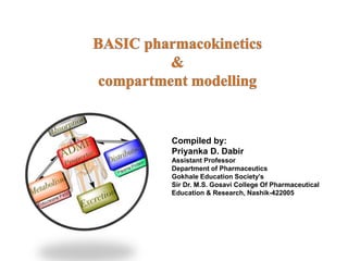
- 1. Compiled by: Priyanka D. Dabir Assistant Professor Department of Pharmaceutics Gokhale Education Society’s Sir Dr. M.S. Gosavi College Of Pharmaceutical Education & Research, Nashik-422005
- 2. Dosage Regimen: The frequency of administration of a drug in a particular dose is called as dosage regimen. Pharmacokinetics: Pharmacokinetics is defined as the kinetics of drug absorption, distribution, metabolism and excretion (KADME) and their relationship with the pharmacological, therapeutic or toxicological response in man and animals. There are two aspects of pharmacokinetic studies – Theoretical aspect – which involves development of pharmacokinetic models to predict drug disposition after its administration. Statistical methods are commonly applied to interpret data and assess various parameters. Experimental aspect – which involves development of biological sampling techniques, analytical methods for measurement of drug (and metabolites) concentration in biological samples and data collection and evaluation.
- 3. Plasma Drug Concentration-Time Profile A direct relationship exists between the concentration of drug at the biophase (site of action) and the concentration of drug in plasma. Two categories of parameters can be evaluated from a plasma concentration time profile – Pharmacokinetic parameters, and Pharmacodynamic parameters.
- 4. Pharmacokinetic Parameters 1. Peak Plasma Concentration (Cmax) The peak plasma level depends upon – The administered dose Rate of absorption, and Rate of elimination. 2. Time of Peak Concentration (tmax) 3. Area Under the Curve (AUC) Pharmacodynamic Parameters 1. Minimum Effective Concentration (MEC) 2. Maximum Safe Concentration (MSC) 3. Onset of Action 4. Onset Time 5. Duration of Action 6. Intensity of Action 7. Therapeutic Range 8. Therapeutic Index
- 5. Rate, Rate Constants and Orders of Reactions Rate: The velocity with which a reaction or a process occurs is called as its rate. Order of reaction: The manner in which the concentration of drug (or reactants) influences the rate of reaction or process is called as the order of reaction or order of process. Consider the following chemical reaction: Drug A Drug B The rate of forward reaction is expressed as – -dA/dt Negative sign indicates that the concentration of drug A decreases with time t. As the reaction proceeds, the concentration of drug B increases and the rate of reaction can also be expressed as: dB/dt dC/dt = -KCn K = rate constant n = order of reaction
- 6. Zero-Order Kinetics (Constant Rate Processes) If n = 0, equation becomes: dC/dt = -Ko Co where Ko = zero-order rate constant (in mg/min) Zero-order process can be defined as the one whose rate is independent of the concentration of drug undergoing reaction i.e. the rate of reaction cannot be increased further by increasing the concentration of reactants. Finally C = Co – Ko t
- 7. Zero-Order Half-Life Half-life (t½) or half-time is defined as the time period required for the concentration of drug to decrease by one-half. t1/2 = Co / 2 Ko Examples of zero-order processes are – 1. Metabolism/protein-drug binding/enzyme or carrier-mediated transport under saturated conditions. The rate of metabolism, binding or transport of drug remains constant as long as its concentration is in excess of saturating concentration. 2. Administration of a drug as a constant rate i.v. infusion. 3. Controlled drug delivery such as that from i.m. implants or osmotic pumps.
- 8. First-Order Kinetics (Linear Kinetics) If n = 1, equation becomes: dC/dt = - K C where K = first-order rate constant (in time–1 or per hour) first-order process is the one whose rate is directly proportional to the concentration of drug undergoing reaction i.e. greater the concentration, faster the reaction. It is because of such proportionality between rate of reaction and the concentration of drug that a first-order process is said to follow linear kinetics.
- 9. ln C = ln Co - K t C = C o – e –Kt log C = log Co – Kt/2.303 The first-order process is also called as monoexponential rate process. Thus, a first-order process is characterized by logarithmic or exponential kinetics i.e. a constant fraction of drug undergoes reaction per unit time.
- 10. t1/2 = 0.693 / K Equation shows that, in contrast to zero-order process, the half-life of a first-order process is a constant and independent of initial drug concentration i.e. irrespective of what the initial drug concentration is, the time required for the concentration to decrease by one-half remains the same. Most pharmacokinetic processes viz. absorption, distribution and elimination follow first- order kinetics.
- 11. Mixed-Order Kinetics (Nonlinear Kinetics) A mixture of both first-order and zero-order kinetics is said to follow mixed-order kinetics. Since deviations from an originally linear pharmacokinetic profile are observed, the rate process of such a drug is called as nonlinear kinetics. Mixed order kinetics is also termed as dose-dependent kinetics as it is observed at increased or multiple doses of some drugs. Nonlinearities in pharmacokinetics have been observed in – Drug absorption (e.g. vitamin C) Drug distribution (e.g. naproxen), and Drug elimination (e.g. riboflavin). The phenomena is seen when a particular pharmacokinetic process involves presence of carriers or enzymes which are substrate specific and have definite capacities and can get saturated at high drug concentrations (i.e. capacity-limited). The kinetics of such capacity- limited processes can be described by the Michaelis-Menten kinetics.
- 12. PHARMACOKINETIC PARAMETERS In practice, pharmacokinetic parameters are determined experimentally from a set of drug concentrations collected over various times known as data. Parameters are also called as variables. Variables are of two types – Independent variables which are not affected by any other parameter, for example time. Dependent variables, which change as the independent variables change, for example, plasma drug concentration.
- 13. PHARMACOKINETIC MODELS Drug movement within the body is a complex process. The major objective is therefore to develop a generalized and simple approach to describe, analyse and interpret the data obtained during in vivo drug disposition studies. The two major approaches in the quantitative study of various kinetic processes of drug disposition in the body are 1. Model approach, and 2. Model-independent approach (also called as non-compartmental analysis).
- 14. Pharmacokinetic Model Approach A model is a hypothesis that employs mathematical terms to concisely describe quantitative relationships. Pharmacokinetic models provide concise means of expressing mathematically or quantitatively, the time course of drug(s) throughout the body and compute meaningful pharmacokinetic parameters. Applications of Pharmacokinetic Models – 1. Characterizing the behaviour of drugs in patients. 2. Predicting the concentration of drug in various body fluids with any dosage regimen. 3. Predicting the multiple-dose concentration curves from single dose experiments. 4. Calculating the optimum dosage regimen for individual patients. 5. Evaluating the risk of toxicity with certain dosage regimens. 6. Correlating plasma drug concentration with pharmacological response. 7. Evaluating the bioequivalence between different formulations of the same drug. 8. Estimating the possibility of drug and/or metabolite(s) accumulation in the body. 9. Determining the influence of altered physiology/disease state on drug ADME. 10. Explaining drug interactions.
- 15. Types of Pharmacokinetic Models Pharmacokinetic models are of three different types – Compartment models – are also called as empirical models, and Physiological models – are realistic models. Distributed parameter models – are also realistic models. Compartment Models Compartmental analysis is the traditional and most commonly used approach to pharmacokinetic characterization of a drug. These models simply interpolate the experimental data and allow an empirical formula to estimate the drug concentration with time. Depending upon whether the compartments are arranged parallel or in a series, compartment models are divided into two categories — 1. Mammillary model 2. Catenary model.
- 16. Since compartments are hypothetical in nature, compartment models are based on certain assumptions – 1. The body is represented as a series of compartments arranged either in series or parallel to each other, that communicate reversibly with each other. 2. Each compartment is not a real physiologic or anatomic region but a fictitious or virtual one and considered as a tissue or group of tissues that have similar drug distribution characteristics (similar blood flow and affinity). This assumption is necessary because if every organ, tissue or body fluid that can get equilibrated with the drug is considered as a separate compartment, the body will comprise of infinite number of compartments and mathematical description of such a model will be too complex. 3. Within each compartment, the drug is considered to be rapidly and uniformly distributed i.e. the compartment is well-stirred. 4. The rate of drug movement between compartments (i.e. entry and exit) is described by first-order kinetics. 5. Rate constants are used to represent rate of entry into and exit from the compartment.
- 17. Mammillary Model This model is the most common compartment model used in pharmacokinetics.
- 18. The number of rate constants which will appear in a particular compartment model is given by R. For intravenous administration, R = 2n – 1 For extravascular administration, R = 2n where n = number of compartments. Catenary Model Physiological Models These models are also known as physiologically-based pharmacokinetic models (PB-PK models) They are drawn based on known anatomic and physiological data and thus present a more realistic picture of drug disposition in various organs and tissues. The number of compartments to be included in the model depends upon the disposition characteristics of the drug. Organs or tissues such as bones that have no drug penetration are excluded.
- 20. The physiological models are further categorized into two types – Blood flow rate-limited models – These models are more popular and commonly used than the second type, and are based on the assumption that the drug movement within a body region is much more rapid than its rate of delivery to that region by the perfusing blood. These models are therefore also called as perfusion rate-limited models. This assumption is however applicable only to the highly membrane permeable drugs i.e. low molecular weight, poorly ionised and highly lipophilic drugs, for example, thiopental, lidocaine, etc. Membrane permeation rate-limited models – These models are more complex and applicable to highly polar, ionised and charged drugs, in which case the cell membrane acts as a barrier for the drug that gradually permeates by diffusion. These models are therefore also called as diffusion-limited models. Owing to the time lag in equilibration between the blood and the tissue, equations for these models are very complicated.
- 21. Noncompartmental Analysis The noncompartmental analysis, also called as the model-independent method, does not require the assumption of specific compartment model. This method is, however, based on the assumption that the drugs or metabolites follow linear kinetics, and on this basis, this technique can be applied to any compartment model. The noncompartmental approach, based on the statistical moments theory, involves collection of experimental data following a single dose of drug. If one considers the time course of drug concentration in plasma as a statistical distribution curve, then: MRT= AUMC/AUC where MRT = mean residence time AUMC = area under the first-moment curve AUC = area under the zero-moment curve MRT is defined as the average amount of time spent by the drug in the body before being eliminated.
- 23. ONE-COMPARTMENT OPEN MODEL (INSTANTANEOUS DISTRIBUTION MODEL) The one-compartment open model is the simplest model. 1. Elimination is a first-order (monoexponential) process with first-order rate constant. 2. Rate of input (absorption) > rate of output (elimination). 3. The anatomical reference compartment is plasma and concentration of drug in plasma is representative of drug concentration in all body tissues i.e. any change in plasma drug concentration reflects a proportional change in drug concentration throughout the body. However, the model does not assume that the drug concentration in plasma is equal to that in other body tissues.
- 24. One-Compartment Open Model : Intravenous Bolus Administration The general expression for rate of drug presentation to the body is: on) (eliminati out Rate - ity) (availabil in Rate dt dX X K dt dX Ε
- 25. Estimation of Pharmacokinetic Parameters Elimination phase can be characterized by 3 parameters— 1. Elimination rate constant 2. Elimination half-life 3. Clearance. ln X = ln Xo – KE t Elimination Rate Constant: X = Xo e–KEt The above equation shows that disposition of a drug that follows one-compartment kinetics is monoexponential. X = Vd C 2.303 t K C log C log E 0 -
- 26. KE = Ke + Km + Kb + Kl + ...... if a drug is eliminated by urinary excretion and metabolism only, then, the fraction of drug excreted unchanged in urine Fe and fraction of drug metabolized Fm can be given as: E e e K K F E m m K K F
- 27. Elimination Half-Life: E 1/2 K 0.693 t T d 1/2 Cl V 0.693 t • Apparent volume of distribution, and • Clearance. Since these parameters are closely related with the physiologic mechanisms in the bo they are called as primary parameters. C X ion concentrat drug Plasma body in the drug of Amount Vd 0 0 0 d C dose bolus i.v. C X V
- 28. Clearance is defined as the theoretical volume of body fluid containing drug (i.e. that fraction of apparent volume of distribution) from which the drug is completely removed in a given period of time. It is expressed in ml/min or liters/hour. C kidney by n eliminatio of Rate ClR C liver by n eliminatio of Rate ClH C organs other by n eliminatio of Rate ClOthers
- 29. For drugs given as i.v. bolus AUC X Cl 0 T For drugs given e.v. AUC X F Cl 0 T 1/2 d T t V 0.693 Cl
- 30. One-Compartment Open Model : Intravenous Infusion X K R dt dX E 0 ) e 1 ( K R X t E K - E 0 ) e 1 ( Cl R ) e 1 ( V K R C t E K - T 0 t E K - d E 0 Clearance rate Infusion i.e. Cl R V K R C T 0 d E 0 ss
- 31. One-Compartment Open Model: Extravascular Administration dX/dt = Rate of absorption – Rate of elimination dt dX dt dX dt dX E ev t K - t K - E a 0 a a E e - e ) K - (K X F K X t K - t K - E a d 0 a a E e - e ) K - (K V X F K C
- 32. At peak plasma concentration, the rate of absorption equals rate of elimination i.e. KaXa = KEX t K - a t K - E E a d 0 a a E e K e K - ) K - (K V X F K dt dC = Zero t -K a t -K E a E e K e K 2.303 t K K log 2.303 t K K log a a E E - - K K ) /K (K log 2.303 t E a E a max - max Et K - d 0 max e V X F C
- 33. Absorption Rate Constant: It can be calculated by the method of residuals. The technique is also known as feathering, peeling and stripping. It is commonly used in pharmacokinetics to resolve a multiexponential curve into its individual components. For a drug that follows one-compartment kinetics and administered e.v., the concentration of drug in plasma is expressed by a biexponential equation. t K - t K - E a d 0 a a E e e ) K - (K V X F K C - t -KE e A C 2.303 t K A log C log E - t -Ka e A C C) C ( - 2.303 t K A log C log a -
- 34. Wagner-Nelson Method for Estimation of Ka The method involves determination of Ka from percent unabsorbed-time plots and does not require the assumption of zero- or first-order absorption. E A X X X t 0 d E E [AUC] V K X t 0 d E d A [AUC] V K C V X 0 d E d A [AUC] V K C V X 0 d E A [AUC] V K X 0 d E 0 d E d A A [AUC] V K [AUC] V K C V X X t 0 E 0 E [AUC] K [AUC] K C t 100 [AUC] K [AUC] K C 1 100 X XA 1 %ARA 0 E 0 E A t -
- 35. Parameters affected Influence when KE is constant Influence when Ka is constant Smaller Ka Larger Ka Smaller KE Larger KE Cmax tmax Long Short Long Short AUC No Change No Change INFLUENCE OF KA AND KE ON CMAX, TMAX AND AUC
- 36. URINARY EXCRETION DATA Criteria for Obtaining Valid Urinary Excretion Data A significant amount of drug must be excreted unchanged in the urine (at least 10%). 1. The analytical method must be specific for the unchanged drug; metabolites should not interfere. 2. Water-loading should be done by taking 400 ml of water after fasting overnight, to promote diuresis and enable collection of sufficient urine samples. 3. Before administration of drug, the bladder must be emptied completely after 1 hour from water- loading and the urine sample taken as blank. The drug should then be administered with 200 ml of water and should be followed by 200 ml given at hourly intervals for the next 4 hours. 4. Volunteers must be instructed to completely empty their bladder while collecting urine samples. 5. Frequent sampling should be done in order to obtain a good curve. 6. During sampling, the exact time and volume of urine excreted should be noted. 7. An individual collection period should not exceed one biological half-life of the drug and ideally should be considerably less. 8. Urine samples must be collected for at least 7 biological half-lives in order to ensure collection of more than 99% of excreted drug. 9. Changes in urine pH and urine volume may alter the urinary excretion rate.
- 37. Determination of KE from Urinary Excretion Data 1. Rate of excretion method, and 2. Sigma-minus method. Rate of Excretion Method: The rate of urinary drug excretion dXu/dt is proportional to the amount of drug in the body X and written as: X K dt dX e u According to first-order disposition kinetics, X = Xo e–KEt t K - 0 e u E e X K dt dX 2.303 t K X K log dt dX log E 0 e u -
- 38. Sigma-Minus Method: A disadvantage of rate of excretion method in estimating KE is that fluctuations in the rate of drug elimination are observed to a high degree and in most instances, the data are so scattered that an estimate of half-life is difficult. These problems can be minimized by using the alternative approach called as sigma-minus method. t K - 0 e u E e X K dt dX ) e (1 K X K X t K - E 0 E u E - Xu = cumulative amount of drug excreted unchanged in urine at any time t. As time approaches infinity i.e. after 6 to 7 half-lives, the value e–KE∞ becomes zero and therefore the cumulative amount excreted at infinite time Xu ∞ can be given by equation K X K X E 0 e u t -K u u u E e X X X - 2.303 t K X log ) X (X log E u u u - -
- 39. (Xu∞ – Xu) = amount remaining to be excreted i.e. ARE at any given time. A semilog plot of ARE versus t yields a straight line with slope -KE/2.303. The method is, therefore, also called as ARE plot method. A disadvantage of this method is that total urine collection has to be carried out until no unchanged drug can be detected in the urine i.e. upto 7 half-lives, which may be tedious for drugs having long t½.
- 40. Pharmacokinetic models- represent drug distribution and elimination in the body. A model should mimic closely the physiologic processes in the body In compartmental models, drug tissue concentration is assumed to be uniform within a given hypothetical compartment. All muscle mass and connective tissues may be lumped into one hypothetical tissue compartment that equilibrates with drug from the central (or plasma) compartment. Multicompartment models were developed to explain and predict plasma and tissue concentrations for the behavior of these drugs. In contrast, a one-compartment model is used when the drug appears to distribute into tissues instantaneously and uniformly. Multicompartment models: intravenous bolus administration
- 41. Central compartment These highly perfused tissues and blood make up the central compartment. Multicompartment drugs Multicompartment drugs are delivered concurrently to one or more peripheral compartments composed of groups of tissues with lower blood perfusion and different affinity for the drug. Many drugs given in a single intravenous bolus dose demonstrate a plasma level–time curve that does not decline as a single exponential (first-order) process. The plasma level–time curve for a drug that follows a two-compartment model shows that the plasma drug concentration declines biexponentially as the sum of two first-order processes—distribution and elimination.
- 42. A drug that follows the pharmacokinetics of a two-compartment model does not equilibrate rapidly throughout the body, as is assumed for a one-compartment model. In this model, the drug distributes into two compartments, the central compartment and the tissue, or peripheral compartment. Central compartment: Represents the blood, extracellular fluid, and highly perfused tissues. The drug distributes rapidly and uniformly in the central compartment. Second compartment, Known as the tissue or peripheral compartment, contains tissues in which the drug equilibrates more slowly. Drug transfer between the two compartments is assumed to take place by first-order processes. Two Compartment Open Model
- 43. General Grouping of Tissue According to Blood Supply
- 44. Distribution phase- represents the initial, more rapid decline of drug from the central compartment into tissue compartment (line a) The decline is 1st order process and called elimination phase or β phase (line b) • Distribution phase- drug elimination and distribution occur concurrently •Net transfer of drug from central to tissue compartment •Fraction of drug in the tissue compartment during distribution phase increases to max. •At max. tissue conc. – rate of drug entry into tissue = rate of drug exit from tissue. •Drug in tissue compartment- equilibrium with drug in central compartment (distribution equilibrium) •Drug conc in both compartment decline in parallel and more slowly compared to distribution phase
- 45. Two Compartment Models There are several possible two-compartment models • compartment 1 is the central compartment and compartment 2 is the tissue compartment. • The rate constants k12 and k21 represent the first-order rate transfer constants for the movement of drug from compartment 1 to compartment 2 (k12) and from compartment 2 to compartment 1 (k21).
- 46. Relationship between drug concentrations in tissue and plasma The maximum tissue drug concentration may be greater or less than the plasma drug concentration. The rate of drug change in and out of the tissues:
- 47. The relationship between the amount of drug in each compartment and the concentration of drug in that compartment is shown by: where, DP = amount of drug in the central compartment, Dt = amount of drug in the tissue compartment, VP = volume of drug in the central compartment, and Vt = volume of drug in the tissue compartment.
- 48. Rate equation:
- 49. Drug concentration in blood and tissue Amount of drug in blood and tissue
- 50. The rate constants for the transfer of drug between compartments are referred to as microconstants or transfer constants, and relate the amount of drug being transferred per unit time from one compartment to the other. The constants a and b are hybrid first-order rate constants for the distribution phase and elimination phase, respectively.
- 51. Equation Constants a and b- rate constant for distribution phase and elimination phase Can be write as The constants A and B are intercepts on the y axis for each exponential segment of the curve Intercepts A and B are hybrid constants
- 52. Method of residuals Method of residual- feathering or peeling, useful for fitting a curve to the experimental data of drug when drug does not follow one compartment model. E.g: 100 mg of drug administered by rapid IV injection to a 70-kg healthy adult male. Blood sample were taken periodically and the following data were obtained:
- 53. When data is plotted, a curved line is observed. The curved-line relationship between logarithm of the plasma conc and time indicates that drug is distributed in more than one compartment. From these data, biexponential equation, may be derived The rate constant and intercepts were calculated by method of residuals As shown in biexponential curve, the decline in initial distribution phase is more rapid than elimination phase. Rapid distribution phase confirmed with constant a being larger than constant b. at some later time Ae-at will approach 0, while Be-bt still have value.
- 54. Therefore, In common logarithms, From equation above, rate constant can be obtained from the slope (-b/2.3) of a straight line representing the terminal exponential phase. The t1/2 for elimination phase (beta half life) can be derived from the following relationship: From Eg. b was found to be 0.21 hr-1. from this info the regression line for terminal exponential or b phase is extrapolated to the y axis; y intercept = B or 15um/mL.
- 55. •Values from the extrapolated line are then substracted from the original experimental data points and a straight line is obtained. This line represents the rapidly distributed phase. •The new line obtained by graphing the logarithm of residual plasma conc (Cp- C’p) against time represents the a phase. The value for a is 1.8 hr-1 and y intercept is 45ug/mL. elimination half life, t1/2 computed from b, has the value of 3.3 hr.
- 56. A no of pharmacokinetic parameters may be derived by proper substitution of rate constants a and b and y intercepts A and B to following equations:
- 57. Three compartment open model Three compartment- two compartment model + deep tissue compartment Central compartment- distributed most rapidly- highly perfused tissues Compartment 2- distributed less rapidly Compartment 3- distributed very slowly- poorly perfused tissues, i.e. bone/ fat
- 58. Rates of flow of drug into and out of the central compartment: A, B and C – y intercept of extrapolated lines for central, tissue and deep tissue compartment a, b and c – 1st order rate constant
- 59. Elimination rate constant, k Volume of central compartment Area
- 60. REFERENCES Remington: The science and practise of Pharmacy, Ed 22 , Pharmaceutical press. Milo Gibaldi, Biopharmceutics and clinical pharmacokinetics, Ed 4. Venkateshwarulu V. Biopharmaceutics and pharmacokinetics, Ed 2, Pharmamed Press, Hyderabad. Bramhankar D. M, Jaiswal S. B, Biopharmaceutics and pharmacokinetics: A Treatise, Vallabh Prakashan.
- 61. THANK YOU