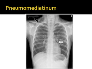
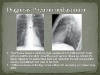
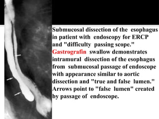
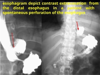
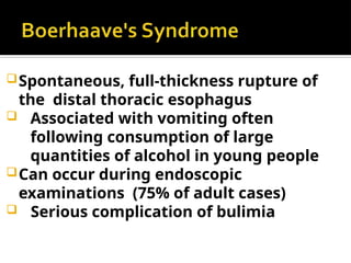
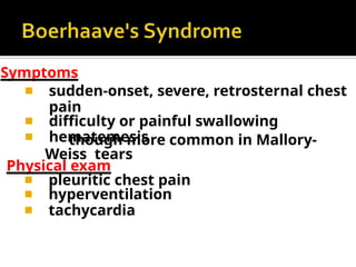
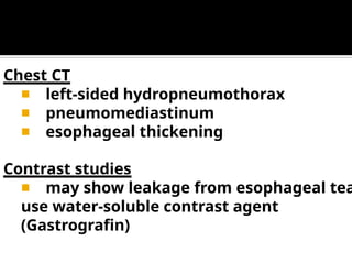
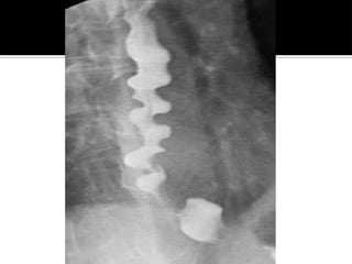
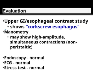
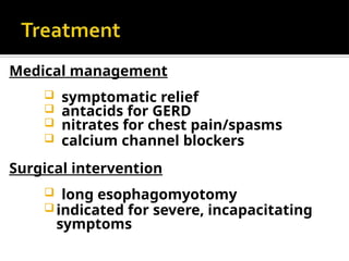
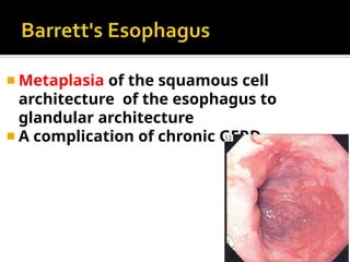
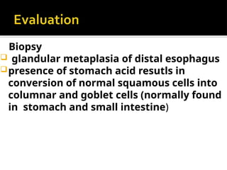
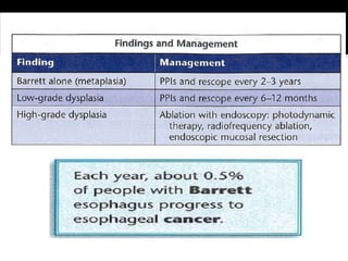
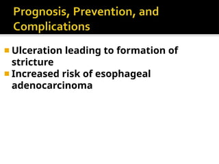
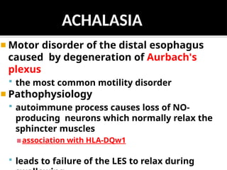
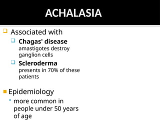
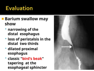
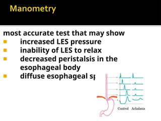
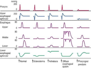
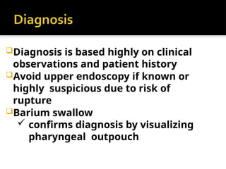
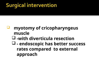
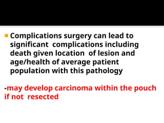
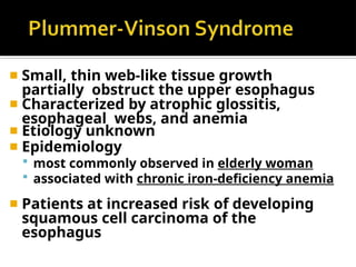
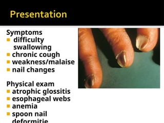
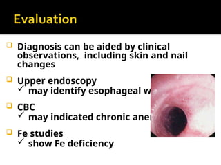
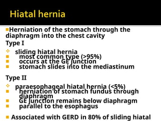
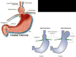
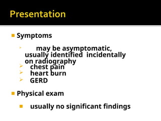
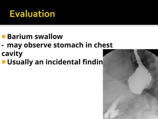
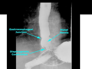
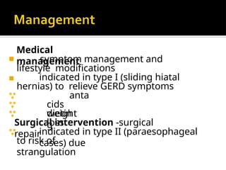
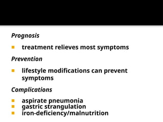
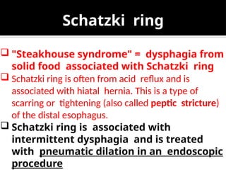
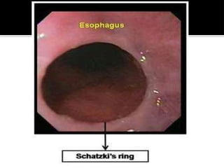
ESOPHAGEAL
DISORDER
DR JABBAR JASIM
2023
A. Esophageal perforation
B. Boerhaave & mallory weiss
syndrome
C. Diffuse esophageal spasm
D. Barett esophagus
E. Achalasia
F. Zenker diverticulum
G. Plummer vinson syndrome
H. Hiatal hernia
I. Esophagitis
J. Esophageal cancer
K. Scleroderma
 is due to the rapid increase in
intraesophageal pressure combined
with negative intrathoracic pressure
caused by vomiting.
 Perforation of the esophagus can
present with:
1. Severe and acute onset of
excruciating retrosternal chest pain
2. Odynophagia
3. Positive Hamman sign, a
crunching heard upon palpation
of the thorax due to
◾ Boerhaave syndrome
◾ is a full thickness tear secondary to
extreme retching and vomiting.
◾ It is most commonly tested in the
setting of
an alcoholic. The most common
location is at the left posterolateral
aspect of the distal esophagus.
◾ Mallory-Weiss syndrome
◾ is a mucosal tear and is also due to
vomiting.
◾ It is not a perforation. The most
◾ Surgical exploration with debridement of
the mediastinum and closure of the
perforation is an absolute emergency.
◾ Mediastinitis is a complication that
carries a very high mortality rate
Submucosal dissection of the esophagus
in patient with endoscopy for ERCP
and "difficulty passing scope."
Gastrografin swallow demonstrates
intramural dissection of the esophagus
from submucosal passage of endoscope
with appearance similar to aortic
dissection and "true and false lumen."
Arrows point to "false lumen" created
by passage of endoscope.
esophagram depict contrast extravasation from
the distal esophagus in a patient with
spontaneous perforation of the esophagus
Spontaneous, full-thickness rupture of
the distal thoracic esophagus
 Associated with vomiting often
following consumption of large
quantities of alcohol in young people
Can occur during endoscopic
examinations (75% of adult cases)
 Serious complication of bulimia
Symptoms
◾ sudden-onset, severe, retrosternal chest
pain
◾ difficulty or painful swallowing
◾ hematemesis
though more common in Mallory-
Weiss tears
Physical exam
◾ pleuritic chest pain
◾ hyperventilation
◾ tachycardia
Chest CT
◾ left-sided hydropneumothorax
◾ pneumomediastinum
◾ esophageal thickening
Contrast studies
◾ may show leakage from esophageal tea
use water-soluble contrast agent
(Gastrografin)
Medical management
conservative therapy
indicated in mild cases with stable patient and
includes
 intravenous resuscitation
 nasogastric suction
 NPO
 prophylactic antibiotics- usually broad-
coverage to prevent mediastinal infection-
imipenim or cilastin
Surgical intervention
surgical repair of perforation considered
standard of care
 indicated depending on severity of tear and
timing of diagnosis
◾ The most accurate test is an esophogram
(Gastrografin;); it will show leakage of
contrast outside of the esophagus.
◾
Barium cannot be used because it is
caustic to the tissues.
◾ Mallory-Weiss tear presents with upper
gastrointestinal bleeding after prolonged or
severe vomiting or retching.
◾ Repeated retching is followed by hematemesis
of
bright red blood, or by black stool.
◾ Mallory Weiss does not present with dysphagia.
There is no specific therapy, and it will resolve
spontaneously.
◾ Severe cases with persistent bleeding are
managed
with an injection of epinephrine to stop bleeding
or the use of electrocautery.
Boerhaave syndrome is full penetration of the
Strong, non-peristaltic contractions of the
esophageal body
Often precipitated by by ingestion of hot
and cold liquids
 Patients have normal sphincter function
 Associated with GERD
• Symptoms
• symptoms may occur following ingestion of
cold liquids and include
• difficulty swallowing
• painful swallowing
• sudden onset chest pain not related to exertion
• spontaneous and radiated to back, ears, and neck
• Physical exam
• symptomatic relief with nitroglycerin
Evaluation
•Upper GI/esophageal contrast study
• shows "corkscrew esophagus"
•Manometry
• may show high-amplitude,
simultaneous contractions (non-
peristaltic)
•Endoscopy - normal
•ECG - normal
•Stress test - normal
Medical management
 symptomatic relief
 antacids for GERD
 nitrates for chest pain/spasms
 calcium channel blockers
Surgical intervention
 long esophagomyotomy
indicated for severe, incapacitating
symptoms
◾ Metaplasia of the squamous cell
architecture of the esophagus to
glandular architecture
◾ A complication of chronic GERD
Biopsy
 glandular metaplasia of distal esophagus
presence of stomach acid resutls in
conversion of normal squamous cells into
columnar and goblet cells (normally found
in stomach and small intestine)
◾ Ulceration leading to formation of
stricture
◾ Increased risk of esophageal
adenocarcinoma
◾ Motor disorder of the distal esophagus
caused by degeneration of Aurbach's
plexus
 the most common motility disorder
◾ Pathophysiology
 autoimmune process causes loss of NO-
producing neurons which normally relax the
sphincter muscles
▪association with HLA-DQw1
 leads to failure of the LES to relax during
ACHALASIA
 Associated with
 Chagas' disease
amastigotes destroy
ganglion cells
 Scleroderma
presents in 70% of these
patients
◾ Epidemiology
 more common in
people under 50 years
of age
ACHALASIA
◾ Symptoms
 dysphagia for solids and liquids
▪usually worse for liquids
 weight loss
◾ Barium swallow may
show
 narrowing of the
distal esophagus
 loss of peristalsis in the
distal two thirds
 dilated proximal
esophagus
 classic "bird's beak"
tapering at the
esophageal sphincter
most accurate test that may show
◾ increased LES pressure
◾ inability of LES to relax
◾ decreased peristalsis in the
esophageal body
◾ diffuse esophageal spasm
◾
useful in excluding secondary causes of
achalasia (i.e. malignancy)
◾ use to rule out malignancy
◾ shows normal mucosa
 medications to reduce LES tone
1. nifedipine
2. nitrates
3. CCBs
4. botulinum toxin injections
▪wears off in approximately 3-6 months
▪requires reinjection
 endoscopic balloon dilation of LES
▪cures 80%
▪leads to perforation in < 3% of patients
 myotomy with fundoplication
▪more effective and dangerous than
pneumatic dilation
◾ Prognosis
 medical and surgical outcomes are similar
 often require multiple treatments
◾ Prevention
 no preventive measures are available at this
time
◾ Complications
 esophageal malignancy secondary to
Barrett's esophagus secondary to chronic
GERD
◾ Pharyngeal pouch that develops in the
proximal esophageal wall
◾ Pulsion diverticula involving only the
mucosa
 located between thyropharyngeal and
cricopharyngeus muscle
◾ Etiology remains unknown, however, some
have
suggested the causes to be related to
structural or physiological abnormalities of
the cricopharyngeus
◾ Epidemiology
◾ Symptoms
◾ dysphagia
◾ regurgitation
◾ choking
◾ chronic cough
◾ bad breath (halitosis)
◾ Physical exam
◾
palpable, fluctuant neck mass may be
appreciable
Diagnosis is based highly on clinical
observations and patient history
Avoid upper endoscopy if known or
highly suspicious due to risk of
rupture
Barium swallow
 confirms diagnosis by visualizing
pharyngeal outpouch
 myotomy of cricopharyngeus
muscle
 -with diverticula resection
 - endoscopic has better success
rates compared to external
approach
◾ Complications surgery can lead to
significant complications including
death given location of lesion and
age/health of average patient
population with this pathology
-may develop carcinoma within the pouch
if not resected
◾ Small, thin web-like tissue growth
partially obstruct the upper esophagus
◾ Characterized by atrophic glossitis,
esophageal webs, and anemia
◾ Etiology unknown
◾ Epidemiology
 most commonly observed in elderly woman
 associated with chronic iron-deficiency anemia
◾ Patients at increased risk of developing
squamous cell carcinoma of the
esophagus
Symptoms
◾ difficulty
swallowing
◾ chronic cough
◾ weakness/malaise
◾ nail changes
Physical exam
◾ atrophic glossitis
◾ esophageal webs
◾ anemia
◾ spoon nail
 Diagnosis can be aided by clinical
observations, including skin and nail
changes
 Upper endoscopy
 may identify esophageal webs
 CBC
 may indicated chronic anemia
 Fe studies
 show Fe deficiency
◾ Fe supplementation
 indicated to treat chronic anemia state
◾ esophageal dilation

can be performed concurrently
with upper GI endoscopy or
manometry

most commonly done with radial
expansion balloon method
◾ Prognosis
 most patients respond to treatment
◾ Prevention
 Fe supplementation in patients with known
anemia may prevent web development
◾ Complications
 bleeding may occur secondary to esophageal
tear during dilation
 esophageal carcinoma
◾Herniation of the stomach through the
diaphragm into the chest cavity
Type I
 sliding hiatal hernia
◾ most common type (>95%)
◾ occurs at the GE junction
◾ stomach slides into the mediastinum
Type II
 paraesophageal hiatal hernia (<5%)
◾ herniation of stomach fundus through
diaphragm
◾ GE junction remains below diaphragm
◾ parallel to the esophagus
◾ Associated with GERD in 80% of sliding hiatal
◾ Symptoms

may be asymptomatic,
usually identified incidentally
on radiography
 chest pain
 heart burn
 GERD
◾ Physical exam
◾ usually no significant findings
◾ Barium swallow
- may observe stomach in chest
cavity
◾ Usually an incidental finding
Medical
management
◾
◾ symptom management and
lifestyle modifications
indicated in type I (sliding hiatal
hernias) to relieve GERD symptoms
anta
cids
weight
loss


 dietin
g
Surgical intervention -surgical
repair
 indicated in type II (paraesophageal
cases) due
to risk of
strangulation
Prognosis
◾ treatment relieves most symptoms
Prevention
◾ lifestyle modifications can prevent
symptoms
Complications
◾ aspirate pneumonia
◾ gastric strangulation
◾ iron-deficiency/malnutrition
Schatzki ring
 "Steakhouse syndrome" = dysphagia from
solid food associated with Schatzki ring
 Schatzki ring is often from acid reflux and is
associated with hiatal hernia. This is a type of
scarring or tightening (also called peptic stricture)
of the distal esophagus.
 Schatzki ring is associated with
intermittent dysphagia and is treated
with pneumatic dilation in an endoscopic
procedure
Corrosive Esophagitis
Caused by ingestion of strongly acidic or
basic chemical
◾ Lye, HCl
Results in
◾ esophageal perforation
◾
esophageal stricture formation
Often seen in suicide attempts or in
the
pediatric population
Infectious Esophagitis
•Commonly seen in AIDS patients
and the Immunocompromised
•May be viral or fungal
• HSV (punched out lesions on
EGD)
• CMV (large solitary ulcers
or erosions on EGD)
• Candida (white mucosal
plaque- like lesions on EGD)
These pills cause esophagitis if in prolonged
contact:
• Doxycycline
• Alendronate
• KCI
Pills esophagitis
"What Is the Most Likely Diagnosis?"
Look for:
 Age 50 or older
 Dysphagia first for solids, followed
later (progressing) to dysphagia for
liquids
 Association with prolonged alcohol
and tobacco use
 More than 5-10 years of GERD
symptoms
1.Endoscopy is indispensible, since only a
biopsy can diagnose cancer.
2.Barium might be the "best initial test," but no
radiologic test can diagnose cancer.
3.CT and MRI scans are not enough to diagnose
esophageal cancer; they are used to determine the
extent of spread into the surrounding tissues.
4.PET scan is used to determine the contents of
anatomic lesions if you are not certain whether
they contain cancer. PET scan is often used to
determine whether a cancer is resectable. Local
disease is resectable, and widely metastatic
disease is not.
1.No resection (removal) = no cure.
Surgical resection is always the thing to
try.
2.Chemotherapy and radiation are used
in addition to surgical removal.
3.Stent placement is used for lesions
that cannot be resected surgically just
to
keep the esophagus open for palliation
◾ These patients present with symptoms of
reflux and have a clear history of
scleroderma, or progressive systemic
sclerosis.
◾ Manometry shows decreased lower
esophageal sphincter pressure from an
inability to close the LES.
◾ The management is with PPis as it would
be for any person with reflux symptoms.
◾ The disorder is simply one of
◾ Esophageal smooth muscle
atrophydecrease LES pressure and
dysmotilityacid reflux and dysphagia
stricture,Barrett esophagus, and
aspiration.
◾ Part of CREST syndrome.
Manometry is the answer for:
1. Achalasia
2. Spasm
3. Scleroderma
4-GIT lecture about esophageal disease.pptx

4-GIT lecture about esophageal disease.pptx

  • 1.
  • 2.
    A. Esophageal perforation B.Boerhaave & mallory weiss syndrome C. Diffuse esophageal spasm D. Barett esophagus E. Achalasia F. Zenker diverticulum G. Plummer vinson syndrome H. Hiatal hernia I. Esophagitis J. Esophageal cancer K. Scleroderma
  • 3.
     is dueto the rapid increase in intraesophageal pressure combined with negative intrathoracic pressure caused by vomiting.  Perforation of the esophagus can present with: 1. Severe and acute onset of excruciating retrosternal chest pain 2. Odynophagia 3. Positive Hamman sign, a crunching heard upon palpation of the thorax due to
  • 4.
    ◾ Boerhaave syndrome ◾is a full thickness tear secondary to extreme retching and vomiting. ◾ It is most commonly tested in the setting of an alcoholic. The most common location is at the left posterolateral aspect of the distal esophagus. ◾ Mallory-Weiss syndrome ◾ is a mucosal tear and is also due to vomiting. ◾ It is not a perforation. The most
  • 5.
    ◾ Surgical explorationwith debridement of the mediastinum and closure of the perforation is an absolute emergency. ◾ Mediastinitis is a complication that carries a very high mortality rate
  • 8.
    Submucosal dissection ofthe esophagus in patient with endoscopy for ERCP and "difficulty passing scope." Gastrografin swallow demonstrates intramural dissection of the esophagus from submucosal passage of endoscope with appearance similar to aortic dissection and "true and false lumen." Arrows point to "false lumen" created by passage of endoscope.
  • 9.
    esophagram depict contrastextravasation from the distal esophagus in a patient with spontaneous perforation of the esophagus
  • 10.
    Spontaneous, full-thickness ruptureof the distal thoracic esophagus  Associated with vomiting often following consumption of large quantities of alcohol in young people Can occur during endoscopic examinations (75% of adult cases)  Serious complication of bulimia
  • 11.
    Symptoms ◾ sudden-onset, severe,retrosternal chest pain ◾ difficulty or painful swallowing ◾ hematemesis though more common in Mallory- Weiss tears Physical exam ◾ pleuritic chest pain ◾ hyperventilation ◾ tachycardia
  • 12.
    Chest CT ◾ left-sidedhydropneumothorax ◾ pneumomediastinum ◾ esophageal thickening Contrast studies ◾ may show leakage from esophageal tea use water-soluble contrast agent (Gastrografin)
  • 13.
    Medical management conservative therapy indicatedin mild cases with stable patient and includes  intravenous resuscitation  nasogastric suction  NPO  prophylactic antibiotics- usually broad- coverage to prevent mediastinal infection- imipenim or cilastin Surgical intervention surgical repair of perforation considered standard of care  indicated depending on severity of tear and timing of diagnosis
  • 18.
    ◾ The mostaccurate test is an esophogram (Gastrografin;); it will show leakage of contrast outside of the esophagus. ◾ Barium cannot be used because it is caustic to the tissues.
  • 19.
    ◾ Mallory-Weiss tearpresents with upper gastrointestinal bleeding after prolonged or severe vomiting or retching. ◾ Repeated retching is followed by hematemesis of bright red blood, or by black stool. ◾ Mallory Weiss does not present with dysphagia. There is no specific therapy, and it will resolve spontaneously. ◾ Severe cases with persistent bleeding are managed with an injection of epinephrine to stop bleeding or the use of electrocautery. Boerhaave syndrome is full penetration of the
  • 20.
    Strong, non-peristaltic contractionsof the esophageal body Often precipitated by by ingestion of hot and cold liquids  Patients have normal sphincter function  Associated with GERD
  • 22.
    • Symptoms • symptomsmay occur following ingestion of cold liquids and include • difficulty swallowing • painful swallowing • sudden onset chest pain not related to exertion • spontaneous and radiated to back, ears, and neck • Physical exam • symptomatic relief with nitroglycerin
  • 23.
    Evaluation •Upper GI/esophageal contraststudy • shows "corkscrew esophagus" •Manometry • may show high-amplitude, simultaneous contractions (non- peristaltic) •Endoscopy - normal •ECG - normal •Stress test - normal
  • 24.
    Medical management  symptomaticrelief  antacids for GERD  nitrates for chest pain/spasms  calcium channel blockers Surgical intervention  long esophagomyotomy indicated for severe, incapacitating symptoms
  • 25.
    ◾ Metaplasia ofthe squamous cell architecture of the esophagus to glandular architecture ◾ A complication of chronic GERD
  • 26.
    Biopsy  glandular metaplasiaof distal esophagus presence of stomach acid resutls in conversion of normal squamous cells into columnar and goblet cells (normally found in stomach and small intestine)
  • 28.
    ◾ Ulceration leadingto formation of stricture ◾ Increased risk of esophageal adenocarcinoma
  • 29.
    ◾ Motor disorderof the distal esophagus caused by degeneration of Aurbach's plexus  the most common motility disorder ◾ Pathophysiology  autoimmune process causes loss of NO- producing neurons which normally relax the sphincter muscles ▪association with HLA-DQw1  leads to failure of the LES to relax during ACHALASIA
  • 30.
     Associated with Chagas' disease amastigotes destroy ganglion cells  Scleroderma presents in 70% of these patients ◾ Epidemiology  more common in people under 50 years of age ACHALASIA
  • 31.
    ◾ Symptoms  dysphagiafor solids and liquids ▪usually worse for liquids  weight loss
  • 32.
    ◾ Barium swallowmay show  narrowing of the distal esophagus  loss of peristalsis in the distal two thirds  dilated proximal esophagus  classic "bird's beak" tapering at the esophageal sphincter
  • 33.
    most accurate testthat may show ◾ increased LES pressure ◾ inability of LES to relax ◾ decreased peristalsis in the esophageal body ◾ diffuse esophageal spasm
  • 35.
    ◾ useful in excludingsecondary causes of achalasia (i.e. malignancy) ◾ use to rule out malignancy ◾ shows normal mucosa
  • 36.
     medications toreduce LES tone 1. nifedipine 2. nitrates 3. CCBs 4. botulinum toxin injections ▪wears off in approximately 3-6 months ▪requires reinjection
  • 37.
     endoscopic balloondilation of LES ▪cures 80% ▪leads to perforation in < 3% of patients  myotomy with fundoplication ▪more effective and dangerous than pneumatic dilation
  • 38.
    ◾ Prognosis  medicaland surgical outcomes are similar  often require multiple treatments ◾ Prevention  no preventive measures are available at this time ◾ Complications  esophageal malignancy secondary to Barrett's esophagus secondary to chronic GERD
  • 39.
    ◾ Pharyngeal pouchthat develops in the proximal esophageal wall ◾ Pulsion diverticula involving only the mucosa  located between thyropharyngeal and cricopharyngeus muscle ◾ Etiology remains unknown, however, some have suggested the causes to be related to structural or physiological abnormalities of the cricopharyngeus ◾ Epidemiology
  • 41.
    ◾ Symptoms ◾ dysphagia ◾regurgitation ◾ choking ◾ chronic cough ◾ bad breath (halitosis) ◾ Physical exam ◾ palpable, fluctuant neck mass may be appreciable
  • 42.
    Diagnosis is basedhighly on clinical observations and patient history Avoid upper endoscopy if known or highly suspicious due to risk of rupture Barium swallow  confirms diagnosis by visualizing pharyngeal outpouch
  • 43.
     myotomy ofcricopharyngeus muscle  -with diverticula resection  - endoscopic has better success rates compared to external approach
  • 44.
    ◾ Complications surgerycan lead to significant complications including death given location of lesion and age/health of average patient population with this pathology -may develop carcinoma within the pouch if not resected
  • 45.
    ◾ Small, thinweb-like tissue growth partially obstruct the upper esophagus ◾ Characterized by atrophic glossitis, esophageal webs, and anemia ◾ Etiology unknown ◾ Epidemiology  most commonly observed in elderly woman  associated with chronic iron-deficiency anemia ◾ Patients at increased risk of developing squamous cell carcinoma of the esophagus
  • 46.
    Symptoms ◾ difficulty swallowing ◾ chroniccough ◾ weakness/malaise ◾ nail changes Physical exam ◾ atrophic glossitis ◾ esophageal webs ◾ anemia ◾ spoon nail
  • 47.
     Diagnosis canbe aided by clinical observations, including skin and nail changes  Upper endoscopy  may identify esophageal webs  CBC  may indicated chronic anemia  Fe studies  show Fe deficiency
  • 48.
    ◾ Fe supplementation indicated to treat chronic anemia state ◾ esophageal dilation  can be performed concurrently with upper GI endoscopy or manometry  most commonly done with radial expansion balloon method
  • 49.
    ◾ Prognosis  mostpatients respond to treatment ◾ Prevention  Fe supplementation in patients with known anemia may prevent web development ◾ Complications  bleeding may occur secondary to esophageal tear during dilation  esophageal carcinoma
  • 50.
    ◾Herniation of thestomach through the diaphragm into the chest cavity Type I  sliding hiatal hernia ◾ most common type (>95%) ◾ occurs at the GE junction ◾ stomach slides into the mediastinum Type II  paraesophageal hiatal hernia (<5%) ◾ herniation of stomach fundus through diaphragm ◾ GE junction remains below diaphragm ◾ parallel to the esophagus ◾ Associated with GERD in 80% of sliding hiatal
  • 52.
    ◾ Symptoms  may beasymptomatic, usually identified incidentally on radiography  chest pain  heart burn  GERD ◾ Physical exam ◾ usually no significant findings
  • 53.
    ◾ Barium swallow -may observe stomach in chest cavity ◾ Usually an incidental finding
  • 55.
    Medical management ◾ ◾ symptom managementand lifestyle modifications indicated in type I (sliding hiatal hernias) to relieve GERD symptoms anta cids weight loss    dietin g Surgical intervention -surgical repair  indicated in type II (paraesophageal cases) due to risk of strangulation
  • 56.
    Prognosis ◾ treatment relievesmost symptoms Prevention ◾ lifestyle modifications can prevent symptoms Complications ◾ aspirate pneumonia ◾ gastric strangulation ◾ iron-deficiency/malnutrition
  • 57.
    Schatzki ring  "Steakhousesyndrome" = dysphagia from solid food associated with Schatzki ring  Schatzki ring is often from acid reflux and is associated with hiatal hernia. This is a type of scarring or tightening (also called peptic stricture) of the distal esophagus.  Schatzki ring is associated with intermittent dysphagia and is treated with pneumatic dilation in an endoscopic procedure
  • 59.
    Corrosive Esophagitis Caused byingestion of strongly acidic or basic chemical ◾ Lye, HCl Results in ◾ esophageal perforation ◾ esophageal stricture formation Often seen in suicide attempts or in the pediatric population
  • 60.
    Infectious Esophagitis •Commonly seenin AIDS patients and the Immunocompromised •May be viral or fungal • HSV (punched out lesions on EGD) • CMV (large solitary ulcers or erosions on EGD) • Candida (white mucosal plaque- like lesions on EGD)
  • 61.
    These pills causeesophagitis if in prolonged contact: • Doxycycline • Alendronate • KCI Pills esophagitis
  • 62.
    "What Is theMost Likely Diagnosis?" Look for:  Age 50 or older  Dysphagia first for solids, followed later (progressing) to dysphagia for liquids  Association with prolonged alcohol and tobacco use  More than 5-10 years of GERD symptoms
  • 64.
    1.Endoscopy is indispensible,since only a biopsy can diagnose cancer. 2.Barium might be the "best initial test," but no radiologic test can diagnose cancer. 3.CT and MRI scans are not enough to diagnose esophageal cancer; they are used to determine the extent of spread into the surrounding tissues. 4.PET scan is used to determine the contents of anatomic lesions if you are not certain whether they contain cancer. PET scan is often used to determine whether a cancer is resectable. Local disease is resectable, and widely metastatic disease is not.
  • 65.
    1.No resection (removal)= no cure. Surgical resection is always the thing to try. 2.Chemotherapy and radiation are used in addition to surgical removal. 3.Stent placement is used for lesions that cannot be resected surgically just to keep the esophagus open for palliation
  • 66.
    ◾ These patientspresent with symptoms of reflux and have a clear history of scleroderma, or progressive systemic sclerosis. ◾ Manometry shows decreased lower esophageal sphincter pressure from an inability to close the LES. ◾ The management is with PPis as it would be for any person with reflux symptoms. ◾ The disorder is simply one of
  • 67.
    ◾ Esophageal smoothmuscle atrophydecrease LES pressure and dysmotilityacid reflux and dysphagia stricture,Barrett esophagus, and aspiration. ◾ Part of CREST syndrome.
  • 68.
    Manometry is theanswer for: 1. Achalasia 2. Spasm 3. Scleroderma