Atherothrombosis
•Download as PPT, PDF•
1 like•679 views

SHAPE Society

Recommended
More Related Content
What's hot
What's hot (17)
Similar to Atherothrombosis
Similar to Atherothrombosis (20)
More from Society for Heart Attack Prevention and Eradication
More from Society for Heart Attack Prevention and Eradication (20)
Recently uploaded
Recently uploaded (20)
Atherothrombosis
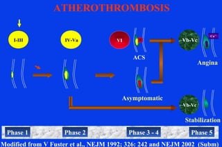
- 1. I-III IV-Va Asymptomatic Modified from V Fuster et al., NEJM 1992; 326: 242 and NEJM 2002 (Subm) VI Angina Vb-Vc Ca++ Phase 3 - 4Phase 1 Phase 2 Phase 5 Stabilization Vb-Vc ATHEROTHROMBOSIS ACS
- 2. TF MMPs CAMs Macrophages Pro-Adhesion/Migration TXA2 PAI-1 Prothrombotic Platelet Aggregation Fibrinolysis Flow Reversal Mechanical & Biohumoral Risk Factors LDL ET Extracellular Matrix Fibroblasts Vasa Vasorum SMC contraction migration proliferation PDGF V Fuster et al NEJM 2002 (Subm)
- 4. G. Helft, S. Worthley, V. Fuster, J.J. Badimon et al. Circ 2002;105:993] Progression and Regression of Atherosclerotic Lesions Monitoring With Serial Noninvasive Magnetic Resonance Imaging
- 5. CORONARY REMODELING IN NON OBSRTUCTIVE CAD W.Y.Kim et al Circ 2002;106:296 (Multicenter)
- 6. A.M. Varnava Circ 2002;105;939 (London) Plaque Vulnerability, Remodeling, Adventitia Thickness
- 7. TYPE VI PLAQUE WITH RUPTURED IEL P.R. Moreno, K-R Purushothaman, V.Fuster et al., Circulation 2002;105:2504
- 8. TYPE VI PLAQUE WITH IMFLAMMATION & RUPTURED IEL P.R.Moreno, K-R Purushothaman, V. Fuster et al Circulation. 2002;105:2504
- 9. Early Plaques Advanced, Non-Disrupted Disrupted Rupture of IEL Medial Inflammation Medial Fibrosis Medial Atrophy P=0.0001 P=0.0001 P=0.0001 P=0.008 598 Human Aortic Plaques By AHA Classification Moreno PR, et al. Circulation 2002;105:2504
- 10. Diabetic Atherosclerotic Microangiopathy Moreno PR, Purushothaman KR, O’Connor WN, Fuster V. 2002 (Subm) Diabetic Non- Diabetic P=0.02 420 440 460 480 500 520 540 No Diabetes Diabetes Total Neovessels 200 220 240 260 280 300 320 340 360 No Diabetes Diabetes Media Neovessels P=0.02 P=0.002
- 11. Sprouting Angiogenesis in Human Atherosclerosis Moreno PR, Purushothaman KR, Echeverri D, Fuster V, 2002 (Subm) CD-34 (Purple) Double Immunohistochemestry α-actin (Brown)
- 12. TF MMPs CAMs Macrophages Pro-Adhesion/Migration TXA2 PAI-1 Prothrombotic Platelet Aggregation Fibrinolysis Flow Reversal Mechanical & Biohumoral Risk Factors LDL ET Extracellular Matrix Fibroblasts Vasa Vasorum SMC contraction migration proliferation PDGF V Fuster et al NEJM 2002 (Subm)
- 13. I-III IV-Va No Sx Modified from V Fuster et al., NEJM 1992; 326: 242 and NEJM 2002 (Subm) VI Angina Vb-Vc Ca++ Phase 3 - 4Phase 1 Phase 2 Phase 5 Stabilization Vb-Vc ATHEROTHROMBOSIS ACS
- 14. Independent Predictors for Plaque Disruption P Value Number of Vasa Vasorum at Plaque Base 0.0001 Rupture of Internal Elastic Lamina (%) 0.01 Fibrous Cap Thickness (microns) 0.02 Percent Lipid Area (%) 0.025 Total Intimal Plaque Area (mm2) 0.031 Multiple Regression Analysis
- 15. LIPID CORE PROGRESSION / REGRESSION AND NEOVASCULARIZATION Progression Regression High-Risk1 Disrupted2 Fibro-Calcific3 P (n=89) (n=55) (n=18) Value Lipid Area (mm2 ) 3.5 ± 3.5 6.6 ± 3.5 2.03 ± 1.7 0.0001 Plaque Neovessels 41 ± 23 66 ± 19 22 ± 11 0.0001 Media Neovessels 284 ± 87 412 ± 133 201 ± 99 0.0001 Adventitia Neovessels 118 ± 69 194 ± 83 100 ± 42 0.0001 Total Neovessels 444 ± 134 671 ± 188 324 ± 140 0.0001 1 Type IV-Va 2 Type VI 3 Type Vb-Vc P Moreno, K-R Puroshothaman, V Fuster et al., Circ 2002 (In Press)
- 16. M.Shinnar, J.A.Fallon, J.J.Badimon, V.Fuster, ATVB 1999;19:2756 – Sensitivity & Specificity
- 18. Baseline 6 months Aortic Atherosclerotic Plaque Evaluated by MR Image Matching over Time Pulmonary veins LAD Corti R, Fuster V, Fayad ZA, Badimon JJ, et al.Circ 2002;106:2884 Ascending Aorta Descending Aorta
- 19. Effects of Lipid Lowering on Human Aortic and Carotid Plaques Study Design Hypercholesterolemic patients Carotid Plaques ≥ 2mm and/or Thoracic Aortic Plaque ≥ 4mm + LDL-Cholesterol ≥ 130 mg/dl or HDL < 45 mg/dl Simvastatin 20 Simvastatin 80 MRI Follow-up 6, 12, 18, 24 mo. MRI & RANDOMIZATION Corti R, Fayad ZA, Fuster V, et al. Circ. 2001;104:249-252
- 20. -6 0 6 12 18 24 30 36 42 48 54 0 50 100 150 200 250 300 350 Follow-up (Weeks) Plasmalipidlevels(mg/dl) HDL: increase - 5% LDL: decrease - 30-35% N=21 patients Total Cholesterol LDL Cholesterol HDL Cholesterol LIPID-LOWERING ON HUMAN ATHEROSCLEROSIS COURSE OF PLASMA LIPID LEVELS R Corti, V Fuster, ZA Fayad, JJ Badimon et al., Circ 2002; 106:2884
- 21. MRI-LIPID LOWERING (SIMVASTATIN) AND REGRESSION OF ATHEROSCLEROSIS AORTA (n=44 plaques) CAROTID ARTERY (n=32 plaques) LUMEN AREA(mm2) ANOVA p<0.001 0 100 600 500 300 200 400 BL 6 12 18 24 R Corti, V Fuster, ZA Fayad et al., Circ 2002; 106:2884 ANOVA p<0.001 0 10 30 20 40 BL 6 12 18 24 0 50 350 250 150 100 200 300 BL 6 12 18 24 VESSELWALL AREA(mm2) ANOVA p<0.001 ANOVA p<0.001 0 10 60 50 30 20 40 BL 6 12 18 24
- 22. LIPID-LOWERING ON HUMAN ATHEROSCLEROSIS CHANGES IN VESSEL WALL DIMENSIONS (CONTROLS) R Corti, V Fuster, ZA Fayad, JJ Badimon et al Circ 2002;106:2884 -6 -4 -2 0 2 4 6 Dimension(mm) Baseline 12 Mo. 24 Mo. Baseline vs. 24 Mo.
- 23. Baseline 24 months follow up R Corti, J J Wentzel, Z A Fayad, J J Badimon, V Fuster 2002 (Subm)
- 24. LIPID CORE PROGRESSION / REGRESSION AND NEOVASCULARIZATION Progression Regression High-Risk1 Disrupted2 Fibro-Calcific3 P (n=89) (n=55) (n=18) Value Lipid Area (mm2 ) 3.5 ± 3.5 6.6 ± 3.5 2.03 ± 1.7 0.0001 Plaque Neovessels 41 ± 23 66 ± 19 22 ± 11 0.0001 Media Neovessels 284 ± 87 412 ± 133 201 ± 99 0.0001 Adventitia Neovessels 118 ± 69 194 ± 83 100 ± 42 0.0001 Total Neovessels 444 ± 134 671 ± 188 324 ± 140 0.0001 1 Type IV-Va 2 Type VI 3 Type Vb-Vc P Moreno, K-R Puroshothaman, V Fuster et al., Circ 2002 (In Press)
- 25. HIGH CHOLESTEROL, SIMVASTATIN VASA VASORUM S. H. Wilson, et al. Circ 2002:105:415
- 26. 5 mm2 (5%) 46 mm2 (84%) 6 mm2 (10%) 0.7 mm2 (1%) 49 mm2 (77%) 2 mm2 (3%) 3 mm2 (3%) 10.7 mm2 (17%) Treated (n=8) Total plaque area = 58 mm2 Untreated (n=8) Total plaque area = 64 mm2 IN VIVO CAROTID PLAQUES AT MRI - INTENSIVE LIPID LOWERING1 Fibrous tissue area, mm2 (% of total plaque) Lipid plus calcium area, mm2 (% of total plaque) Lipid deposits area, mm2 (% of total plaque) Calcium cluster area, mm2 (% of total plaque) 1 Three Drugs X-Q Zhao et al., ATVB 2001;21:1623
- 27. I-III IV-Va Asymptomatic Modified from V Fuster et al., NEJM 1992; 326: 242 and NEJM 2002 (Subm) VI Angina Vb-Vc Ca++ Phase 3 - 4Phase 1 Phase 2 Phase 5 Stabilization Vb-Vc ATHEROTHROMBOSIS ACS
- 29. A. Tedgui et al. Thromb Haemost 2001;86;422 EXPRESSION OF CASPASE 3 IN MACROPHAGES (LIPID CORE , HUMAN CAROTID PLAQUE)
- 30. Co-localization of Caspase-3 and Tissue Factor AntigenCo-localization of Caspase-3 and Tissue Factor Antigen in Lipid-Rich Area of Human Carotid Atheromain Lipid-Rich Area of Human Carotid Atheroma R. HutterR. Hutter et al., Circ 2002 (Subm)et al., Circ 2002 (Subm)
- 31. Apoptosis and Tissue Factor Expression inApoptosis and Tissue Factor Expression in Macrophages of Human Coronary Atheroma (n=5)Macrophages of Human Coronary Atheroma (n=5) R Hutter et al Circ 2002 (Subm) – Apo-E -/-
- 32. 20 40 60 80 20 40 60 TissueFactorExpressionTissueFactorExpression Caspase-3 ExpressionCaspase-3 Expression R = 0.72R = 0.72 PP < .01< .01 %% %% Correlation Between Intimal Apoptosis and Tissue FactorCorrelation Between Intimal Apoptosis and Tissue Factor In Lipid-Rich Areas of Coronary and Carotid AtheromaIn Lipid-Rich Areas of Coronary and Carotid Atheroma R. HutterR. Hutter et al., Circ 2002 (Subm)et al., Circ 2002 (Subm)
- 33. Co-Expression of Tissue-Factor and Active Caspase-3Co-Expression of Tissue-Factor and Active Caspase-3 In Neointimal Foam Cells of ApoE-/- MouseIn Neointimal Foam Cells of ApoE-/- Mouse ApoE-/-ApoE-/-
- 34. 20 40 60 20 40 60 TissueFactorExpressionTissueFactorExpression Caspase-3 ExpressionCaspase-3 Expression R = 0.81R = 0.81 PP < .01< .01 % Correlation Between Apoptosis and Tissue FactorCorrelation Between Apoptosis and Tissue Factor Expression in Neointima of Apo-E -/- MiceExpression in Neointima of Apo-E -/- Mice Randolph Hutter et al 2002 %
- 35. CELLULAR CHOLESTEROL ACCUMULATIONS AND CELL DEATH I. Tabas JCI 2002;110:905
- 36. Ultrastructural Detection of Apoptosis and NecrosisUltrastructural Detection of Apoptosis and Necrosis in Human Atheromain Human Atheroma
- 37. Enhanced Reverse Cholesterol Transp. HDL Anti-Atherothrombotic Effect Anti-inflammatory Anti-thrombotic Pro-fibrinolytic Anti-oxidant HDL AS ANTI-ATHEROTHROMBOTIC PK Shah et al., Circ 2001; 104:2376
- 39. J.X. Rong et al. Circ 2001;104:2447-2452 ELEVATING HDL IN APO-E DEFICIENT MICE Macrophage Content
- 40. J.X. Rong et al. Circ 2001;104:2447-2452 ELEVATING HDL IN APO-E DEFICIENT MICE Smooth Muscle α-Actin Content
- 41. Background Peroxisome proliferator-activated receptors (PPARs) are a subfamilly of nuclear receptors which control a variety of cellular functions. PPAR-γ agonist have potential pleiotropic effects required to induce plaque regression and stabilization. Reverse Cholesterol Transport Free cholesterol scavegenreceptor CLA-1 HDL Vessel Wall Liver ET-1, PAI-1 Thrombosis Blood HDL Hepatic cell Apo A-1 ABC-1 receptor MCP-1, VCAM, chemokines ET-1 Recruitment, adherence and homingof macrophages Vasoconstriction MMPs SMCMigration Roberto Corti JACC 2002;248A
- 42. EFFECT OF SIMVASTATIN AND PPARγ AGONIST ON RABBIT ATHEROSCLEROTIC LESIONS: BIOLOGICAL CHANGES Infrarenal Aorta Immunohistochemistry Plaque size (correlation to MRI) Frozen + Formalin-fixed section Aortic Arch MMP1&3-activ. / Caspase-activ. / TF Fresh-frozen section
- 43. Rabbit Aorta plaque size -25 -20 -15 -10 -5 0 5 10 15 20 * * * *p=0.045 p=0.012 PPARγ regression diet progression simvastatin simvastatin+ PPARγ p<0.01 vs. progression * Changes in Vessel Wall Area vs. AT-Baseline (%)Changes in Vessel Wall Area vs. AT-Baseline (%) Comparison of the treatmentsComparison of the treatments R Corti et al JACC 2002;248A
- 44. Rabbit Aorta plaque composition 0 10 20 30 40 50 60 70 80 90 **** plaque area (%) RAM-11 +plaque area (%) RAM-11 + 0 10 20 30 40 50 60 0 10 20 30 40 50 60 70 vessel wall area (%)vessel wall area (%) αα-actin +-actin + vessel wall area (%)vessel wall area (%) αα-actin/collagen +-actin/collagen + regression diet progression control simvastatin+PPARγ simvastatin PPARγ p<0.05 vs. control * * * * * * * R Corti et al JACC 2002;248A
- 45. Results: MMP-activity 0 5 10 15 20 25 30 35 40 45 50 * * * regression diet progression control simvastatin+PPARγ simvastatin PPARγ p<0.05 vs. progression * Optical Density (arbitrary units)Optical Density (arbitrary units) 72kDa -- MMP-2 Roberto Corti et al JACC 2002;248A
- 46. Active 6-week wash- out period Patient Identification Consent Form Screening and Enrollment Qualifying baseline labs baseline MRI & UFCT Patient Randomization low-dose Statin low-dose Statin + Fibrate high-dose Statin 1 year-post MRI & UFCT 2 year-post MRI & UFCT 3 year-post MRI & UFCT ACTIVE TREATMENT ACTIVE TREATMENT 3 YEARS FOLLOW-UP DATA ANALYSIS MRI - DIABETES ATHEROSCLEROSIS LIPID ALTERING STUDY
- 47. S. Achenbach et al 2002 MSCT – SENSATION 16
- 50. Dynamic Contrast Enhanced MRA Global Evaluation of the Arterial and Venous Systems M.Poon, Z.A.Fayad, V.Fuster 2002
- 51. LAD Wall Fayad ZA; Fuster V et al. Circ. 2000;102;506-510 RCA Wall LAD Wall Black-Blood Coronary Plaque MR Eccentric (“lipid-rich”) Concentric (“fibrotic”) Ectatic (“remodeled”)
- 52. Fayad ZA, Fuster V, Nikolaou K, Becker C. Circ. 2002 ( In Press) Carotid Aortic Arch Desc. Aorta Complex Plaques – Systemic Disease
- 53. I-III IV-Va Asymptomatic Modified from V Fuster et al., NEJM 1992; 326: 242 and NEJM 2002 (Subm) VI Angina Vb-Vc Ca++ Phase 3 - 4Phase 1 Phase 2 Phase 5 Stabilization Vb-Vc ATHEROTHROMBOSIS ACS
- 54. CVMR-ISL Zahi Fayad, PhD Gilbert Aguinaldo, MD Vitalii Itskovich, PhD Gabor Mizsei, MS Dan Samber, MS Frank Macalusso, RT Karen Metroka Paul Wisdom, RT Cardiology Valentin Fuster, MD, PhD Juan Badimon, PhD Michael Poon, MD Stella Palentia, RN Don Smith, MD Meir Shinnar, MD, PhD Pedro R Moreno MD Pathology John Fallon, MD, PhD Molecular Biology Yale Nemerson, MD Mark Taubman, MD Edward Fisher, MD, PhD Ernane Reis, MD Robin Choudhury, MD Funding NIH-HL 94013 NIH-HL 61801 NIH-HL 07208 BMS Inv. award Merck Cardiology Fellows Ursula Rauch MD Roberto Corti, MD Julio Osende, MD Antonia Sambola, MD Stephen Worthley, MD Gerard Helft, MD Randolph Hutter MD The Mount Sinai Medical Center The Cardiovascular Institute Radiology Burton Drayer, MD Jeff Goldman, MD Neurology Jessey Weinberger, MD
- 60. ATHEROTHROMBOSIS: APPROACH IN 2002 Aggressive Intervention1 Effective Prevention 3 Coronary Atherothrombosis Atherothrombosis Subclinical Atherothrombosis Low Risk Modified from V Fuster, Circulation 1999; 99:1132 Multiple Risk F. Acute Coronary Syndromes Early Detection 2 1 Secondary Prevention , 2,3 Primary Prevention
- 61. Reproducibility of Vessel Wall Measurement 3.51.52CAROTID ARTERY (N = 4 plaques) 2.64.56AORTA (N = 5 plaques) Percent error (%) Mean of 5 contiguous slices Error (mm2) Slice Specific Error (mm2)N=6 patients 14 Analysis Courtesy of Leslie Shaw Corti R, Fuster V, Fayad ZA, Badimon JJ et al. Circ 2002; 106:2884
- 62. CHD RISK IN WOMEN ACCORDING TO FRAMINGHAM SCORING - 10 y Age, y HDL cholesterol < 35 -9 ≥ 60 -3 35-39 -4 50-59 0 40-44 0 45-49 1 45-49 3 35-44 2 50-54 6 < 35 5 55-59 7 Syst BP 60-64 8 < 120 -3 65-69 8 120-129 0 70-74 8 130-139 1 Cholesterol 140-149 2 < 160 -2 > 160 3 169-199 0 Diabetes 200-239 1 No 0 240-279 2 Yes 4 ≥ 280 3 Smoking No 0 Yes 2 Points 0 1 2 3 4 5 6 7 8 9 10 11 12 13 >14 Total CHD (%) 2 3 4 5 7 8 10 13 16 20 25 31 37 45 > 53 Hard CHD (%) 2 2 3 4 5 6 7 9 13 16 20 25 30 35 > 45 Grundy SM, Pasternak R, Greenland P, Smith S, Fuster V, Circ 1999; 100:1481 ATP III - Aggressive Rx: Framingham, Diabetes, Metab. Synd (obes, BP, HDL, TC, Gluc) JAMA 2001; 285:2475
- 63. HDL AND CRP AS RISK FACTORS FOR CAD P. Libby, PM Ridker, A. Maseri Circ 2002;105:1135
- 64. Effect of Lipid-lowering by Simvastatin onEffect of Lipid-lowering by Simvastatin on Human Atherosclerotic Aortic LesionsHuman Atherosclerotic Aortic Lesions Corti R, Fuster V, ZA Fayad, Badimon JJ et al., Circulation 2002;106:2284Corti R, Fuster V, ZA Fayad, Badimon JJ et al., Circulation 2002;106:2284
- 65. 5 cm A B C D E F
- 66. Co-localization of Caspase-3 and Tissue-Factor AntigenCo-localization of Caspase-3 and Tissue-Factor Antigen in Cultured Monocytes Treated with oxLDLin Cultured Monocytes Treated with oxLDL Hutter R. et al 2002 (Subm)
Editor's Notes
- We decided to investigate the effects of lipid lowering on human aortic and carotid plaque by serially MR imaging in hypercholesterolemic patients. This ongoing study is also aiming to evaluate the importance of aggressive versus non-aggressive approaches by using 2 different regimes (Simvastatin 20 and 80 mg).
- .
- 7772 Dubin @ 3/26/01 S9I13 T2W