Aspiration pneumonia occurs when a large volume of oropharyngeal or gastric contents are aspirated into the lungs, depositing a large bacterial inoculum. This can overwhelm normal lung defenses and cause pneumonia. Risk factors include dysphagia, altered mental status, vomiting, enteral feeding, and oropharyngeal colonization with more virulent bacteria. Aspiration is common but often does not cause pneumonia due to protective mechanisms; however, large volume macroaspiration can lead to aspiration pneumonia.

![ Given this broad use of the term aspiration, classifying the
majority
of bacterial pneumonias as a consequence of aspiration is
strictly
correct based on known pathophysiology of community-
acquired
(CAP) and hospital-acquired pneumonia (HAP) [2–5].
However, when
a clinician uses the term aspiration pneumonia, he or she is
typically
implying a subset of bacterial pneumonia that, although
sharing the
common pathophysiologic mechanism with most other
pneumonias,
represents a unique entity of a macroaspiration event resulting
in](https://image.slidesharecdn.com/2-230304034504-f19b8d23/85/2-3-Aspiration-Pneumonia-pptx-2-320.jpg)
![ Animal experiments helped differentiate the pathophysiology
of
chemical pneumonitis from subclinical aspiration based on the
pH
and volume of gastric material needed to stimulate an
immediate and
severe inflammatory reaction. Based on experiments using
human
gastric secretions and rabbit lungs, a pH less than 2.4 was
required to
cause vigorous inflammation. At higher pH, the reaction seen
microscopically was more similar to the changes caused by
the
instillation of water into the lungs [21]. In terms of quantity,
experiments inducing chemical pneumonitis in a dog model
required
2 mL of hydrochloric acid solution per kilogram to induce the
clinical](https://image.slidesharecdn.com/2-230304034504-f19b8d23/85/2-3-Aspiration-Pneumonia-pptx-8-320.jpg)
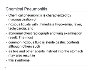
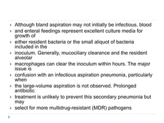
![Bland aspiration
Not all noninfectious macroaspirations cause an inflammatory
response in the lung; and therefore, to label these as
pneumonitis
would be inappropriate. Probably the 2 most common
examples are
aspiration of blood as a complication of severe epistaxis or
hematemesis and the aspiration of enteral feedings. Twenty
percent
of patients undergoing esophagogastroduodenoscopy will
have an
infiltrate immediately after the procedure in the dependent lung
[24,25]. Most resolve without antibiotic changes. Most
episodes of
aspiration with enteral nutrition are also uncomplicated](https://image.slidesharecdn.com/2-230304034504-f19b8d23/85/2-3-Aspiration-Pneumonia-pptx-9-320.jpg)
![CAP & HAP
Microaspiration has long been known to be the dominant
pathophysiologic mechanism behind CAP. Supporting evidence
includes the finding that most common CAP-causing microorganisms
colonize the oropharynx or nasopharynx in nonhospitalized patients
[2,27,28]. Similarly, the pathophysiology underlying HAP, including
ventilator-associated pneumonia (VAP), has proved to be microaspiration
of oropharyngeal, upper gastrointestinal, or subglottic
contents [3,5,29–32]. The distinct microbiology of HAP stems from
microaspiration occurring after hospitalized patients become colonized
with the virulent organisms found in intensive care unit and
hospital environments [4,33–36].
Given the above evidence of aspiration as a common event,
development of a parenchymal lung infection depends largely on host
defense factors [12,37] and the virulence of the aspirated pathogen.
This interaction helps explain the phenomenon of subclinical
aspiration without subsequent pneumonia described mostly in
young healthy volunteers and surgical candidates](https://image.slidesharecdn.com/2-230304034504-f19b8d23/85/2-3-Aspiration-Pneumonia-pptx-11-320.jpg)
![Aspiration Pneumonia
Current use of this term most commonly refers to an acute
lung
infection developing after a large-volume aspiration of
oropharyngeal
or upper gastrointestinal contents with a high enough pH to
avoid
chemical pneumonitis (likely pH much greater than 2.5). This
type of
aspiration deposits a large bacterial load of pathogens from
the oral
cavity or upper gastrointestinal tract into the lungs. The
possibility of
infection with these normally nonvirulent, predominantly
anaerobic
organisms is partly because of the large inoculum
[2,17,21,39–41].
Confusion surrounding this terminology and the exact
definition](https://image.slidesharecdn.com/2-230304034504-f19b8d23/85/2-3-Aspiration-Pneumonia-pptx-12-320.jpg)
![ Macroaspiration is the unique pathophysiologic
component of
what most clinicians call aspiration pneumonia. The
challenge in
specifically diagnosing aspiration pneumonia is that, for
many
patients in the community who are at risk for
macroaspiration, the
events in the days leading up to presentation with fever,
cough, and
chest radiograph infiltrate are unclear. A common risk for
macroaspiration
is decreased mental status, but this can be the result of
CAP
rather than the cause [43]. Because of this reality,
substantial
diagnostic overlap exists between aspiration, HAP, and](https://image.slidesharecdn.com/2-230304034504-f19b8d23/85/2-3-Aspiration-Pneumonia-pptx-13-320.jpg)
![ Aspiration pneumonia represents 5% to 15% of
pneumonias in the
hospitalized population. The ICD-9 code–based
reviews suggest an
increasing incidence, making it the second most
common diagnosis in
Medicare patients who are hospitalized [2,44].
However, higher
reimbursement rates for this ICD-9 code than for
CAP ICD-9 codes may
falsely increase the frequency in this population.](https://image.slidesharecdn.com/2-230304034504-f19b8d23/85/2-3-Aspiration-Pneumonia-pptx-14-320.jpg)
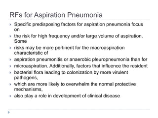
![RFs for Aspiration Pneumonia
Dysphagia/swallowing dysfunction
It is important to remember that dysphagia itself is not
definitive
evidence of aspiration. Many high-risk patients will not
complain of
dysphagia but still aspirate based on advanced testing [51].
The
poststroke population certainly has a higher prevalence of
pneumonia
with or without symptomatic dysphagia [52,53]. A lag time of
more
than 5 seconds between noxious stimuli and cough, as well as
an
increasing stimuli needed to produce a cough, has been linked
to
pneumonia in poststroke patients regardless of dysphagia](https://image.slidesharecdn.com/2-230304034504-f19b8d23/85/2-3-Aspiration-Pneumonia-pptx-17-320.jpg)
![RFs for Aspiration Pneumonia
Dysphagia/swallowing dysfunction
Certain medications interfere with the swallow reflex
and may
potentially lead to aspiration [58]. Although sedatives
may suppress
the patient’s mental status sufficiently to lead to
aspiration,
antipsychotic medications may actually affect the
swallowing mechanism
by inhibiting dopamine and therefore lead to
aspiration.
Accordingly, these drugs have been linked to
pneumonia in a fairly
large retrospective study](https://image.slidesharecdn.com/2-230304034504-f19b8d23/85/2-3-Aspiration-Pneumonia-pptx-19-320.jpg)
![RFs for Aspiration Pneumonia
Enteral feeding
Exact risk is difficult to characterize given the wide
variety of incidences reported, small sample sizes,
and lack of
standard definitions regarding aspiration and
aspiration pneumonia
[65–75]. Regardless of the deficiencies in
epidemiologic data,
aspiration pneumonia is common enough in this
population that it
should be a consideration for all patients on tube
feeds.](https://image.slidesharecdn.com/2-230304034504-f19b8d23/85/2-3-Aspiration-Pneumonia-pptx-26-320.jpg)
![RFs for Aspiration Pneumonia
Enteral feeding
Certain
patients appear to be at greater risk. Intuitively, GERD and
decreased
gastric motility are implicated when tube feeds are aspirated
Decreased gastric motility, typically defined by high gastric
residual volume, has also been suggested as a risk factor for
aspiration
in tube-fed patients [65,67]. However, the criteria for high
gastric
residual volume vary widely between studies from 50 to
greater than
500 mL at every 4-hour checks. A potentially independent risk
factor
is that patients with high gastric residuals may also be at
increased
risk of vomiting](https://image.slidesharecdn.com/2-230304034504-f19b8d23/85/2-3-Aspiration-Pneumonia-pptx-27-320.jpg)
![RFs for Aspiration Pneumonia
Oropharyngeal colonization
An independent association of poor oral hygiene with
aspiration
pneumonia is also supported by the literature [56,77]. The
microbial
density is increased in patients with gingival disease even if
the
spectrum has not shifted, increasing the likelihood of
pneumonia
developing in association with an episode of aspiration due to
the
greater inoculum. This suggests that edentulous patients are
at lower
risk for aspiration pneumonia. In edentulous patients, the
tongue is
more important as a focal point for colonization. Abe et al [78]
associated tongue-coating scores in an edentulous elderly](https://image.slidesharecdn.com/2-230304034504-f19b8d23/85/2-3-Aspiration-Pneumonia-pptx-29-320.jpg)
![RFs for Aspiration Pneumonia
Other Risks
Other risks
General risk factors like male sex and smoking may
increase risk
for aspiration pneumonia based on case-controlled and
cohort studies
[48]. Diabetes mellitus has been repeatedly associated
with pneumonia
in patients who have had an acute stroke [48].
Much has been discussed regarding the increased risk of
pneumonia as a whole in patients being treated with
proton pump
inhibitors and/or histamine receptor–2 antagonists
[80,81].](https://image.slidesharecdn.com/2-230304034504-f19b8d23/85/2-3-Aspiration-Pneumonia-pptx-31-320.jpg)
![Diagnosis
Clinical features can help distinguish aspiration pneumonia
from
chemical pneumonitis and other lung infections. As opposed to
chemical pneumonitis, the aspiration event in aspiration
pneumonia
is rarely witnessed [17]. The large volume of stomach contents
required to cause chemical pneumonitis usually makes it a
more
obvious event. Furthermore, the clinical course of chemical
pneumonitis
is hyperacute hypoxemia, occurring almost immediately (within
hours) and resulting in either devastating lung injury or
resolution
within 48 hours. These patients are likely to also have
bronchospasm,
frothy sputum, and chest radiographs with bilateral patchy
infiltrates
including nondependent areas](https://image.slidesharecdn.com/2-230304034504-f19b8d23/85/2-3-Aspiration-Pneumonia-pptx-34-320.jpg)
![Diagnosis
Because of this difficulty, efforts have been made to
use biomarkers
to distinguish aspiration pneumonia from other
aspiration syndromes.
El-Solh et al [85] attempted to use procalcitonin to
distinguish aspiration pneumonitis from aspiration
pneumonia in
the intensive care unit setting, given data to suggest
that procalcitonin
is a helpful marker for bacterial causes of sepsis](https://image.slidesharecdn.com/2-230304034504-f19b8d23/85/2-3-Aspiration-Pneumonia-pptx-35-320.jpg)
![Diagnosis
Biomarkers more specific to aspiration have also been
studied.
Pepsinogen in tracheal secretions or BAL was very suggestive
of
aspiration as part of the pathogenesis of posttransplant BO
and VAP.
Bronchoalveolar lavage amylase levels have been
demonstrated to
correlate with clinical risk factors for aspiration, as well as with
positive cultures [87–89]. This relationship may even be true in
patients with VAP [90]. Bronchoalveolar lavage amylase can
also
function as an end point for studies of interventions to
decrease risk of
aspiration in ventilated patients](https://image.slidesharecdn.com/2-230304034504-f19b8d23/85/2-3-Aspiration-Pneumonia-pptx-37-320.jpg)
![ As homogenous as these initial results appeared, evidence
accumulated
that aspiration pneumonia occurring after hospitalization had a
microbiologic
spectrum that included more Staphylococcus aureus, aerobes,
and gram-negative bacilli [17,84,92,94]. The pathogens that
dominate aspiration
pneumonia microbiology after a macroaspiration event after
hospitalization are similar to those of many nosocomial
infections.
Although very limited, data fromreliable cultures in
nonintubated patients
do suggest a higher frequency of anaerobes than in intubated
patients; but
the frequency is substantially lower than that of the prior](https://image.slidesharecdn.com/2-230304034504-f19b8d23/85/2-3-Aspiration-Pneumonia-pptx-42-320.jpg)
![ Recent studies reveal much different results even for
patients
presenting from the community. El-Solh et al [97]
reported a series of
patients with suspected aspiration pneumonia who
underwent
bronchial sampling after intubation. Of the 54
patients with a
bacterial diagnosis, 20% grew only anaerobes, with
an additional
11% that included anaerobes as part of mixed flora.](https://image.slidesharecdn.com/2-230304034504-f19b8d23/85/2-3-Aspiration-Pneumonia-pptx-43-320.jpg)
![ In contrast,
common causative organisms in this study were
Escherichia coli,
S aureus, and Klebsiella pneumoniae. Tokuyasu et al [98]
described this
trend further in a series of elderly Japanese patients with
clinically
diagnosed aspiration pneumonia. Of 111 organisms
isolated in 62
individuals, only 22 (20%) were anaerobes. Anaerobes
were heavily
outweighed by gram-negative bacilli (almost all enteric
gramnegatives),
found in 51.6% of patients](https://image.slidesharecdn.com/2-230304034504-f19b8d23/85/2-3-Aspiration-Pneumonia-pptx-44-320.jpg)
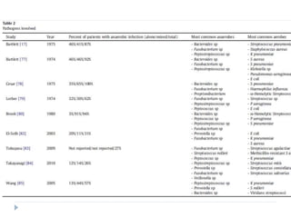
![ Even the etiology in patients with lung abscess has changed.
Takayanagi et al [99] reported bacterial etiologies in 122
patients
diagnosed with community-acquired lung abscess, likely a
result of
untreated aspiration. In this population, 74% grew aerobes
only, 12%
grew anaerobes only, and 14% grew mixed flora. Of the 107
aerobic
cases, 79% were Streptococcus species. In a very similar
study, Wang et
al [100] reported that only 40 (44%) of 90 community-acquired
lung
abscesses grew any anaerobes, with only 13% purely
anaerobic. Of the
remaining cases, 33% were caused by K pneumoniae (almost
all being
pure K pneumoniae isolates).](https://image.slidesharecdn.com/2-230304034504-f19b8d23/85/2-3-Aspiration-Pneumonia-pptx-45-320.jpg)
![Treatment
As one would expect, empirical treatment of aspiration
pneumonia
has evolved, given the above changes in the microbiology of
the
infection [102]. Intravenous penicillin was the drug of choice in
the
past, as anaerobes constituted the vast majority of infections
with few
penicillinase-producing bacterial strains [103,104]. A
randomized
controlled trial (RCT) of 39 patients with lung abscesses
compared
penicillin with clindamycin in the early 1980s [105]. Although a
small
group of patients, the treatment failure rate and cure rate were
much
better for clindamycin, with all 13 followed patients being cured
vs 8](https://image.slidesharecdn.com/2-230304034504-f19b8d23/85/2-3-Aspiration-Pneumonia-pptx-48-320.jpg)
![ The failure of penicillin to cure anaerobic
infections was better characterized several years
later in a Spanish
RCT of confirmed anaerobic lung infections [106]. In
this cohort of 37
patients, 47 anaerobes were isolated. Ten of these
47, all Bacteroides
species, were penicillin resistant, whereas none
were clindamycin
resistant. None of the 5 patients with penicillin-
resistant bacteria
randomized to penicillin responded to therapy.](https://image.slidesharecdn.com/2-230304034504-f19b8d23/85/2-3-Aspiration-Pneumonia-pptx-49-320.jpg)
![ Metronidazole has also been studied in anaerobic lungs
infections.
Sanders et al [107] described a poor cure rate in 13
patients with
pleuropulmonary (11 of 13 being lung abscesses)
infections with
confirmed anaerobic bacteria. Similarly, Perlino [108]
reported higher
cure rates with clindamycin when compared to
metronidazole in
cases of lung abscess and pneumonia with confirmed
anaerobic flora
in a small RCT of 13 patients.](https://image.slidesharecdn.com/2-230304034504-f19b8d23/85/2-3-Aspiration-Pneumonia-pptx-50-320.jpg)
![ Recent studies have focused on pneumonia in patients with
risk
factors for aspiration. Kadowaki et al [109] randomly assigned
100
elderly Japanese patients with suspected aspiration
pneumonia to
clindamycin, a carbapenem (penipenem/betamiprom), low-
dose
ampicillin/sulbactam (1.5 g twice daily), or high-dose
ampicillin/
sulbactam (3 g twice daily). The investigators found little
variance in
efficacy (N75% cure rate in all groups) and adverse events.
Interestingly,
no anaerobes were actually cultured. Of note, clindamycin was
the](https://image.slidesharecdn.com/2-230304034504-f19b8d23/85/2-3-Aspiration-Pneumonia-pptx-52-320.jpg)
![ Another study of elderly Japanese
with aspiration pneumonia [98] demonstrated a clinical
efficacy rate of
61.3% with another carbapenem, meropenem. This lower
efficacy than
that found by Kadowaki et al [109] may be due to greater
severity of
illness in the study patients. Once again, nosocomial
pathogens rather
than anaerobes were the most common documented
etiologies; and 33
of these 62 patients had MRSA growing in their
postantibiotic sputum
culture.](https://image.slidesharecdn.com/2-230304034504-f19b8d23/85/2-3-Aspiration-Pneumonia-pptx-53-320.jpg)
![ A recent randomized German study compared high-dose
ampicillin/sulbactam (3 g thrice times daily) to the standard
CAP
antibiotic moxifloxacin for the treatment of aspiration
pneumonia and
lung abscess in 96 elderly patients [110]. Clinical response
rates were
identical at 66.7%, and adverse reaction rates were very
similar.
Microbiology was consistent with the more recent data
described
above, with less than 10% of bacteria cultured being
anaerobes. Of note,
higher (although not statistically significant) mortality was seen
in the
ampicillin/sulbactamgroup, with 14 patients dying compared to
6 in the
moxifloxacin group.](https://image.slidesharecdn.com/2-230304034504-f19b8d23/85/2-3-Aspiration-Pneumonia-pptx-54-320.jpg)
![Prevention
Dietary Changes
Dietary interventions have been studied in patients with dysphagia.
In a small study involving patients with dysphagia secondary to
neurodegenerative disease (pseudobulbar dysphagia) [111], more
aspiration pneumonia occurred in those on a pureed diet compared
to
a mechanical soft diet with thickened liquids. However, the utility of
dietary intervention has been questioned. Depippo et al [112]
randomized 115 poststroke patients to 3 groups according to speech
therapist intervention: Group A was given advice based on swallow
testing, but the ultimate dietwas determined by the patient and
family;
Group B was prescribed a specific diet based on swallow testing;
and
Group C was prescribed a specific diet and directly observed for
compliance daily. No statistically significant differences between the
groups were found in any end point.](https://image.slidesharecdn.com/2-230304034504-f19b8d23/85/2-3-Aspiration-Pneumonia-pptx-58-320.jpg)
![Prevention
Drugs to Protect the Airway
A number of small studies have reviewed
pharmacologic intervention
to protect the airway via the cough reflex. The most
interesting drugs studied are angiotensin-converting
enzyme inhibitors
(ACEIs) because of their role in degrading substance
P and
bradykinin, stimulants of the cough reflex. A
reduction in aspiration
pneumonia in patients on an ACEI has been
suggested in one casecontrol
study [113] in elderly Japanese patients](https://image.slidesharecdn.com/2-230304034504-f19b8d23/85/2-3-Aspiration-Pneumonia-pptx-59-320.jpg)
![Prevention
Enteric Feeding Tubes
Two small prospective
trials have found no difference [115,116] in
pneumonia rates. To the
contrary, a very small randomized trial, with almost
no cases of
aspiration pneumonia, and another prospective trial
found advantages
to jejunal feeds [117,118]. Comparisons have also
been made between
nasogastric tube and percutaneous endoscopic
gastrostomy tube feeds
in a variety of clinical settings.](https://image.slidesharecdn.com/2-230304034504-f19b8d23/85/2-3-Aspiration-Pneumonia-pptx-62-320.jpg)
![Prevention
Enteric Feeding Residual Volumes
Many institutions monitor residual volumes from tube
feeds to
know when aspiration risk is increased. Residual
volumes of 500 mL are considered high enough to hold
tube feeds [65]. However, the
inaccuracy of this method has been well documented
[122].
Furthermore, results from a recent randomized clinical
trial suggest
that using strict residual volumes (250 mL) to understand
when to
hold nasogastric tube feeds does not affect the incidence
of VAP](https://image.slidesharecdn.com/2-230304034504-f19b8d23/85/2-3-Aspiration-Pneumonia-pptx-64-320.jpg)
![Prevention
Prophylactic Antibiotics
For patients at risk of aspiration around the time of
endotracheal
intubation, several studies have shown that a short
course (≤24
hours) of “prophylactic” β-lactam antibiotics may
decrease the risk of
subsequent VAP [125,126]. An extremely elevated
BAL amylase may
better select patients for this intervention.](https://image.slidesharecdn.com/2-230304034504-f19b8d23/85/2-3-Aspiration-Pneumonia-pptx-66-320.jpg)
![Prevention
Head Elevation
The same investigators then confirmed
the importance of this phenomenon by demonstrating
drastically
reduced rates of HAP in mechanically ventilated patients
in the
semirecumbent position compared to supine [128]. A
subsequent
study did not show a benefit of semirecumbant position
when
compared to elevation of as little as 10° from supine
[129]. However,
the risk benefit ratio of elevation of the head of the bed in
ventilated
patients is so favorable that it has become standard
practice](https://image.slidesharecdn.com/2-230304034504-f19b8d23/85/2-3-Aspiration-Pneumonia-pptx-68-320.jpg)