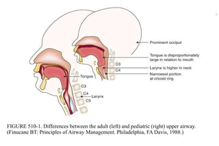
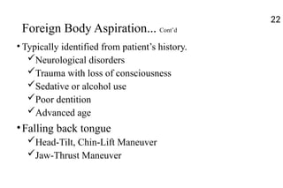
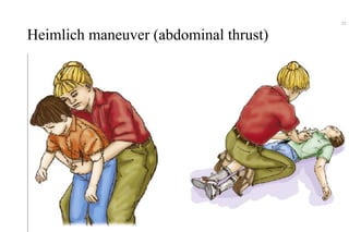
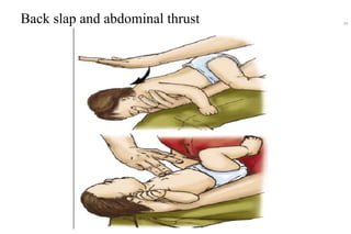
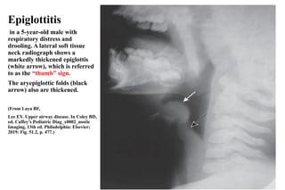
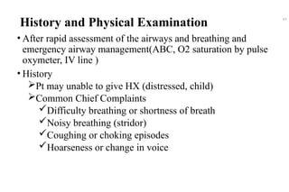
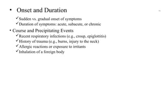
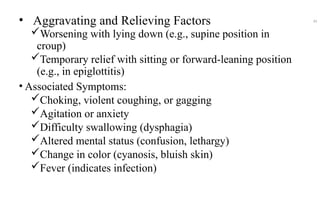
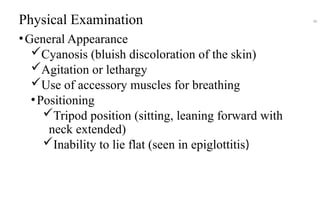
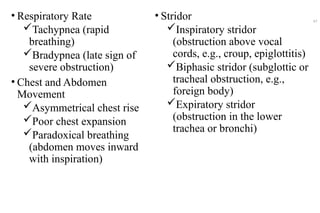
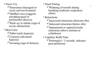
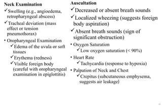
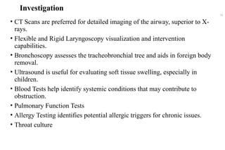
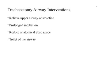
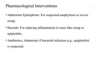
Approach to a patient with acute upper
airway obstruction
Prepared by GIRM ABDISSA (C-1)
& AHEMD KITAW (C-1)
Moderator: Dr. Anwar M. (MD, Assistant Professor of
Surgery)
Yekatit 12 Hospital Medical College
Department of surgery
Seminar
Outlines
• Objectives
• Introduction
• Etiology
• Classification
• History and Physical examination
• Investigations
• Management Principles
2
Objectives
• Understand the causes of acute upper airway obstruction
• Describe the clinical manifestations, assessment
approaches, diagnostic methods and its principle of
management.
3
Introduction
• Acute upper airway obstruction is a life-threatening emergency
requiring immediate assessment and intervention B/c it can leads
do to
respiratory failure
 hypoxia, and brain damage
4
Incidence and Prevalence
Children:
 UAO is notably prevalent in paediatric populations,
accounting for up to 15% of emergency department visits due
to respiratory distress.
The most common cause croup, with an annual incidence of
approximately 18 per 1,000 children in the USA. The peak
incidence occurs in children aged 1 to 2 years
5
Infants
A study highlighted that 58% of cases of UAO in infants occur
between 1 to 6 months of age.
 laryngomalacia (70%)
 subglottic stenosis (18%)
Adults
most frequent cause of chronic UAO is obstructive sleep
apnea (OSA),
26% of the adult population in the U.S.
Other include inflammation, infection, and trauma.
6
Surgical Anatomy
• The upper airway extends from the nose/mouth down to the
larynx, including the nasal cavity, oral cavity, and pharynx
(naso-, oro-, and laryngopharynx), and ends at the glottis in the
larynx.
• Key physiological narrowing points in the upper airway:
Nostrils
Velopharyngeal valve (between nasopharynx and oropharynx)
Glottis
7
Respiratory Airway Anatomy
8
FIGURE 510-1. Differences between the adult (left) and pediatric (right) upper airway.
(Finucane BT: Principles of Airway Management. Philadelphia, FA Davis, 1988.)
9
Types of Airway Obstruction
• Upper airway obstruction (UAO): Blockage from the mouth to
the trachea, including the nasopharynx and larynx. CAO and UAO
can coexist in certain conditions.
• Central airway obstruction (CAO): Blockage in the trachea and
main stem bronchi.
• Lower airway obstruction: Blockage in smaller bronchi from
chronic diseases like asthma or COPD, usually unrelated to CAO or
UAO.
10
Upper Vs Lower Airway Obstruction
• Ventilation-perfusion mismatch, Hypercarbia, Hypoxemia
is rare unless upper airway obstruction is severe
• Upper airway obstruction does not improve with
bronchodilators, unlike asthma or COPD.
• Upper airway obstruction present as inspiratory stridor
• Lower airway obstructive processes present as expiratory
wheezing.
11
• Normal loop: The expiratory curve rises quickly to the peak,
then falls linearly. The inspiratory curve is symmetrical and
saddle-shaped.
• Extrathoracic obstruction: Inspiratory limb shows flow
limitation and flattening.
• Intrathoracic obstruction: Expiratory limb shows flow
limitation and flattening.
• Fixed upper airway obstruction: Both inspiratory and
expiratory limbs show flow limitation and flattening.
• Lower airway obstruction: Expiratory limb appears concave
upward, with a "scooped-out" pattern.
Distinguish the site of airway narrowing.
12
13
FIGURE 285-4 Flow-volume loops. A. Normal. B. Airflow obstruction. C. Fixed central airway obstruction
(either above or below the thoracic inlet). D. Variable upper airway obstruction (above the thoracic inlet) E.
Variable lower airway obstruction (below the thoracic inlet). RV, residual volume; TLC, total lung capacity.
Classification of upper airway obstruction
Based on
Anatomical based Classification
Age-based based Classification
Functional based Classification
Clinical presentation based Classification
Etiological Based classification
14
15
Anatomical Classification 16
• Nasal Cavity/Nasopharynx: foreign bodies, structural
anomalies like choanal atresia, or growths such as nasal polyps
and adenoid hypertrophy.
• Oropharynx: Enlarged tonsils, peritonsillar abscesses, and
retropharyngeal abscesses c
• Larynx: Inflammation (laryngitis), swelling (edema), vocal
cord paralysis, and tumors
• Trachea: Narrowing due to stenosis or malacia, tumors, and
foreign bodies
Age-based Classification
17
• Pediatric: Common conditions include croup, epiglottitis,
foreign bodies, and congenital anomalies.
• Adult: Tumors, vocal cord paralysis, anaphylaxis, and
infectious causes are prevalent.
• Elderly: Laryngeal tumors, aspiration, and neurological
causes are typical concerns.
Functional Classification 18
• Obstructive: Physical blockages such as foreign bodies,
tumors, and swelling can obstruct the airway.
• Restrictive: Conditions that limit airway expansion, like
tracheal stenosis and neck trauma, fall under this category.
• Central: Issues related to central control, such as CNS
depression and brainstem injuries, can affect breathing.
Clinical presentation-based blassification 19
● Gradual Onset: Conditions such as tumors, chronic
infections, and subglottic stenosis develop slowly over
time.
● Sudden Onset: Rapidly occurring conditions include
anaphylaxis, foreign body aspiration, acute infections, and
trauma.
Etiological classification
20
• Foreign Bodies/Inflammatory/Edematous: Inhaled objects, food
particles, allergic reactions, and inflammatory edema.
• Infectious: epiglottitis and croup
• Traumatic: Neck trauma and post-surgical edema
• Allergic/Immunological: Anaphylaxis and angioedema.
• Neoplastic: laryngeal carcinoma and thyroid tumors.
• Congenital: laryngomalacia and tracheoesophageal fistula.
• Neurological: vocal cord paralysis and CNS injuries
Foreign Body Aspiration
• Sudden choking, violent coughing, inspiratory stridor, no
fever
• Occurs after playing with food or small objects
• Common in toddlers (1 to 3 years)
• History of choking episode, localized wheezing or stridor
21
Foreign Body Aspiration... Cont’d
• Typically identified from patient’s history.
Neurological disorders
Trauma with loss of consciousness
Sedative or alcohol use
Poor dentition
Advanced age
•Falling back tongue
Head-Tilt, Chin-Lift Maneuver
Jaw-Thrust Maneuver
22
Heimlich maneuver (abdominal thrust)
23
Back slap and abdominal thrust 24
Deep cervical space infections
• life-threatening upper airway obstruction.
• Symptoms: sore throat, odynophagia, neck swelling, pain,
fever, dyspnea.
• Severe cases show stridor and profound respiratory difficulty.
• Common sites: parapharyngeal, peritonsillar, submandibular,
and retropharyngeal abscesses.
25
Treatment of deep cervical infections
• Maintenance of oxygenation and ventilation by securing an
adequate airway,
• administration of appropriate antibiotics,
• when indicated, use of surgical drainage.
26
Ludwig’s angina
• Ludwig’s angina an infection of the submandibular space and
the floor of the mouth is potentially lethal and is commonly
associated with significant upper airway obstruction.
• A cellulitic process and can affect the submandibular spaces
bilaterally.
• 75 percent of the cases with true Ludwig’s angina required
tracheostomy.
27
Ludwig’s angina 28
Lemierre’s syndrome
• Starts from nasopharyngitis or peritonsillar abscess.
• Leads to internal jugular vein thrombophlebitis, septicemia, and
metastatic abscesses.
• Fusobacterium necrophorum is the main causative agent.
• Symptoms: sore throat, fever, neck swelling, tender lymph
nodes, dysphagia, trismus, and airway obstruction.
• Treatment: early antibiotics (penicillin with metronidazole or
clindamycin) to prevent high mortality.
29
Epiglottitis
• Caused by various organisms in adults,
including Haemophilus influenzae,
pneumococci, and group A streptococci.
• Initial treatment: third-generation
cephalosporin or extended-spectrum
penicillin.
• Corticosteroids
• Close observation and readiness for
airway management (intubation or
surgery) are essential.
30
Epiglottitis
in a 5-year-old male with
respiratory distress and
drooling. A lateral soft tissue
neck radiograph shows a
markedly thickened epiglottis
(white arrow), which is referred
to as the “thumb” sign.
The aryepiglottic folds (black
arrow) also are thickened.
(From Laya BF,
Lee EY. Upper airway disease. In Coley BD,
ed. Caffey’s Pediatric Diag_x0002_nostic
Imaging, 13th ed. Philadelphia: Elsevier;
2019: Fig. 51.2, p. 477.)
31
Laryngotracheobronchitis
• Croup is an acute viral illness in children, causing
subglottic narrowing, stridor, barking cough, and
hoarseness.
• Rare in adults,
32
Fig. 433.1 Croup. A, Frontal soft
tissue neck radiograph
demon_x0002_strates a
“Steeple” appearance of the
subglottic trachea (arrows). B,
Lateral soft tissue neck radiograph
in another patient shows a dilated
hypopharynx (asterisk), along with
haziness and narrowing of the
sub_x0002_glottic region (arrows).
(From Laya BF, Lee EY. Upper
airway disease. In
Coley BD, ed. Caffey’s Pediatric
Diagnostic Imaging, 13th ed.
Philadel_x0002_phia: Elsevier;
2019: Fig. 51.8, p. 481.)
33
Facial Trauma
• Emergency airway access is needed in up to 6% of facial
trauma cases.
• If intubation is difficult, consider cricothyroidotomy or
tracheostomy.
• Common signs: stridor, wheezing, dysphonia, hemoptysis,
neurological deficits, cervical crepitus, subcutaneous
emphysema, ecchymoses, hematomas,
pneumomediastinum, pneumothorax.
34
Facial Trauma... Cont’d
• Diagnostic tools: flexible fiberoptic laryngoscopy,
bronchoscopy, CT imaging.
• Management: secure airway promptly, avoid blind intubation,
consider tracheostomy, use awake fiberoptic intubation.
• High mortality rate (20-40%).
35
Inhalation Injuries
• Serious airway obstruction from thermal/chemical injuries.
• Signs: cough, dyspnea, hoarseness, singed nasal hairs,
carbonaceous sputum, facial burns.
• Early fiberoptic bronchoscopy for assessment and management.
• Trans-laryngeal intubation standard; early tracheostomy
possible.
• Tracheal stenosis risk.
36
Neuromuscular Disorders
• Myasthenia gravis and motor neuron disease affect bulbar muscles,
increasing airway resistance.
• Inspiratory flow plateau and "saw tooth pattern" in flow-volume loops are
seen, indicating extrathoracic obstruction.
• Extrapyramidal disorders cause vocal cord tremors, leading to flow
oscillations.
• Muscle denervation in motor neuron diseases results in upper airway
muscle tremor and irregular airflow.
37
Vocal Cord Dysfunction
• Vocal cord dysfunction causes paradoxical vocal cord
closure during inspiration, mimicking asthma and leading to
wheezing and stridor.
• Often misdiagnosed as asthma, it may have psychogenic
causes and is linked to psychiatric disorders or past trauma.
• Speech therapy or psychotherapy is recommended for
management.
38
Allergic Reaction (Anaphylaxis)
• Stridor, swelling of lips, tongue, face (angioedema), hives,
wheezing
• Rapid onset after allergen exposure, systemic reaction
(rash, hypotension)
• Affects any age group
• Recent allergen exposure (e.g., food, insect sting), history
of allergies or asthma
39
• Laryngotracheal Trauma
 Hoarseness, painful swallowing, stridor, hemoptysis
 Neck swelling or bruising
 Any age
 Recent neck trauma or intubation
• Retropharyngeal Abscess
 Difficulty swallowing, muffled voice, high fever, neck stiffness
 Unilateral neck swelling
 Mostly children < 5 years
 Recent bacterial throat infection
40
Peri tonsillar Abscess
 Severe sore throat,
 uvula deviation
 Adolescents and adults
 Recent fever,
 throat infection
41
Angioedema
• Causes swelling of the face, lips, tongue, and throat,
risking fatal airway obstruction.
• Triggers include drugs, NSAIDs, and ACE inhibitors;
most are non-allergic.
• Hereditary angioedema involves C1 esterase inhibitor
deficiency, leading to painless swelling.
• Treatment: secure airway, corticosteroids, antihistamines,
and epinephrine.
42
History and Physical Examination
• After rapid assessment of the airways and breathing and
emergency airway management(ABC, O2 saturation by pulse
oxymeter, IV line )
• History
Pt may unable to give HX (distressed, child)
Common Chief Complaints
Difficulty breathing or shortness of breath
Noisy breathing (stridor)
Coughing or choking episodes
Hoarseness or change in voice
43
• Onset and Duration
Sudden vs. gradual onset of symptoms
Duration of symptoms: acute, subacute, or chronic
• Course and Precipitating Events
Recent respiratory infections (e.g., croup, epiglottitis)
History of trauma (e.g., burns, injury to the neck)
Allergic reactions or exposure to irritants
Inhalation of a foreign body
44
• Aggravating and Relieving Factors
Worsening with lying down (e.g., supine position in
croup)
Temporary relief with sitting or forward-leaning position
(e.g., in epiglottitis)
• Associated Symptoms:
Choking, violent coughing, or gagging
Agitation or anxiety
Difficulty swallowing (dysphagia)
Altered mental status (confusion, lethargy)
Change in color (cyanosis, bluish skin)
Fever (indicates infection)
45
Physical Examination
•General Appearance
Cyanosis (bluish discoloration of the skin)
Agitation or lethargy
Use of accessory muscles for breathing
•Positioning
Tripod position (sitting, leaning forward with
neck extended)
Inability to lie flat (seen in epiglottitis)
46
• Respiratory Rate
Tachypnea (rapid
breathing)
Bradypnea (late sign of
severe obstruction)
• Chest and Abdomen
Movement
Asymmetrical chest rise
Poor chest expansion
Paradoxical breathing
(abdomen moves inward
with inspiration)
• Stridor
Inspiratory stridor
(obstruction above vocal
cords, e.g., croup, epiglottitis)
Biphasic stridor (subglottic or
tracheal obstruction, e.g.,
foreign body)
Expiratory stridor
(obstruction in the lower
trachea or bronchi)
47
• Voice/ Cry
Hoarseness (laryngeal or
vocal cord involvement)
Muffled voice (suggests
retropharyngeal or
peritonsillar abscess)
Weak cry in infants (sign of
severe obstruction)
• Skin Color
Pallor (early hypoxia)
Cyanosis (advanced
hypoxia)
Sweating (sign of distress)
• Nasal Flaring
Widening of nostrils during
breathing (indicates respiratory
effort)
• Retractions
Intercostal retractions (between ribs)
Subcostal retractions (below ribs)
Suprasternal or supraclavicular
retractions (above sternum or
collarbone)
• Capillary Refill Time
Prolonged (> 3 seconds, indicates
poor perfusion)
48
Neck Examination
Swelling (e.g., angioedema,
retropharyngeal abscess)
Tracheal deviation (mass
effect or tension
pneumothorax)
• Oropharyngeal Examination
Edema of the uvula or soft
tissues
Erythema (redness)
Visible foreign body
(careful with oropharyngeal
examination in epiglottitis)
Auscultation
Decreased or absent breath sounds
Localized wheezing (suggests foreign
body aspiration)
Absent breath sounds (sign of
significant obstruction)
• Oxygen Saturation
Low oxygen saturation (< 90%)
• Heart Rate
Tachycardia (response to hypoxia)
• Palpation of Neck and Chest
Crepitus (subcutaneous emphysema,
suggests air leakage)
49
Severity signs of upper airway obstruction
• Tachypnea
• Change in Voice or Cry
• Barking Cough
• Hoarseness
• Inspiratory Stridor
• Poor Chest Rise on Inspiration
• Nasal Flaring: Widening of the nostrils.
Recognizing these signs promptly is crucial for managing upper airway
obstruction
50
Severity based on Clinical Features
1. Mild: Stridor only with activity, mild cough,
normal oxygenation.
2. Moderate: Stridor at rest, moderate
retractions, oxygen saturation 90-95%, nasal
flaring.
3. Severe: Continuous stridor, severe
retractions, cyanosis, oxygen saturation <
90%, altered mental status.
51
Investigation
• CT Scans are preferred for detailed imaging of the airway, superior to X-
rays.
• Flexible and Rigid Laryngoscopy visualization and intervention
capabilities.
• Bronchoscopy assesses the tracheobronchial tree and aids in foreign body
removal.
• Ultrasound is useful for evaluating soft tissue swelling, especially in
children.
• Blood Tests help identify systemic conditions that may contribute to
obstruction.
• Pulmonary Function Tests
• Allergy Testing identifies potential allergic triggers for chronic issues.
• Throat culture
52
Emergency Management Principles
• Immediate rapid Assessment with simultanous Airway
Managment Before History taking and P/E
Rapid Airway Assessment: Quickly evaluate airway
patency and breathing.
Assess for Signs of Severe Obstruction: Look for stridor,
cyanosis, use of accessory muscles, and altered
consciousness.
Check Consciousness Level: Determine if the patient is
alert, lethargic, or unresponsive.
53
Emergency Airway Management
Ensure Airway Patency:
Head-Tilt, Chin-Lift: If the
patient is unconscious or unable
to maintain their airway.
Jaw-Thrust Maneuver:
Alternative if cervical spine
injury is suspected.
Positioning:
Supine: For patients with
suspected croup or tracheitis.
Sitting Forward: For suspected
epiglottitis to optimize airway
position.
Comfortable Position: For
children, hold in a parent's lap if
possible.
54
Positioning
Supine: For patients with suspected croup or tracheitis.
Sitting Forward: For suspected epiglottitis to optimize
airway position.
Comfortable Position: For children, hold in a parent's lap if
possible.
55
Provide Supplemental Oxygen:
Hand Position: The little,
ring, and index fingers are
spread over the mandible from
the angle of the jaw forward
towards the chin in the shape
of an "E".
●Jaw Lift: The jaw is lifted,
pulling the face into the mask.
●Mask Seal: The thumb and
forefinger are placed over the
mask in the shape of a "C",
forming a seal between the
mask and the skin.
Advanced Airway Interventions
• Suctioning: Clear visible obstructions or
secretions.
• Endotracheal Intubation: Prepare for intubation
if the airway is compromised and other
measures are insufficient.
• Tracheostomy: Consider if intubation fails or is
not possible, especially in severe cases.
57
Tracheostomy Airway Interventions
• Relieve upper airway obstruction
• Prolonged intubation
• Reduce anatomical dead space
• Toilet of the airway
58
Pharmacological Interventions
• Administer Epinephrine: For suspected anaphylaxis or severe
croup.
• Steroids: For reducing inflammation in cases like croup or
epiglottitis.
• Antibiotics: Administer if bacterial infection (e.g., epiglottitis)
is suspected.
59
References
1. Schwartz's Principles of Surgery (11th Edition, McGraw-Hill
Education)
2. Bailey & Love's Short Practice of Surgery (28th Edition, CRC Press)
3. Nelson Textbook of Pediatrics (21st Edition, Elsevier)
4. Rosen's Emergency Medicine: Concepts and Clinical Practice (9th
Edition, Elsevier Saunders)
5. Up-to-date
60
61
THANK YOU !!

approch to UPPER AIR ways obstraction.pptx

  • 1.
    Approach to apatient with acute upper airway obstruction Prepared by GIRM ABDISSA (C-1) & AHEMD KITAW (C-1) Moderator: Dr. Anwar M. (MD, Assistant Professor of Surgery) Yekatit 12 Hospital Medical College Department of surgery Seminar
  • 2.
    Outlines • Objectives • Introduction •Etiology • Classification • History and Physical examination • Investigations • Management Principles 2
  • 3.
    Objectives • Understand thecauses of acute upper airway obstruction • Describe the clinical manifestations, assessment approaches, diagnostic methods and its principle of management. 3
  • 4.
    Introduction • Acute upperairway obstruction is a life-threatening emergency requiring immediate assessment and intervention B/c it can leads do to respiratory failure  hypoxia, and brain damage 4
  • 5.
    Incidence and Prevalence Children: UAO is notably prevalent in paediatric populations, accounting for up to 15% of emergency department visits due to respiratory distress. The most common cause croup, with an annual incidence of approximately 18 per 1,000 children in the USA. The peak incidence occurs in children aged 1 to 2 years 5
  • 6.
    Infants A study highlightedthat 58% of cases of UAO in infants occur between 1 to 6 months of age.  laryngomalacia (70%)  subglottic stenosis (18%) Adults most frequent cause of chronic UAO is obstructive sleep apnea (OSA), 26% of the adult population in the U.S. Other include inflammation, infection, and trauma. 6
  • 7.
    Surgical Anatomy • Theupper airway extends from the nose/mouth down to the larynx, including the nasal cavity, oral cavity, and pharynx (naso-, oro-, and laryngopharynx), and ends at the glottis in the larynx. • Key physiological narrowing points in the upper airway: Nostrils Velopharyngeal valve (between nasopharynx and oropharynx) Glottis 7
  • 8.
  • 9.
    FIGURE 510-1. Differencesbetween the adult (left) and pediatric (right) upper airway. (Finucane BT: Principles of Airway Management. Philadelphia, FA Davis, 1988.) 9
  • 10.
    Types of AirwayObstruction • Upper airway obstruction (UAO): Blockage from the mouth to the trachea, including the nasopharynx and larynx. CAO and UAO can coexist in certain conditions. • Central airway obstruction (CAO): Blockage in the trachea and main stem bronchi. • Lower airway obstruction: Blockage in smaller bronchi from chronic diseases like asthma or COPD, usually unrelated to CAO or UAO. 10
  • 11.
    Upper Vs LowerAirway Obstruction • Ventilation-perfusion mismatch, Hypercarbia, Hypoxemia is rare unless upper airway obstruction is severe • Upper airway obstruction does not improve with bronchodilators, unlike asthma or COPD. • Upper airway obstruction present as inspiratory stridor • Lower airway obstructive processes present as expiratory wheezing. 11
  • 12.
    • Normal loop:The expiratory curve rises quickly to the peak, then falls linearly. The inspiratory curve is symmetrical and saddle-shaped. • Extrathoracic obstruction: Inspiratory limb shows flow limitation and flattening. • Intrathoracic obstruction: Expiratory limb shows flow limitation and flattening. • Fixed upper airway obstruction: Both inspiratory and expiratory limbs show flow limitation and flattening. • Lower airway obstruction: Expiratory limb appears concave upward, with a "scooped-out" pattern. Distinguish the site of airway narrowing. 12
  • 13.
    13 FIGURE 285-4 Flow-volumeloops. A. Normal. B. Airflow obstruction. C. Fixed central airway obstruction (either above or below the thoracic inlet). D. Variable upper airway obstruction (above the thoracic inlet) E. Variable lower airway obstruction (below the thoracic inlet). RV, residual volume; TLC, total lung capacity.
  • 14.
    Classification of upperairway obstruction Based on Anatomical based Classification Age-based based Classification Functional based Classification Clinical presentation based Classification Etiological Based classification 14
  • 15.
  • 16.
    Anatomical Classification 16 •Nasal Cavity/Nasopharynx: foreign bodies, structural anomalies like choanal atresia, or growths such as nasal polyps and adenoid hypertrophy. • Oropharynx: Enlarged tonsils, peritonsillar abscesses, and retropharyngeal abscesses c • Larynx: Inflammation (laryngitis), swelling (edema), vocal cord paralysis, and tumors • Trachea: Narrowing due to stenosis or malacia, tumors, and foreign bodies
  • 17.
    Age-based Classification 17 • Pediatric:Common conditions include croup, epiglottitis, foreign bodies, and congenital anomalies. • Adult: Tumors, vocal cord paralysis, anaphylaxis, and infectious causes are prevalent. • Elderly: Laryngeal tumors, aspiration, and neurological causes are typical concerns.
  • 18.
    Functional Classification 18 •Obstructive: Physical blockages such as foreign bodies, tumors, and swelling can obstruct the airway. • Restrictive: Conditions that limit airway expansion, like tracheal stenosis and neck trauma, fall under this category. • Central: Issues related to central control, such as CNS depression and brainstem injuries, can affect breathing.
  • 19.
    Clinical presentation-based blassification19 ● Gradual Onset: Conditions such as tumors, chronic infections, and subglottic stenosis develop slowly over time. ● Sudden Onset: Rapidly occurring conditions include anaphylaxis, foreign body aspiration, acute infections, and trauma.
  • 20.
    Etiological classification 20 • ForeignBodies/Inflammatory/Edematous: Inhaled objects, food particles, allergic reactions, and inflammatory edema. • Infectious: epiglottitis and croup • Traumatic: Neck trauma and post-surgical edema • Allergic/Immunological: Anaphylaxis and angioedema. • Neoplastic: laryngeal carcinoma and thyroid tumors. • Congenital: laryngomalacia and tracheoesophageal fistula. • Neurological: vocal cord paralysis and CNS injuries
  • 21.
    Foreign Body Aspiration •Sudden choking, violent coughing, inspiratory stridor, no fever • Occurs after playing with food or small objects • Common in toddlers (1 to 3 years) • History of choking episode, localized wheezing or stridor 21
  • 22.
    Foreign Body Aspiration...Cont’d • Typically identified from patient’s history. Neurological disorders Trauma with loss of consciousness Sedative or alcohol use Poor dentition Advanced age •Falling back tongue Head-Tilt, Chin-Lift Maneuver Jaw-Thrust Maneuver 22
  • 23.
  • 24.
    Back slap andabdominal thrust 24
  • 25.
    Deep cervical spaceinfections • life-threatening upper airway obstruction. • Symptoms: sore throat, odynophagia, neck swelling, pain, fever, dyspnea. • Severe cases show stridor and profound respiratory difficulty. • Common sites: parapharyngeal, peritonsillar, submandibular, and retropharyngeal abscesses. 25
  • 26.
    Treatment of deepcervical infections • Maintenance of oxygenation and ventilation by securing an adequate airway, • administration of appropriate antibiotics, • when indicated, use of surgical drainage. 26
  • 27.
    Ludwig’s angina • Ludwig’sangina an infection of the submandibular space and the floor of the mouth is potentially lethal and is commonly associated with significant upper airway obstruction. • A cellulitic process and can affect the submandibular spaces bilaterally. • 75 percent of the cases with true Ludwig’s angina required tracheostomy. 27
  • 28.
  • 29.
    Lemierre’s syndrome • Startsfrom nasopharyngitis or peritonsillar abscess. • Leads to internal jugular vein thrombophlebitis, septicemia, and metastatic abscesses. • Fusobacterium necrophorum is the main causative agent. • Symptoms: sore throat, fever, neck swelling, tender lymph nodes, dysphagia, trismus, and airway obstruction. • Treatment: early antibiotics (penicillin with metronidazole or clindamycin) to prevent high mortality. 29
  • 30.
    Epiglottitis • Caused byvarious organisms in adults, including Haemophilus influenzae, pneumococci, and group A streptococci. • Initial treatment: third-generation cephalosporin or extended-spectrum penicillin. • Corticosteroids • Close observation and readiness for airway management (intubation or surgery) are essential. 30
  • 31.
    Epiglottitis in a 5-year-oldmale with respiratory distress and drooling. A lateral soft tissue neck radiograph shows a markedly thickened epiglottis (white arrow), which is referred to as the “thumb” sign. The aryepiglottic folds (black arrow) also are thickened. (From Laya BF, Lee EY. Upper airway disease. In Coley BD, ed. Caffey’s Pediatric Diag_x0002_nostic Imaging, 13th ed. Philadelphia: Elsevier; 2019: Fig. 51.2, p. 477.) 31
  • 32.
    Laryngotracheobronchitis • Croup isan acute viral illness in children, causing subglottic narrowing, stridor, barking cough, and hoarseness. • Rare in adults, 32
  • 33.
    Fig. 433.1 Croup.A, Frontal soft tissue neck radiograph demon_x0002_strates a “Steeple” appearance of the subglottic trachea (arrows). B, Lateral soft tissue neck radiograph in another patient shows a dilated hypopharynx (asterisk), along with haziness and narrowing of the sub_x0002_glottic region (arrows). (From Laya BF, Lee EY. Upper airway disease. In Coley BD, ed. Caffey’s Pediatric Diagnostic Imaging, 13th ed. Philadel_x0002_phia: Elsevier; 2019: Fig. 51.8, p. 481.) 33
  • 34.
    Facial Trauma • Emergencyairway access is needed in up to 6% of facial trauma cases. • If intubation is difficult, consider cricothyroidotomy or tracheostomy. • Common signs: stridor, wheezing, dysphonia, hemoptysis, neurological deficits, cervical crepitus, subcutaneous emphysema, ecchymoses, hematomas, pneumomediastinum, pneumothorax. 34
  • 35.
    Facial Trauma... Cont’d •Diagnostic tools: flexible fiberoptic laryngoscopy, bronchoscopy, CT imaging. • Management: secure airway promptly, avoid blind intubation, consider tracheostomy, use awake fiberoptic intubation. • High mortality rate (20-40%). 35
  • 36.
    Inhalation Injuries • Seriousairway obstruction from thermal/chemical injuries. • Signs: cough, dyspnea, hoarseness, singed nasal hairs, carbonaceous sputum, facial burns. • Early fiberoptic bronchoscopy for assessment and management. • Trans-laryngeal intubation standard; early tracheostomy possible. • Tracheal stenosis risk. 36
  • 37.
    Neuromuscular Disorders • Myastheniagravis and motor neuron disease affect bulbar muscles, increasing airway resistance. • Inspiratory flow plateau and "saw tooth pattern" in flow-volume loops are seen, indicating extrathoracic obstruction. • Extrapyramidal disorders cause vocal cord tremors, leading to flow oscillations. • Muscle denervation in motor neuron diseases results in upper airway muscle tremor and irregular airflow. 37
  • 38.
    Vocal Cord Dysfunction •Vocal cord dysfunction causes paradoxical vocal cord closure during inspiration, mimicking asthma and leading to wheezing and stridor. • Often misdiagnosed as asthma, it may have psychogenic causes and is linked to psychiatric disorders or past trauma. • Speech therapy or psychotherapy is recommended for management. 38
  • 39.
    Allergic Reaction (Anaphylaxis) •Stridor, swelling of lips, tongue, face (angioedema), hives, wheezing • Rapid onset after allergen exposure, systemic reaction (rash, hypotension) • Affects any age group • Recent allergen exposure (e.g., food, insect sting), history of allergies or asthma 39
  • 40.
    • Laryngotracheal Trauma Hoarseness, painful swallowing, stridor, hemoptysis  Neck swelling or bruising  Any age  Recent neck trauma or intubation • Retropharyngeal Abscess  Difficulty swallowing, muffled voice, high fever, neck stiffness  Unilateral neck swelling  Mostly children < 5 years  Recent bacterial throat infection 40
  • 41.
    Peri tonsillar Abscess Severe sore throat,  uvula deviation  Adolescents and adults  Recent fever,  throat infection 41
  • 42.
    Angioedema • Causes swellingof the face, lips, tongue, and throat, risking fatal airway obstruction. • Triggers include drugs, NSAIDs, and ACE inhibitors; most are non-allergic. • Hereditary angioedema involves C1 esterase inhibitor deficiency, leading to painless swelling. • Treatment: secure airway, corticosteroids, antihistamines, and epinephrine. 42
  • 43.
    History and PhysicalExamination • After rapid assessment of the airways and breathing and emergency airway management(ABC, O2 saturation by pulse oxymeter, IV line ) • History Pt may unable to give HX (distressed, child) Common Chief Complaints Difficulty breathing or shortness of breath Noisy breathing (stridor) Coughing or choking episodes Hoarseness or change in voice 43
  • 44.
    • Onset andDuration Sudden vs. gradual onset of symptoms Duration of symptoms: acute, subacute, or chronic • Course and Precipitating Events Recent respiratory infections (e.g., croup, epiglottitis) History of trauma (e.g., burns, injury to the neck) Allergic reactions or exposure to irritants Inhalation of a foreign body 44
  • 45.
    • Aggravating andRelieving Factors Worsening with lying down (e.g., supine position in croup) Temporary relief with sitting or forward-leaning position (e.g., in epiglottitis) • Associated Symptoms: Choking, violent coughing, or gagging Agitation or anxiety Difficulty swallowing (dysphagia) Altered mental status (confusion, lethargy) Change in color (cyanosis, bluish skin) Fever (indicates infection) 45
  • 46.
    Physical Examination •General Appearance Cyanosis(bluish discoloration of the skin) Agitation or lethargy Use of accessory muscles for breathing •Positioning Tripod position (sitting, leaning forward with neck extended) Inability to lie flat (seen in epiglottitis) 46
  • 47.
    • Respiratory Rate Tachypnea(rapid breathing) Bradypnea (late sign of severe obstruction) • Chest and Abdomen Movement Asymmetrical chest rise Poor chest expansion Paradoxical breathing (abdomen moves inward with inspiration) • Stridor Inspiratory stridor (obstruction above vocal cords, e.g., croup, epiglottitis) Biphasic stridor (subglottic or tracheal obstruction, e.g., foreign body) Expiratory stridor (obstruction in the lower trachea or bronchi) 47
  • 48.
    • Voice/ Cry Hoarseness(laryngeal or vocal cord involvement) Muffled voice (suggests retropharyngeal or peritonsillar abscess) Weak cry in infants (sign of severe obstruction) • Skin Color Pallor (early hypoxia) Cyanosis (advanced hypoxia) Sweating (sign of distress) • Nasal Flaring Widening of nostrils during breathing (indicates respiratory effort) • Retractions Intercostal retractions (between ribs) Subcostal retractions (below ribs) Suprasternal or supraclavicular retractions (above sternum or collarbone) • Capillary Refill Time Prolonged (> 3 seconds, indicates poor perfusion) 48
  • 49.
    Neck Examination Swelling (e.g.,angioedema, retropharyngeal abscess) Tracheal deviation (mass effect or tension pneumothorax) • Oropharyngeal Examination Edema of the uvula or soft tissues Erythema (redness) Visible foreign body (careful with oropharyngeal examination in epiglottitis) Auscultation Decreased or absent breath sounds Localized wheezing (suggests foreign body aspiration) Absent breath sounds (sign of significant obstruction) • Oxygen Saturation Low oxygen saturation (< 90%) • Heart Rate Tachycardia (response to hypoxia) • Palpation of Neck and Chest Crepitus (subcutaneous emphysema, suggests air leakage) 49
  • 50.
    Severity signs ofupper airway obstruction • Tachypnea • Change in Voice or Cry • Barking Cough • Hoarseness • Inspiratory Stridor • Poor Chest Rise on Inspiration • Nasal Flaring: Widening of the nostrils. Recognizing these signs promptly is crucial for managing upper airway obstruction 50
  • 51.
    Severity based onClinical Features 1. Mild: Stridor only with activity, mild cough, normal oxygenation. 2. Moderate: Stridor at rest, moderate retractions, oxygen saturation 90-95%, nasal flaring. 3. Severe: Continuous stridor, severe retractions, cyanosis, oxygen saturation < 90%, altered mental status. 51
  • 52.
    Investigation • CT Scansare preferred for detailed imaging of the airway, superior to X- rays. • Flexible and Rigid Laryngoscopy visualization and intervention capabilities. • Bronchoscopy assesses the tracheobronchial tree and aids in foreign body removal. • Ultrasound is useful for evaluating soft tissue swelling, especially in children. • Blood Tests help identify systemic conditions that may contribute to obstruction. • Pulmonary Function Tests • Allergy Testing identifies potential allergic triggers for chronic issues. • Throat culture 52
  • 53.
    Emergency Management Principles •Immediate rapid Assessment with simultanous Airway Managment Before History taking and P/E Rapid Airway Assessment: Quickly evaluate airway patency and breathing. Assess for Signs of Severe Obstruction: Look for stridor, cyanosis, use of accessory muscles, and altered consciousness. Check Consciousness Level: Determine if the patient is alert, lethargic, or unresponsive. 53
  • 54.
    Emergency Airway Management EnsureAirway Patency: Head-Tilt, Chin-Lift: If the patient is unconscious or unable to maintain their airway. Jaw-Thrust Maneuver: Alternative if cervical spine injury is suspected. Positioning: Supine: For patients with suspected croup or tracheitis. Sitting Forward: For suspected epiglottitis to optimize airway position. Comfortable Position: For children, hold in a parent's lap if possible. 54
  • 55.
    Positioning Supine: For patientswith suspected croup or tracheitis. Sitting Forward: For suspected epiglottitis to optimize airway position. Comfortable Position: For children, hold in a parent's lap if possible. 55
  • 56.
    Provide Supplemental Oxygen: HandPosition: The little, ring, and index fingers are spread over the mandible from the angle of the jaw forward towards the chin in the shape of an "E". ●Jaw Lift: The jaw is lifted, pulling the face into the mask. ●Mask Seal: The thumb and forefinger are placed over the mask in the shape of a "C", forming a seal between the mask and the skin.
  • 57.
    Advanced Airway Interventions •Suctioning: Clear visible obstructions or secretions. • Endotracheal Intubation: Prepare for intubation if the airway is compromised and other measures are insufficient. • Tracheostomy: Consider if intubation fails or is not possible, especially in severe cases. 57
  • 58.
    Tracheostomy Airway Interventions •Relieve upper airway obstruction • Prolonged intubation • Reduce anatomical dead space • Toilet of the airway 58
  • 59.
    Pharmacological Interventions • AdministerEpinephrine: For suspected anaphylaxis or severe croup. • Steroids: For reducing inflammation in cases like croup or epiglottitis. • Antibiotics: Administer if bacterial infection (e.g., epiglottitis) is suspected. 59
  • 60.
    References 1. Schwartz's Principlesof Surgery (11th Edition, McGraw-Hill Education) 2. Bailey & Love's Short Practice of Surgery (28th Edition, CRC Press) 3. Nelson Textbook of Pediatrics (21st Edition, Elsevier) 4. Rosen's Emergency Medicine: Concepts and Clinical Practice (9th Edition, Elsevier Saunders) 5. Up-to-date 60
  • 61.

Editor's Notes

  • #7 The upper airway comprises the portion of the conducting airways extending from the nose during nasopharyngeal breathing or the mouth during oropharyngeal breathing, down to the main carina at the distal trachea. Physiological points of narrowing are the nostrils, the velopharyngeal valve (at the passage between the nasopharynx and oropharynx), and the glottis. Malignant etiologies and benign strictures related to airway interventions are becoming more prevalent. Physiological points of narrowing are the nostrils, the velopharyngeal valve (at the passage between the nasopharynx and oropharynx), and the glottis.
  • #10 For example, large tumors or significant swelling can cause both UAO and CAO by involving multiple sections of the airway. Coexistence of these conditions can complicate management, requiring careful assessment and intervention to ensure proper airway function.
  • #11 High V/Q ratio (ventilation without adequate perfusion): This can occur in conditions like pulmonary embolism, where blood flow to certain parts of the lung is reduced, but ventilation remains normal. Low V/Q ratio (perfusion without adequate ventilation): This happens in conditions like asthma, chronic obstructive pulmonary disease (COPD), or pneumonia, where airflow is obstructed or reduced.
  • #12 The configuration of the flow-volume loop can help distinguish the site of airway narrowing. The airways are divided into intrathoracic and extrathoracic components by the thoracic inlet. (A) Normal flow-volume loop: the expiratory portion of the flow-volume curve is characterized by a rapid rise to the peak flow rate, followed by a nearly linear fall in flow. The inspiratory curve is a relatively symmetrical, saddle-shaped curve. (B) Dynamic (or variable, nonfixed) extrathoracic obstruction: flow limitation and flattening are noted on the inspiratory limb of the loop. (C) Dynamic (or variable, nonfixed) intrathoracic obstruction: flow limitation and flattening are noted on the expiratory limb of the loop. (D) Fixed upper airway obstruction (can be intrathoracic or extrathoracic): flow limitation and flattening are noted in both the inspiratory and expiratory limbs of the flow-volume loop. (E) Peripheral or lower airways obstruction: expiratory limb demonstrates concave upward, also called "scooped-out" or "coved" pattern.
  • #29 ●Lemierre’s syndrome arises from nasopharyngitis or peritonsillar abscess. ●Causes suppurative thrombophlebitis of the internal jugular vein, septicemia, and metastatic abscesses (lungs, joints). ●Fusobacterium necrophorum is the usual causative agent, cultured from blood in >80% of cases. ●Symptoms: sore throat, fever, painful neck swelling, tender lymphadenopathy, tenderness along the sternocleidomastoid muscle. ●Dysphagia, trismus, and upper airway obstruction may occur due to lateral pharyngeal space swelling. ●Diagnosis: contrast-enhanced CT scan of the neck to detect internal jugular vein thrombosis, soft-tissue abscesses, fasciitis, and myositis. ●Treatment: early and appropriate antibiotics (high-dose penicillin with metronidazole or clindamycin monotherapy) to prevent near 100% mortality.
  • #37 Bulbar muscles refer to the muscles controlled by the motor neurons of the brainstem (bulb), which include: Tongue muscles (for speech and swallowing). Pharyngeal muscles (important for swallowing). Laryngeal muscles (critical for voice production and protecting the airway). Soft palate muscles (for speech and preventing food from entering the nasal passage). "saw tooth pattern" The "saw tooth pattern" refers to an abnormal flow pattern seen on a flow-volume loop during inspiration, where the flow curve appears jagged, resembling the teeth of a saw. It is caused by oscillations or interruptions in airflow due to irregular muscle activity or vibrations in the upper airway. Commonly observed in conditions like sleep apnea, extrapyramidal disorders, myasthenia gravis, and motor neuron diseases. It reflects upper airway obstruction, often due to vocal cord tremors or fasciculations of airway muscles.
  • #51 Prompt recognition and grading of airway obstruction are critical for timely and effective treatment to prevent progression to complete airway obstruction or respiratory failure.
  • #52 CT Scans are preferred for detailed imaging of the airway, superior to X-rays. Flexible and Rigid Laryngoscopy provide direct visualization and intervention capabilities. Bronchoscopy assesses the tracheobronchial tree and aids in foreign body removal. Ultrasound is useful for evaluating soft tissue swelling, especially in children. Blood Tests help identify systemic conditions that may contribute to obstruction. Pulmonary Function Tests measure the impact of obstruction on respiratory performance. Allergy Testing identifies potential allergic triggers for chronic issues. Laryngoscopy The evaluation of stridor, problems with vocalization, and other upper airway abnormalities usually requires direct inspection. Although indi_x0002_rect (mirror) laryngoscopy may be reasonable in older children and adults, it is rarely feasible in infants and small children. Direct laryn_x0002_goscopy may be performed with either a rigid or a flexible instrument. The safe use of the rigid scope for examining the upper airway requires topical anesthesia and either sedation or general anesthesia, whereas the flexible laryngoscope can often be used in the office setting with or without sedation. Further advantages to the flexible scope include the ability to assess the airway without the distortion that may be intro_x0002_duced by the use of the rigid scope and the ability to assess airway dynamics more accurately. Because there is a relatively high incidence of concomitant lesions in the upper and lower airways, it is often pru_x0002_dent to examine the airways above and below the glottis, even when the primary indication is in the upper airway (stridor).