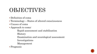
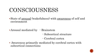
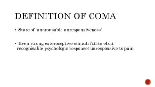
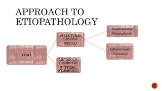
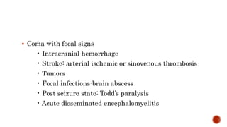
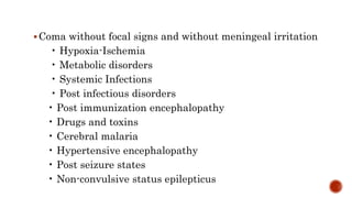
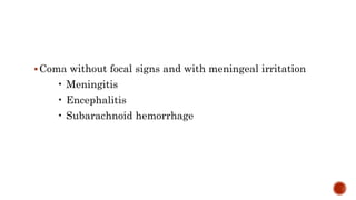
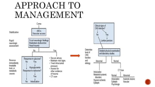
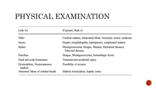
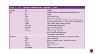
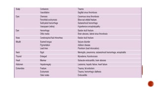
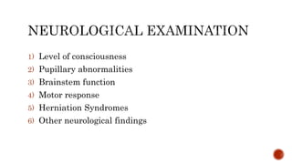
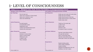
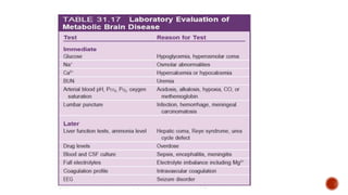
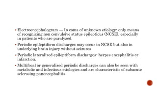
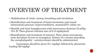
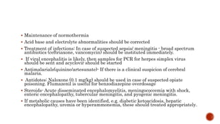
PRESENTER: DR. MUFEEDA ALUNGAL
MOHAMED
MODERATOR: DR. RATHIKA. D. SHENOY
 Definition of coma
 Terminology – States of altered consciousness
 Causes of coma
 Approach to coma:
Rapid assessment and stabilisation
History
Examination and neurological assessment
Investigations
Management
 Prognosis
State of arousal (wakefulness) with awareness of self and
environment
Arousal mediated by - Brainstem
- Subcortical structure
- Cerebral cortex
 Awareness primarily mediated by cerebral cortex with
subcortical connections
 State of ‘unarousable unresponsiveness’
 Even strong exteroceptive stimuli fail to elicit
recognisable psychologic response; unresponsive to pain
 Stupor – Spontaneous unarousability interruptive only by
vigorous, direct external stimulation ; responsive only to
pain
 Delirium – An acute or subacute reduction in awareness,
attention, orientation and perception (clouding of
consciousness), associated with abnormal sleep/wake
patterns and often psychomotor disturbances
 Syncope – Brief loss of consciousness caused by global
failure of cerebrovascular perfusion
 Dementia – A sustained multidimensional or global
decline in cognitive function
 Vegetative state – A sustained, complete loss of cognition,
with sleep/wake cycles and other autonomic functions
being relatively intact; either follow acute, severe bilateral
cerebral damage or develop gradually as the end stage of
progressive dementia
 A state of preserved arousal and awareness
 Preservation of intellectual activity accompanied by
severe or total incapacity to express voluntary responses ,
vertical eye movement is present
 Due to damage to or dysfunction of descending motor
pathways in the brain or peripheral motor nerves
 Normal EEG activity
 Sparing midbrain – Caused by severe neuromuscular
disease
COMA
STRUCTURAL
LESIONS
(FOCAL)
Supratentorial
(Hemispheric)
Infratentorial
(Brainstem)
METABOLIC
DISORDERS
(DIFFUSE ,
SYMMETRIC)
Clinically categorized causes of coma :
1) Coma with focal signs
2) Coma without focal signs and without meningeal
irritation
3) Coma without focal signs and with meningeal irritation
 Coma with focal signs
• Intracranial hemorrhage
• Stroke: arterial ischemic or sinovenous thrombosis
• Tumors
• Focal infections-brain abscess
• Post seizure state: Todd’s paralysis
• Acute disseminated encephalomyelitis
Coma without focal signs and without meningeal irritation
• Hypoxia-Ischemia
• Metabolic disorders
• Systemic Infections
• Post infectious disorders
• Post immunization encephalopathy
• Drugs and toxins
• Cerebral malaria
• Hypertensive encephalopathy
• Post seizure states
• Non-convulsive status epilepticus
Coma without focal signs and with meningeal irritation
• Meningitis
• Encephalitis
• Subarachnoid hemorrhage
 Establish and maintain Airway
 Ventilation, Oxygenation as indicated
 Circulation : Establish IV access, send investigations,
check blood glucose levels
 Identify signs of cerebral herniation or raised ICP
 Temperature: treat fever & hypothermia
 History
 Physical examination
 Rapid neurological examination
1) Level of consciousness
2) Pupillary abnormalities
3) Brainstem function
4) Motor response
5) Herniation Syndromes
6) Other neurological findings
Light reaction: Absent - deep sedative poisoning or acute
or chronic structural brainstem damage
Equality: 15% of normal patients have mild anisocoria,
but new or >2-mm dilation means parasympathetic (3rd
nerve) palsy
Extraocular movements: Absent - deep drug poisoning,
severe brainstem damage, polyneuropathy, or botulism
Dysconjugate deviation: Acute 3rd, 4th, or 6th nerve palsy
or internuclear ophthalmoplegia
Spontaneous eye movements: Nystagmus, bobbing, and
independently moving eyes - brainstem damage
Oculocephalic or oculovestibular responses: Absence of
responses means drug overdose or severe brainstem
disease
- dysconjugate responses with equal pupils-
internuclear ophthalmoplegia
- unequal pupils - 3rd nerve disease
 Observe the respiratory pattern, assessing corneal
reflexes, and testing oculocephalic (doll’s eye) or
oculovestibular (cold caloric) reflexes
Cheyne–Stokes respiration - bilateral hemispheric or
diencephalic dysfunction. It may also precede
transtentorial herniation. The hyperpneic periods have a
characteristic smooth, crescendo-decrescendo pattern
 Central neurogenic hyperventilation- midbrain
dysfunction (tachypneic and hyperpneic)
 Apneustic breathing- damage in the middle to lower
pontine region (prolonged pause at full inspiration)
Clusters of breaths separated by periods of apnea- low
pontine to upper medullary lesions
Ataxic or irregular breathing, slow regular breathing, or
agonal respiration- Medullary lesions
 Assessing muscle tone, observe spontaneous and stimulus-elicited
movements, and test deep tendon reflexes.
 Painful stimuli- to elicit motor responses in an unconscious child.
 Asymmetries suggest involvement of the corticospinal tracts in the
cerebral hemisphere or brainstem.
 Presence of spontaneous limb movements (eg, withdrawing from or
pushing away a painful stimulus) suggests a lighter depth of coma.
 Decerebrate posturing includes extension and internal rotation of the arms
and legs- brainstem involvement from a compressive or destructive process
 Decorticate posturing produces adduction and flexion at the elbows,
wrists, and fingers, with leg extension and rotation- implies a more rostral
and potentially less dire insult
 Brain tissue deforms intracranially and moves from higher to lower
pressure when there is asymmetric, unilateral or generalized
increased intracranial pressure.
 Signs of herniation tend to progress in a rostrocaudal manner.
 The importance lies in recognition and prompt treatment, before
the damage becomes irreversible
 Neuro-ophthalmologic examination.
Funduscopy - Papilledema , Haemorrhages
 Signs of meningeal irritation- Neck rigidity -- meningitis, tonsillar
herniation or cranio cervical trauma
- The Kernig’s and Brudzinski’s
signs are more reliable signs of meningeal irritation
 Basic laboratory investigations:
Immediate blood glucose by reagent strips
CBC + differential, Peripheral smear, RDT for malarial
parasite Electrolytes Coagulation profiles Liver Function
test Renal function test Calcium, Magnesium Lactate
Arterial blood gas Urine for reducing substance and ketone
 CT - intracranial hemorrhage, cranial trauma, stroke,
herniation and cerebral edema. A contrast study may reveal
features of infection in the form of meningeal enhancement,
brain abscess or neurocysticercosis
 Lumbar puncture: CSF should be tested for cell count, glucose,
protein, Gram stain, Ziehl-Neelsen stain, bacterial culture, viral
polymerase chain reaction for Herpes Simplex virus, Latex
agglutination test, and additional cultures guided by clinical
suspicion (fungal or tubercular)
 MRI: to evaluate parenchymal abnormalities. Imaging may reveal
subtle signs of edema, ischemia, or demyelination before these signs
are visible on CT scan
 If MRI results are normal, the most likely causes of coma are
intoxication, psychologic factors, or related to seizure
 Electroencephalogram — In coma of unknown etiology- only means
of recognizing non convulsive status epilepticus (NCSE), especially
in patients who are paralyzed.
 Periodic epileptiform discharges may occur in NCSE but also in
underlying brain injury without seizures
 Periodic lateralized epileptiform discharges- herpes encephalitis or
infarction.
 Multifocal or generalized periodic discharges can also be seen with
metabolic and infectious etiologies and are characteristic of subacute
sclerosing panencephalitis
 Stabilization of vitals: airway, breathing and circulation
 Identification and treatment of brain herniation and raised
intracranial pressure: hyperventilation, mannitol/3% saline etc.
 Identify and treat hypoglycemia with intravenous dextrose (2 ml/kg
10% D, Then glucose infusion rate of 6–8 mg/kg/min)
 Identification and treatment of seizures: Tonic clonic movements,
tonic deviation of eyes or nystagmus, history of a seizure preceding
the encephalopathy - anticonvulsant should be administered.
Lorazepam should be given (0.1 mg/kg), followed by phenytoin
loading (20 mg/kg)
 Maintenance of normothermia
 Acid base and electrolyte abnormalities should be corrected
 Treatment of infections: In case of suspected sepsis/ meningitis - broad spectrum
antibiotics (ceftriaxone, vancomycin) should be instituted immediately.
 If viral encephalitis is likely, then samples for PCR for herpes simplex virus
should be sent and acyclovir should be started
 Antimalarials(quinine/artesunate)- If there is a clinical suspicion of cerebral
malaria.
 Antidotes: Naloxone (0.1 mg/kg) should be used in case of suspected opiate
poisoning. Flumazenil is useful for benzodiazepine overdosage
 Steroids- Acute disseminated encephalomyelitis, meningococcemia with shock,
enteric encephalopathy, tubercular meningitis, and pyogenic meningitis.
 If metabolic causes have been identified, e.g. diabetic ketoacidosis, hepatic
encephalopathy, uremia or hyperammonemia, these should treated appropriately.
 The outcome of coma depends on the etiology, depth and duration of
impaired consciousness
 In a study by Wong CP et al , of the 283 episodes of pediatric coma
(defined as GCS <12 for at least 6 h), mortality at 1yr ranged from 3%
(intoxication) to 84% (accident), depending on etiology (3).
 Prolonged coma after a hypoxic-ischemic insult carries a poor prognosis(4).
 Most children surviving infectious encephalopathies have a comparatively
better outcomes, often surviving with mild or moderate difficulties only(5,6).
 Worse outcome in younger children with lower GCS score on presentation,
or had absent brainstem reflexes, worse motor responses, hypothermia, or
hypotension (7).
1) Nelson pediatric symptom based diagnosis,ISBN 978-0-323-39956-2, Philadelphia,
PA : Elsevier, 2018
2) S. Sharma:G. S. Kochar:N. Sankhyan:S. Gulati et al. Approach to the Child with
Coma, Indian J Pediatr (2010) 77:1279–1287 DOI 10.1007/s12098-010-0191-1
3) Wong CP, Forsyth RJ, Kelly TP, et al. Incidence, aetiology, and outcome of non-
traumatic coma: a population based study. Arch Dis Child. 2001;84:193–9
4) Kriel RL, Krach LE, Luxenberg MG, et al. Outcome of severe anoxic/ischemic brain
damage in children. Pediatr Neurol. 1994;10:207–12.
5) Grimwood K, Andersen P, Andersen V, et al. Twelve year outcomes following
bacterial meningitis: further evidence for persisting effects. Arch Dis Child.
2000;83:111–6
6) Lahat E, Barr J, Barkai J, et al. Long term neurological outcome of herpes
encephalitis. Arch Dis Child. 1999;80:69–71
7) Johnston B, Seshia SS. Prediction of outcome in nontraumatic coma in childhood.
Acta Neurol Scand. 1984;69:417–27
APPROACH

APPROACH

  • 1.
    PRESENTER: DR. MUFEEDAALUNGAL MOHAMED MODERATOR: DR. RATHIKA. D. SHENOY
  • 2.
     Definition ofcoma  Terminology – States of altered consciousness  Causes of coma  Approach to coma: Rapid assessment and stabilisation History Examination and neurological assessment Investigations Management  Prognosis
  • 3.
    State of arousal(wakefulness) with awareness of self and environment Arousal mediated by - Brainstem - Subcortical structure - Cerebral cortex  Awareness primarily mediated by cerebral cortex with subcortical connections
  • 4.
     State of‘unarousable unresponsiveness’  Even strong exteroceptive stimuli fail to elicit recognisable psychologic response; unresponsive to pain
  • 5.
     Stupor –Spontaneous unarousability interruptive only by vigorous, direct external stimulation ; responsive only to pain  Delirium – An acute or subacute reduction in awareness, attention, orientation and perception (clouding of consciousness), associated with abnormal sleep/wake patterns and often psychomotor disturbances  Syncope – Brief loss of consciousness caused by global failure of cerebrovascular perfusion
  • 6.
     Dementia –A sustained multidimensional or global decline in cognitive function  Vegetative state – A sustained, complete loss of cognition, with sleep/wake cycles and other autonomic functions being relatively intact; either follow acute, severe bilateral cerebral damage or develop gradually as the end stage of progressive dementia
  • 7.
     A stateof preserved arousal and awareness  Preservation of intellectual activity accompanied by severe or total incapacity to express voluntary responses , vertical eye movement is present  Due to damage to or dysfunction of descending motor pathways in the brain or peripheral motor nerves  Normal EEG activity  Sparing midbrain – Caused by severe neuromuscular disease
  • 8.
  • 9.
    Clinically categorized causesof coma : 1) Coma with focal signs 2) Coma without focal signs and without meningeal irritation 3) Coma without focal signs and with meningeal irritation
  • 10.
     Coma withfocal signs • Intracranial hemorrhage • Stroke: arterial ischemic or sinovenous thrombosis • Tumors • Focal infections-brain abscess • Post seizure state: Todd’s paralysis • Acute disseminated encephalomyelitis
  • 11.
    Coma without focalsigns and without meningeal irritation • Hypoxia-Ischemia • Metabolic disorders • Systemic Infections • Post infectious disorders • Post immunization encephalopathy • Drugs and toxins • Cerebral malaria • Hypertensive encephalopathy • Post seizure states • Non-convulsive status epilepticus
  • 12.
    Coma without focalsigns and with meningeal irritation • Meningitis • Encephalitis • Subarachnoid hemorrhage
  • 15.
     Establish andmaintain Airway  Ventilation, Oxygenation as indicated  Circulation : Establish IV access, send investigations, check blood glucose levels  Identify signs of cerebral herniation or raised ICP  Temperature: treat fever & hypothermia
  • 16.
     History  Physicalexamination  Rapid neurological examination
  • 20.
    1) Level ofconsciousness 2) Pupillary abnormalities 3) Brainstem function 4) Motor response 5) Herniation Syndromes 6) Other neurological findings
  • 24.
    Light reaction: Absent- deep sedative poisoning or acute or chronic structural brainstem damage Equality: 15% of normal patients have mild anisocoria, but new or >2-mm dilation means parasympathetic (3rd nerve) palsy Extraocular movements: Absent - deep drug poisoning, severe brainstem damage, polyneuropathy, or botulism Dysconjugate deviation: Acute 3rd, 4th, or 6th nerve palsy or internuclear ophthalmoplegia
  • 25.
    Spontaneous eye movements:Nystagmus, bobbing, and independently moving eyes - brainstem damage Oculocephalic or oculovestibular responses: Absence of responses means drug overdose or severe brainstem disease - dysconjugate responses with equal pupils- internuclear ophthalmoplegia - unequal pupils - 3rd nerve disease
  • 27.
     Observe therespiratory pattern, assessing corneal reflexes, and testing oculocephalic (doll’s eye) or oculovestibular (cold caloric) reflexes Cheyne–Stokes respiration - bilateral hemispheric or diencephalic dysfunction. It may also precede transtentorial herniation. The hyperpneic periods have a characteristic smooth, crescendo-decrescendo pattern  Central neurogenic hyperventilation- midbrain dysfunction (tachypneic and hyperpneic)
  • 28.
     Apneustic breathing-damage in the middle to lower pontine region (prolonged pause at full inspiration) Clusters of breaths separated by periods of apnea- low pontine to upper medullary lesions Ataxic or irregular breathing, slow regular breathing, or agonal respiration- Medullary lesions
  • 31.
     Assessing muscletone, observe spontaneous and stimulus-elicited movements, and test deep tendon reflexes.  Painful stimuli- to elicit motor responses in an unconscious child.  Asymmetries suggest involvement of the corticospinal tracts in the cerebral hemisphere or brainstem.  Presence of spontaneous limb movements (eg, withdrawing from or pushing away a painful stimulus) suggests a lighter depth of coma.  Decerebrate posturing includes extension and internal rotation of the arms and legs- brainstem involvement from a compressive or destructive process  Decorticate posturing produces adduction and flexion at the elbows, wrists, and fingers, with leg extension and rotation- implies a more rostral and potentially less dire insult
  • 33.
     Brain tissuedeforms intracranially and moves from higher to lower pressure when there is asymmetric, unilateral or generalized increased intracranial pressure.  Signs of herniation tend to progress in a rostrocaudal manner.  The importance lies in recognition and prompt treatment, before the damage becomes irreversible
  • 35.
     Neuro-ophthalmologic examination. Funduscopy- Papilledema , Haemorrhages  Signs of meningeal irritation- Neck rigidity -- meningitis, tonsillar herniation or cranio cervical trauma - The Kernig’s and Brudzinski’s signs are more reliable signs of meningeal irritation
  • 36.
     Basic laboratoryinvestigations: Immediate blood glucose by reagent strips CBC + differential, Peripheral smear, RDT for malarial parasite Electrolytes Coagulation profiles Liver Function test Renal function test Calcium, Magnesium Lactate Arterial blood gas Urine for reducing substance and ketone  CT - intracranial hemorrhage, cranial trauma, stroke, herniation and cerebral edema. A contrast study may reveal features of infection in the form of meningeal enhancement, brain abscess or neurocysticercosis
  • 37.
     Lumbar puncture:CSF should be tested for cell count, glucose, protein, Gram stain, Ziehl-Neelsen stain, bacterial culture, viral polymerase chain reaction for Herpes Simplex virus, Latex agglutination test, and additional cultures guided by clinical suspicion (fungal or tubercular)  MRI: to evaluate parenchymal abnormalities. Imaging may reveal subtle signs of edema, ischemia, or demyelination before these signs are visible on CT scan  If MRI results are normal, the most likely causes of coma are intoxication, psychologic factors, or related to seizure
  • 39.
     Electroencephalogram —In coma of unknown etiology- only means of recognizing non convulsive status epilepticus (NCSE), especially in patients who are paralyzed.  Periodic epileptiform discharges may occur in NCSE but also in underlying brain injury without seizures  Periodic lateralized epileptiform discharges- herpes encephalitis or infarction.  Multifocal or generalized periodic discharges can also be seen with metabolic and infectious etiologies and are characteristic of subacute sclerosing panencephalitis
  • 40.
     Stabilization ofvitals: airway, breathing and circulation  Identification and treatment of brain herniation and raised intracranial pressure: hyperventilation, mannitol/3% saline etc.  Identify and treat hypoglycemia with intravenous dextrose (2 ml/kg 10% D, Then glucose infusion rate of 6–8 mg/kg/min)  Identification and treatment of seizures: Tonic clonic movements, tonic deviation of eyes or nystagmus, history of a seizure preceding the encephalopathy - anticonvulsant should be administered. Lorazepam should be given (0.1 mg/kg), followed by phenytoin loading (20 mg/kg)
  • 41.
     Maintenance ofnormothermia  Acid base and electrolyte abnormalities should be corrected  Treatment of infections: In case of suspected sepsis/ meningitis - broad spectrum antibiotics (ceftriaxone, vancomycin) should be instituted immediately.  If viral encephalitis is likely, then samples for PCR for herpes simplex virus should be sent and acyclovir should be started  Antimalarials(quinine/artesunate)- If there is a clinical suspicion of cerebral malaria.  Antidotes: Naloxone (0.1 mg/kg) should be used in case of suspected opiate poisoning. Flumazenil is useful for benzodiazepine overdosage  Steroids- Acute disseminated encephalomyelitis, meningococcemia with shock, enteric encephalopathy, tubercular meningitis, and pyogenic meningitis.  If metabolic causes have been identified, e.g. diabetic ketoacidosis, hepatic encephalopathy, uremia or hyperammonemia, these should treated appropriately.
  • 42.
     The outcomeof coma depends on the etiology, depth and duration of impaired consciousness  In a study by Wong CP et al , of the 283 episodes of pediatric coma (defined as GCS <12 for at least 6 h), mortality at 1yr ranged from 3% (intoxication) to 84% (accident), depending on etiology (3).  Prolonged coma after a hypoxic-ischemic insult carries a poor prognosis(4).  Most children surviving infectious encephalopathies have a comparatively better outcomes, often surviving with mild or moderate difficulties only(5,6).  Worse outcome in younger children with lower GCS score on presentation, or had absent brainstem reflexes, worse motor responses, hypothermia, or hypotension (7).
  • 43.
    1) Nelson pediatricsymptom based diagnosis,ISBN 978-0-323-39956-2, Philadelphia, PA : Elsevier, 2018 2) S. Sharma:G. S. Kochar:N. Sankhyan:S. Gulati et al. Approach to the Child with Coma, Indian J Pediatr (2010) 77:1279–1287 DOI 10.1007/s12098-010-0191-1 3) Wong CP, Forsyth RJ, Kelly TP, et al. Incidence, aetiology, and outcome of non- traumatic coma: a population based study. Arch Dis Child. 2001;84:193–9 4) Kriel RL, Krach LE, Luxenberg MG, et al. Outcome of severe anoxic/ischemic brain damage in children. Pediatr Neurol. 1994;10:207–12. 5) Grimwood K, Andersen P, Andersen V, et al. Twelve year outcomes following bacterial meningitis: further evidence for persisting effects. Arch Dis Child. 2000;83:111–6 6) Lahat E, Barr J, Barkai J, et al. Long term neurological outcome of herpes encephalitis. Arch Dis Child. 1999;80:69–71 7) Johnston B, Seshia SS. Prediction of outcome in nontraumatic coma in childhood. Acta Neurol Scand. 1984;69:417–27