Download to read offline


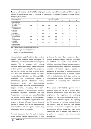

The study investigates the antimicrobial properties of propolis samples from various regions, revealing significant efficacy against both gram-positive and gram-negative bacteria, particularly in samples from Russia and Washington. Various chemical compounds within propolis, including flavonoids, contribute to its antimicrobial activity, with distinct samples exhibiting unique profiles and varying effectiveness. The findings suggest that propolis could be a promising natural alternative to synthetic antibiotics, addressing issues of antibiotic resistance and environmental impact.


