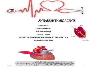
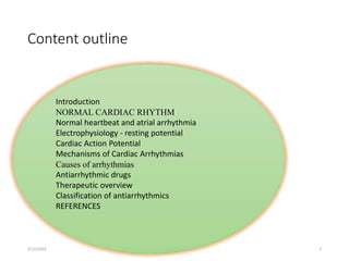
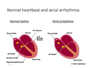
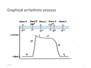
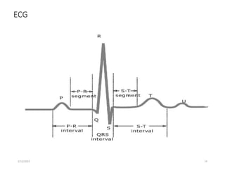
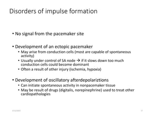
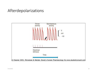
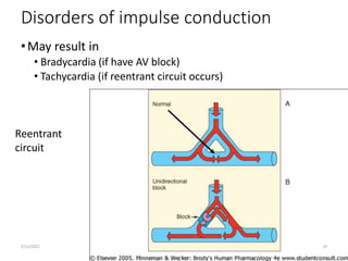
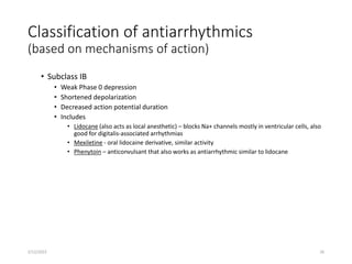
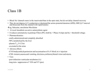
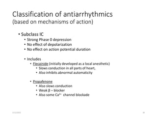
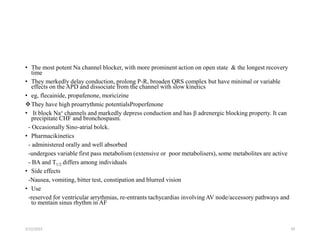
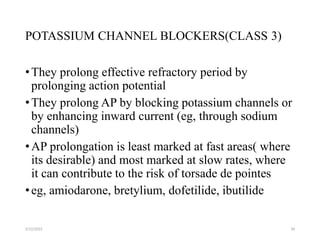
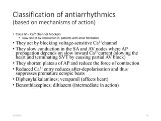
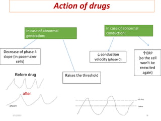
ANTIARRHYTHMICAGENTS
Presented By
Sani Ahmad Dutse
MSc Pharmacology
2020/2021 session
DEPARTMENT OF PHARMACOLOGY & THERAPEUTICS
Bayero University Kano
2/12/2022 1
Content outline
Introduction
NORMAL CARDIAC RHYTHM
Normal heartbeat and atrial arrhythmia
Electrophysiology - resting potential
Cardiac Action Potential
Mechanisms of Cardiac Arrhythmias
Causes of arrhythmias
Antiarrhythmic drugs
Therapeutic overview
Classification of antiarrhythmics
REFERENCES
2/12/2022 2
Arrhythmia
• Abnormality in the site or origin of an impulse, its rate or regularity,
or its conduction
• Heart condition where disturbances in
• Pacemaker impulse formation
• Contraction impulse conduction
• Combination of the two
Results in rate and/or timing of contraction of heart muscle that is insufficient
to maintain normal cardiac output (CO)
To understand how antiarrhythmic drugs work, need to understand
electrophysiology of normal contraction of heart
2/12/2022 3
NORMAL CARDIAC RHYTHM
Physiological sinus
rhythm occurs in a
sequence
• Sinoatrial (SA) node
• Atria
• Atrioventricular (AV) node
• Bundle of His
• Purkinje fibres
• Ventricles
2/12/2022 4
Normal heartbeat and atrial arrhythmia
Normal rhythm Atrial arrhythmia
AV septum
2/12/2022 5
Ventricular Arrhythmia
• Ventricular arrhythmias are common in
most people and are usually not a
problem but…
• VA’s are most common cause of sudden
death
• Majority of sudden death occurs in
people with neither a previously
known heart disease nor history of VA’s
• Medications which decrease incidence
of VA’s do not decrease (and may
increase) the risk of sudden death
treatment may be worse then the
disease!
2/12/2022 6
Supraventricular tachycardia
2/12/2022 7
Electrophysiology - resting potential
• A transmembrane electrical gradient (potential) is
maintained, with the interior of the cell negative with
respect to outside the cell
• Caused by unequal distribution of ions inside vs. outside cell
• Na+ higher outside than inside cell
• Ca+ much higher “ “ “ “
• K+ higher inside cell than outside
• Maintenance by ion selective channels, active pumps and
exchangers
2/12/2022 8
ECG (EKG) showing wave
segments
Contraction of
atria
Contraction of
ventricles
Repolarization of
ventricles
2/12/2022 9
Cardiac Action Potential
• Divided into five phases (0,1,2,3,4)
• Phase 4 - resting phase (resting membrane potential)
• Phase cardiac cells remain in until stimulated
• Associated with diastole portion of heart cycle
• Addition of current into cardiac muscle (stimulation) causes
• Phase 0 – opening of fast Na channels and rapid depolarization
• Drives Na+ into cell (inward current), changing membrane potential
• Transient outward current due to movement of Cl- and K+
• Phase 1 – initial rapid repolarization
• Closure of the fast Na+ channels
• Phase 0 and 1 together correspond to the R and S waves of the ECG
2/12/2022 10
Cardiac Na+ channels
2/12/2022 11
Cardiac Action Potential (con’t)
• Phase 2 - plateau phase
• sustained by the balance between the inward movement of Ca+ and outward
movement of K +
• Has a long duration compared to other nerve and muscle tissue
• Normally blocks any premature stimulator signals (other muscle tissue can
accept additional stimulation and increase contractility in a summation effect)
• Corresponds to ST segment of the ECG.
• Phase 3 – repolarization
• K+ channels remain open,
• Allows K+ to build up outside the cell, causing the cell to repolarize
• K + channels finally close when membrane potential reaches certain level
• Corresponds to T wave on the ECG
2/12/2022 12
Graphical arrhythmic process
2/12/2022 13
ECG
2/12/2022 14
Mechanisms of Cardiac Arrhythmias
• Precipitating factors; ischemia, hypoxia, acidosis or alkalosis,
electrolyte abnormalities, excessive catecholamine exposure,
autonamic influences, drug toxicity (eg, digitalis, antiarrhythmic
drugs), overstretching of cardiac fibres, and the presence of scarred or
otherwise diseased tissue
• Result from disorders of
• impulse formation
• conduction,
• or both
2/12/2022 15
Causes of arrhythmias
• Cardiac ischemia
• Excessive discharge or sensitivity to autonomic transmitters
• Exposure to toxic substances
• Unknown etiology
2/12/2022 16
Disorders of impulse formation
• No signal from the pacemaker site
• Development of an ectopic pacemaker
• May arise from conduction cells (most are capable of spontaneous
activity)
• Usually under control of SA node  if it slows down too much
conduction cells could become dominant
• Often a result of other injury (ischemia, hypoxia)
• Development of oscillatory afterdepolariztions
• Can initiate spontaneous activity in nonpacemaker tissue
• May be result of drugs (digitalis, norepinephrine) used to treat other
cardiopathologies
2/12/2022 17
Afterdepolarizations
2/12/2022 18
Disorders of impulse conduction
•May result in
• Bradycardia (if have AV block)
• Tachycardia (if reentrant circuit occurs)
Reentrant
circuit
2/12/2022 19
Antiarrhythmic drugs
• Biggest problem – antiarrhythmics can cause arrhythmia!
• Example: Treatment of a non-life threatening tachycardia may cause fatal
ventricular arrhythmia
• Must be vigilant in determining dosing, blood levels, and in follow-up when
prescribing antiarrhythmics
2/12/2022 20
Therapeutic overview
• Na+ channel blockade
• β-adrenergic receptor blockade
• Prolong repolarization
• Ca2+ channel blockade
• Adenosine
• Digitalis glycosides
2/12/2022 21
Antyarrhythmic drugs
class mechanism action notes
I Na+ channel blocker
Change the slope of
phase 0
Can abolish
tachyarrhythmia
caused by reentry
circuit
II β blocker
↓heart rate and
conduction velocity
Can indirectly alter K
and Ca conductance
III K+ channel blocker
1. ↑action potential
duration (APD) or
effective refractory
period (ERP).
2. Delay repolarization.
Inhibit reentry
tachycardia
IV Ca++ channel blocker
Slowing the rate of rise in
phase 4 of SA node(slide
12)
↓conduction velocity
in SA and AV node
•Most antiarrhythmic drugs are pro-arrhythmic (promote arrhythmia)
•They are classified according to Vaughan William into four classes according to their effects on
the cardiac action potential
2/12/2022 22
Classification of antiarrhythmics
(based on mechanisms of action)
Class I – blocker’s of fast Na+ channels
• Subclass IA
• They limit the conductance of Na + (and K+) across cell membrane (LA
action)
• Prolong action potential duration (APD) and delay channel recovery. It
dissociate from the channel with intermediate kinetics
• The blockade is greater at high frequency (premature depolarisation)
• procainamide, quinidine, disopyramide
• Cause moderate Phase 0 depression
• Prolong repolarization
• Increased duration of action potential
• Includes
• Quinidine – 1st antiarrhythmic used, treat both atrial and ventricular arrhythmias, increases refractory period
• Procainamide - increases refractory period but side effects
• Disopyramide – extended duration of action, used only for treating ventricular arrthymias
2/12/2022 23
Procainamide:
• A derivative of LA procaine
• Electrophysiological action:
-slowing of 0 phase and impulse conduction
-Prolongation of action potential duration (APD) effective refractory period
(ERP), QRS complex and QT interval.
• Pharmacokinetics
-Oral BA 75%,
-PPCs occours in 1 hr
-metabolised in the liver by acetylation to N-acetyl-procainamide (NAPA)
- T1/2relatively short 3-4 hrs (more frequent dosing)
• -morethan half is excreated unchanged in the urine
2/12/2022 24
Adverse effects
• GIT- Nausea,vomiting
CNS- weakness,mental confusion and hallucination at high doses
CVS-flushing and hypotension at rapid iv injection,cardiac toxicity.
hypersensitivity –fever ,rashes,angioedema
agranulocytosis and aplastic aneamia (rare)
2/12/2022 25
Classification of antiarrhythmics
(based on mechanisms of action)
• Subclass IB
• Weak Phase 0 depression
• Shortened depolarization
• Decreased action potential duration
• Includes
• Lidocane (also acts as local anesthetic) – blocks Na+ channels mostly in ventricular cells, also
good for digitalis-associated arrhythmias
• Mexiletine - oral lidocaine derivative, similar activity
• Phenytoin – anticonvulsant that also works as antiarrhythmic similar to lidocane
2/12/2022 26
Class 1B
• Block Na+ channels more in the inactivated than in the open state, but do not delay channel recovery
• They do not depress A-V conduction or shortened the action potential duration (APD), ERP, Q-T interval
and dissociate from the channel with rapid kinetics
eg, lidocaine, mexiletine Mexiletine
• A Local Aneathetic an active antiarrythmic agent
• It reduces automaticity in purkinje Fibers (PF), both by < Phase-4 slope and by < threshold voltage.
• Pharmacokinetics
-orally administered and completly absorbed
-90% metabolised by the liver
-plasma T1/2 9-12 hrs
-excreated in the urine
• Adverse effects
-CVS bradycardia,hypotension and accentuation of A-V block in iv injection
-CNS- tremors,nausea,and vomiting, dizziness,confusion,blured vision and ataxia.
• Uses
-post-infarction ventricular arrythmias (iv)
-long term suppression of VES and VT (pos)
2/12/2022 27
Classification of antiarrhythmics
(based on mechanisms of action)
• Subclass IC
• Strong Phase 0 depression
• No effect of depolarization
• No effect on action potential duration
• Includes
• Flecainide (initially developed as a local anesthetic)
• Slows conduction in all parts of heart,
• Also inhibits abnormal automaticity
• Propafenone
• Also slows conduction
• Weak β – blocker
• Also some Ca2+ channel blockade
2/12/2022 28
• The most potent Na channel blocker, with more prominent action on open state & the longest recovery
time
• They merkedly delay conduction, prolong P-R, broaden QRS complex but have minimal or variable
effects on the APD and dissociate from the channel with slow kinetics
• eg, flecainide, propafenone, moricizine
They have high proarrythmic potentialsProperfenone
• It block Na+ channels and markedly depress conduction and has β adrenergic blocking property. It can
precipitate CHF and bronchospasm.
- Occasionally Sino-atrial bolck.
• Pharmacikinetics
- administered orally and well absorbed
-undergoes variable first pass metabolism (extensive or poor metabolisers), some metabolites are active
- BA and T1/2 differs among individuals
• Side effects
-Nausea, vomiting, bitter test, constipation and blurred vision
• Use
-reserved for ventricular arrythmias, re-entrants tachycardias involving AV node/accessory pathways and
to mentain sinus rhythm in AF
2/12/2022 29
Classification of antiarrhythmics
(based on mechanisms of action)
• Class II – β–adrenergic blockers
• Based on two major actions
1) blockade of myocardial β–adrenergic receptors
2) Direct membrane-stabilizing effects related to Na+ channel blockade
• Includes
• Propranolol
• causes both myocardial β–adrenergic blockade and membrane-stabilizing effects
• Slows SA node and ectopic pacemaking
• Can block arrhythmias induced by exercise or apprehension
• Other β–adrenergic blockers have similar therapeutic effect
• Metoprolol
• Nadolol
• Atenolol
• Acebutolol
• Pindolol
• Stalol
• Timolol
• Esmolol
2/12/2022 30
CLASS 2 cont.….
Counteracts the arrythmogenic effects of catecholamines
• Adrenaline can cause dysrhythmias by its effect on pacemaker
potential and on the slow inward Ca2+ current
• Many ectopic sites depends on adrenergic drive
• Ventricular dysrhythmias after MI are partly due to increased
sympathetic activity. They reduce motality following MI
• AV conduction depends on sympathetic activity
• β -blockers increase the refractory of AV node and can
therefore prevent recurrent attacks of SVT
• Prevent recurrence of tachyarrhythmias (eg paroxysmal AF
provoked by increased sympathetic activity)
• Propranolol has some class 1 action
2/12/2022 31
cont…..
Propranolol
• At high doses it has a direct membrane stabilizing action. While at
clinical used dosage antiarrhythmic action is adrenargic blockade in
the heart
• It <s the slope of phase-4 depolarization and automaticity in SA node,
PF and other ectopic foci (adrenargic influence)
2/12/2022 32
Classification of antiarrhythmics
(based on mechanisms of action)
•Class III – K+ channel blockers
• Developed because some patients negatively sensitive to Na
channel blockers (they died!)
• Cause delay in repolarization and prolonged refractory
period
• Includes
• Amiodarone – prolongs action potential by delaying K+ efflux but
many other effects characteristic of other classes
• Ibutilide – slows inward movement of Na+ in addition to delaying K +
influx.
• Bretylium – first developed to treat hypertension but found to also
suppress ventricular fibrillation associated with myocardial infarction
• Dofetilide - prolongs action potential by delaying K+ efflux with no
other effects
2/12/2022 33
POTASSIUM CHANNEL BLOCKERS(CLASS 3)
•They prolong effective refractory period by
prolonging action potential
•They prolong AP by blocking potassium channels or
by enhancing inward current (eg, through sodium
channels)
•AP prolongation is least marked at fast areas( where
its desirable) and most marked at slow rates, where
it can contribute to the risk of torsade de pointes
•eg, amiodarone, bretylium, dofetilide, ibutilide
2/12/2022 34
Classification of antiarrhythmics
(based on mechanisms of action)
• Class IV – Ca2+ channel blockers
• slow rate of AV-conduction in patients with atrial fibrillation
• They act by blocking voltage-sensitive Ca2+channel
• They slow conduction in the SA and AV nodes where AP
propagation depends on slow inward Ca2+current (slowing the
heart and terminating SVT by causing partial AV block)
• They shorten plateau of AP and reduce the force of contraction
• Reduced Ca2+ entry reduces after-depolarisation and thus
suppresses premature ectopic beats
• Diphenylalkalamines; verapamil (affects heart)
• Benzothiazepines; diltiazem (intermediate in action)
2/12/2022 35
Action of drugs
In case of abnormal
generation:
Decrease of phase 4
slope (in pacemaker
cells)
Before drug
after
phase4
Raises the threshold
In case of abnormal
conduction:
↓conduction
velocity (phase 0)
↑ERP
(so the cell
won’t be
reexcited
again)
2/12/2022 36
2/12/2022 37
Campbell, T. J. & Williams, K. M. (1998). Therapeutic drug monitoring:
Antiarryhthmic drugs. Br J Clin Pharmacol, 46: 307- 319.
Cardiovascular Pharmacology Concepts (2009) Antiarrhythmic Classes. [online]
Available at: www.cvpharmacology.com [Accessed: October 2012].
Stanfield, C. L, & Germann, W. J. Principles of Human Physiology. 3. London,
England: Benjamin Cummings, 2007. Print.
phm.utoronto.ca/~jeffh/PPT/phm12barr.ppt
www.ksums.net/.../2.%20antiarrhythmic%20dr..
faculty.ksu.edu.sa/.../Antiarrhythmic%20Drugs..
References

ANTIARRYTHMIA

  • 1.
    ANTIARRHYTHMICAGENTS Presented By Sani AhmadDutse MSc Pharmacology 2020/2021 session DEPARTMENT OF PHARMACOLOGY & THERAPEUTICS Bayero University Kano 2/12/2022 1
  • 2.
    Content outline Introduction NORMAL CARDIACRHYTHM Normal heartbeat and atrial arrhythmia Electrophysiology - resting potential Cardiac Action Potential Mechanisms of Cardiac Arrhythmias Causes of arrhythmias Antiarrhythmic drugs Therapeutic overview Classification of antiarrhythmics REFERENCES 2/12/2022 2
  • 3.
    Arrhythmia • Abnormality inthe site or origin of an impulse, its rate or regularity, or its conduction • Heart condition where disturbances in • Pacemaker impulse formation • Contraction impulse conduction • Combination of the two Results in rate and/or timing of contraction of heart muscle that is insufficient to maintain normal cardiac output (CO) To understand how antiarrhythmic drugs work, need to understand electrophysiology of normal contraction of heart 2/12/2022 3
  • 4.
    NORMAL CARDIAC RHYTHM Physiologicalsinus rhythm occurs in a sequence • Sinoatrial (SA) node • Atria • Atrioventricular (AV) node • Bundle of His • Purkinje fibres • Ventricles 2/12/2022 4
  • 5.
    Normal heartbeat andatrial arrhythmia Normal rhythm Atrial arrhythmia AV septum 2/12/2022 5
  • 6.
    Ventricular Arrhythmia • Ventriculararrhythmias are common in most people and are usually not a problem but… • VA’s are most common cause of sudden death • Majority of sudden death occurs in people with neither a previously known heart disease nor history of VA’s • Medications which decrease incidence of VA’s do not decrease (and may increase) the risk of sudden death treatment may be worse then the disease! 2/12/2022 6
  • 7.
  • 8.
    Electrophysiology - restingpotential • A transmembrane electrical gradient (potential) is maintained, with the interior of the cell negative with respect to outside the cell • Caused by unequal distribution of ions inside vs. outside cell • Na+ higher outside than inside cell • Ca+ much higher “ “ “ “ • K+ higher inside cell than outside • Maintenance by ion selective channels, active pumps and exchangers 2/12/2022 8
  • 9.
    ECG (EKG) showingwave segments Contraction of atria Contraction of ventricles Repolarization of ventricles 2/12/2022 9
  • 10.
    Cardiac Action Potential •Divided into five phases (0,1,2,3,4) • Phase 4 - resting phase (resting membrane potential) • Phase cardiac cells remain in until stimulated • Associated with diastole portion of heart cycle • Addition of current into cardiac muscle (stimulation) causes • Phase 0 – opening of fast Na channels and rapid depolarization • Drives Na+ into cell (inward current), changing membrane potential • Transient outward current due to movement of Cl- and K+ • Phase 1 – initial rapid repolarization • Closure of the fast Na+ channels • Phase 0 and 1 together correspond to the R and S waves of the ECG 2/12/2022 10
  • 11.
  • 12.
    Cardiac Action Potential(con’t) • Phase 2 - plateau phase • sustained by the balance between the inward movement of Ca+ and outward movement of K + • Has a long duration compared to other nerve and muscle tissue • Normally blocks any premature stimulator signals (other muscle tissue can accept additional stimulation and increase contractility in a summation effect) • Corresponds to ST segment of the ECG. • Phase 3 – repolarization • K+ channels remain open, • Allows K+ to build up outside the cell, causing the cell to repolarize • K + channels finally close when membrane potential reaches certain level • Corresponds to T wave on the ECG 2/12/2022 12
  • 13.
  • 14.
  • 15.
    Mechanisms of CardiacArrhythmias • Precipitating factors; ischemia, hypoxia, acidosis or alkalosis, electrolyte abnormalities, excessive catecholamine exposure, autonamic influences, drug toxicity (eg, digitalis, antiarrhythmic drugs), overstretching of cardiac fibres, and the presence of scarred or otherwise diseased tissue • Result from disorders of • impulse formation • conduction, • or both 2/12/2022 15
  • 16.
    Causes of arrhythmias •Cardiac ischemia • Excessive discharge or sensitivity to autonomic transmitters • Exposure to toxic substances • Unknown etiology 2/12/2022 16
  • 17.
    Disorders of impulseformation • No signal from the pacemaker site • Development of an ectopic pacemaker • May arise from conduction cells (most are capable of spontaneous activity) • Usually under control of SA node  if it slows down too much conduction cells could become dominant • Often a result of other injury (ischemia, hypoxia) • Development of oscillatory afterdepolariztions • Can initiate spontaneous activity in nonpacemaker tissue • May be result of drugs (digitalis, norepinephrine) used to treat other cardiopathologies 2/12/2022 17
  • 18.
  • 19.
    Disorders of impulseconduction •May result in • Bradycardia (if have AV block) • Tachycardia (if reentrant circuit occurs) Reentrant circuit 2/12/2022 19
  • 20.
    Antiarrhythmic drugs • Biggestproblem – antiarrhythmics can cause arrhythmia! • Example: Treatment of a non-life threatening tachycardia may cause fatal ventricular arrhythmia • Must be vigilant in determining dosing, blood levels, and in follow-up when prescribing antiarrhythmics 2/12/2022 20
  • 21.
    Therapeutic overview • Na+channel blockade • β-adrenergic receptor blockade • Prolong repolarization • Ca2+ channel blockade • Adenosine • Digitalis glycosides 2/12/2022 21
  • 22.
    Antyarrhythmic drugs class mechanismaction notes I Na+ channel blocker Change the slope of phase 0 Can abolish tachyarrhythmia caused by reentry circuit II β blocker ↓heart rate and conduction velocity Can indirectly alter K and Ca conductance III K+ channel blocker 1. ↑action potential duration (APD) or effective refractory period (ERP). 2. Delay repolarization. Inhibit reentry tachycardia IV Ca++ channel blocker Slowing the rate of rise in phase 4 of SA node(slide 12) ↓conduction velocity in SA and AV node •Most antiarrhythmic drugs are pro-arrhythmic (promote arrhythmia) •They are classified according to Vaughan William into four classes according to their effects on the cardiac action potential 2/12/2022 22
  • 23.
    Classification of antiarrhythmics (basedon mechanisms of action) Class I – blocker’s of fast Na+ channels • Subclass IA • They limit the conductance of Na + (and K+) across cell membrane (LA action) • Prolong action potential duration (APD) and delay channel recovery. It dissociate from the channel with intermediate kinetics • The blockade is greater at high frequency (premature depolarisation) • procainamide, quinidine, disopyramide • Cause moderate Phase 0 depression • Prolong repolarization • Increased duration of action potential • Includes • Quinidine – 1st antiarrhythmic used, treat both atrial and ventricular arrhythmias, increases refractory period • Procainamide - increases refractory period but side effects • Disopyramide – extended duration of action, used only for treating ventricular arrthymias 2/12/2022 23
  • 24.
    Procainamide: • A derivativeof LA procaine • Electrophysiological action: -slowing of 0 phase and impulse conduction -Prolongation of action potential duration (APD) effective refractory period (ERP), QRS complex and QT interval. • Pharmacokinetics -Oral BA 75%, -PPCs occours in 1 hr -metabolised in the liver by acetylation to N-acetyl-procainamide (NAPA) - T1/2relatively short 3-4 hrs (more frequent dosing) • -morethan half is excreated unchanged in the urine 2/12/2022 24
  • 25.
    Adverse effects • GIT-Nausea,vomiting CNS- weakness,mental confusion and hallucination at high doses CVS-flushing and hypotension at rapid iv injection,cardiac toxicity. hypersensitivity –fever ,rashes,angioedema agranulocytosis and aplastic aneamia (rare) 2/12/2022 25
  • 26.
    Classification of antiarrhythmics (basedon mechanisms of action) • Subclass IB • Weak Phase 0 depression • Shortened depolarization • Decreased action potential duration • Includes • Lidocane (also acts as local anesthetic) – blocks Na+ channels mostly in ventricular cells, also good for digitalis-associated arrhythmias • Mexiletine - oral lidocaine derivative, similar activity • Phenytoin – anticonvulsant that also works as antiarrhythmic similar to lidocane 2/12/2022 26
  • 27.
    Class 1B • BlockNa+ channels more in the inactivated than in the open state, but do not delay channel recovery • They do not depress A-V conduction or shortened the action potential duration (APD), ERP, Q-T interval and dissociate from the channel with rapid kinetics eg, lidocaine, mexiletine Mexiletine • A Local Aneathetic an active antiarrythmic agent • It reduces automaticity in purkinje Fibers (PF), both by < Phase-4 slope and by < threshold voltage. • Pharmacokinetics -orally administered and completly absorbed -90% metabolised by the liver -plasma T1/2 9-12 hrs -excreated in the urine • Adverse effects -CVS bradycardia,hypotension and accentuation of A-V block in iv injection -CNS- tremors,nausea,and vomiting, dizziness,confusion,blured vision and ataxia. • Uses -post-infarction ventricular arrythmias (iv) -long term suppression of VES and VT (pos) 2/12/2022 27
  • 28.
    Classification of antiarrhythmics (basedon mechanisms of action) • Subclass IC • Strong Phase 0 depression • No effect of depolarization • No effect on action potential duration • Includes • Flecainide (initially developed as a local anesthetic) • Slows conduction in all parts of heart, • Also inhibits abnormal automaticity • Propafenone • Also slows conduction • Weak β – blocker • Also some Ca2+ channel blockade 2/12/2022 28
  • 29.
    • The mostpotent Na channel blocker, with more prominent action on open state & the longest recovery time • They merkedly delay conduction, prolong P-R, broaden QRS complex but have minimal or variable effects on the APD and dissociate from the channel with slow kinetics • eg, flecainide, propafenone, moricizine They have high proarrythmic potentialsProperfenone • It block Na+ channels and markedly depress conduction and has β adrenergic blocking property. It can precipitate CHF and bronchospasm. - Occasionally Sino-atrial bolck. • Pharmacikinetics - administered orally and well absorbed -undergoes variable first pass metabolism (extensive or poor metabolisers), some metabolites are active - BA and T1/2 differs among individuals • Side effects -Nausea, vomiting, bitter test, constipation and blurred vision • Use -reserved for ventricular arrythmias, re-entrants tachycardias involving AV node/accessory pathways and to mentain sinus rhythm in AF 2/12/2022 29
  • 30.
    Classification of antiarrhythmics (basedon mechanisms of action) • Class II – β–adrenergic blockers • Based on two major actions 1) blockade of myocardial β–adrenergic receptors 2) Direct membrane-stabilizing effects related to Na+ channel blockade • Includes • Propranolol • causes both myocardial β–adrenergic blockade and membrane-stabilizing effects • Slows SA node and ectopic pacemaking • Can block arrhythmias induced by exercise or apprehension • Other β–adrenergic blockers have similar therapeutic effect • Metoprolol • Nadolol • Atenolol • Acebutolol • Pindolol • Stalol • Timolol • Esmolol 2/12/2022 30
  • 31.
    CLASS 2 cont.…. Counteractsthe arrythmogenic effects of catecholamines • Adrenaline can cause dysrhythmias by its effect on pacemaker potential and on the slow inward Ca2+ current • Many ectopic sites depends on adrenergic drive • Ventricular dysrhythmias after MI are partly due to increased sympathetic activity. They reduce motality following MI • AV conduction depends on sympathetic activity • β -blockers increase the refractory of AV node and can therefore prevent recurrent attacks of SVT • Prevent recurrence of tachyarrhythmias (eg paroxysmal AF provoked by increased sympathetic activity) • Propranolol has some class 1 action 2/12/2022 31
  • 32.
    cont….. Propranolol • At highdoses it has a direct membrane stabilizing action. While at clinical used dosage antiarrhythmic action is adrenargic blockade in the heart • It <s the slope of phase-4 depolarization and automaticity in SA node, PF and other ectopic foci (adrenargic influence) 2/12/2022 32
  • 33.
    Classification of antiarrhythmics (basedon mechanisms of action) •Class III – K+ channel blockers • Developed because some patients negatively sensitive to Na channel blockers (they died!) • Cause delay in repolarization and prolonged refractory period • Includes • Amiodarone – prolongs action potential by delaying K+ efflux but many other effects characteristic of other classes • Ibutilide – slows inward movement of Na+ in addition to delaying K + influx. • Bretylium – first developed to treat hypertension but found to also suppress ventricular fibrillation associated with myocardial infarction • Dofetilide - prolongs action potential by delaying K+ efflux with no other effects 2/12/2022 33
  • 34.
    POTASSIUM CHANNEL BLOCKERS(CLASS3) •They prolong effective refractory period by prolonging action potential •They prolong AP by blocking potassium channels or by enhancing inward current (eg, through sodium channels) •AP prolongation is least marked at fast areas( where its desirable) and most marked at slow rates, where it can contribute to the risk of torsade de pointes •eg, amiodarone, bretylium, dofetilide, ibutilide 2/12/2022 34
  • 35.
    Classification of antiarrhythmics (basedon mechanisms of action) • Class IV – Ca2+ channel blockers • slow rate of AV-conduction in patients with atrial fibrillation • They act by blocking voltage-sensitive Ca2+channel • They slow conduction in the SA and AV nodes where AP propagation depends on slow inward Ca2+current (slowing the heart and terminating SVT by causing partial AV block) • They shorten plateau of AP and reduce the force of contraction • Reduced Ca2+ entry reduces after-depolarisation and thus suppresses premature ectopic beats • Diphenylalkalamines; verapamil (affects heart) • Benzothiazepines; diltiazem (intermediate in action) 2/12/2022 35
  • 36.
    Action of drugs Incase of abnormal generation: Decrease of phase 4 slope (in pacemaker cells) Before drug after phase4 Raises the threshold In case of abnormal conduction: ↓conduction velocity (phase 0) ↑ERP (so the cell won’t be reexcited again) 2/12/2022 36
  • 37.
    2/12/2022 37 Campbell, T.J. & Williams, K. M. (1998). Therapeutic drug monitoring: Antiarryhthmic drugs. Br J Clin Pharmacol, 46: 307- 319. Cardiovascular Pharmacology Concepts (2009) Antiarrhythmic Classes. [online] Available at: www.cvpharmacology.com [Accessed: October 2012]. Stanfield, C. L, & Germann, W. J. Principles of Human Physiology. 3. London, England: Benjamin Cummings, 2007. Print. phm.utoronto.ca/~jeffh/PPT/phm12barr.ppt www.ksums.net/.../2.%20antiarrhythmic%20dr.. faculty.ksu.edu.sa/.../Antiarrhythmic%20Drugs.. References