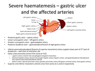
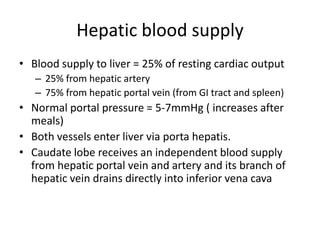
MRCS Revision

           Anatomy
Awesomemedicalnotes.wordpress.com
Severe haematemesis – gastric ulcer
          and the affected arteries


•   Posterior gastric ulcer – splenic artery
•   Lesser curve gastric ulcer – left gastric artery
•   Greater curve gastric ulcer – gastroepiploic artery
•   Posterior duodenal ulcer – gastroduodenal branch of right gastric artery

•   Inferior pancreaticoduodenal (branch of superior mesenteric) artery supplies lower part of 2nd part of
    duodenum – well clear of ulceration site
•   Celiac artery branches (arises from aorta at T12):
     –   L gastric artery ( branch into oesophageal and gastric branches)
     –   Common hepatic artery ( branch into proper hepatic artery, R gastric artery and gastroduodenal artery(branch
         into superior pancreaticoduodenal artery))
     –   Splenic artery (dorsal pancreatic artery, greater pancreatic artery, left gastro-omental artery, short gastric artery)
•   Superior and inferior mesenteric arteries arise from aorta at L1 and L3 respectively
Hepatic blood supply
• Blood supply to liver = 25% of resting cardiac output
   – 25% from hepatic artery
   – 75% from hepatic portal vein (from GI tract and spleen)
• Normal portal pressure = 5-7mmHg ( increases after
  meals)
• Both vessels enter liver via porta hepatis.
• Caudate lobe receives an independent blood supply
  from hepatic portal vein and artery and its branch of
  hepatic vein drains directly into inferior vena cava
Psoas major muscle
• Flexes thigh at hip joint
• It joins illiacus muscle (origin: inner aspect of
  iliac wing of pelvis), and becomes iliopsoas
  muscle and inserts on lesser trochanter of
  femur = flexes thigh at hip joint
Femoral sheath
       • Femoral sheath ends 4cm
         inferior to inguinal ligament
       • Medial wall of fem sheath is
         pierced by great saphenous
         vein and lymphatic vessels
       • It is divided by 2 ventral septa
         into 3 compartments:
          – Lateral (contains fem. artery)
          – Intermediate (contains fem. vein)
          – Medial (a space called fem. canal)
       • Fem. canal contains efferent
         lymphatic vessels and a lymph
         node
       • Entrance of fem canal = fem
         ring = site of fem hernia
Uterine Artery
       • Arises from internal iliac
         artery (may also arise
         from umbilical artery)
       • Lies anterosuperior to
         ureter near lateral
         portion of fornix
          – Risk of ureteric damage
            during hysterectomy
          – Esp. left ureter as it lies
            very close to lat. Aspect of
            cervix
       • Point of crossing b/w
         uterine artery and ureter
         = 2 cm superior to ischial
         spine
Pelvis
Pelvis 2
Uterus
Uterus 2
• Uterus is normally bent anteriorly b/w cervix and body
  of uterus
• Commonly anteverted (inclined anteriorly) but
  frequently retroverted in older woman
• 3 layers: perimetrium, myometrium and endometrium
• Uterine body is enclosed b/w layers of broad ligament
  – freely mobile
• Uterus covered by peritoneum anteriorly and
  superiorly except for vaginal part of cervix
• Uterus is supported by pelvic floor
Hypoglossal Nerve
• Supplies all intrinsic muscles of the tongue but
  not the palate (the palatoglossus muscle supplied
  by vagus nerve)
• No sensory component
• Genioglossus muscle protrudes the tongue
• In paralysis, tongue deviates towards the affected
  side. (e.g. damaged left hypoglossal nerve =
  tongue deviates to left
• Left submandibular salivary gland excision = risk
  of damaging hypoglossal nerve
Inferior/recurrent laryngeal nerve
Inferior/recurrent laryngeal nerve 2
• A branch of vagus nerve
• Supplies all muscles of larynx except cricothroid
  muscle (supplied by superior laryngeal branch of
  vagus nerve)
• Sensory component – supplies larynx inferior to
  vocal cords
• Damage recurrent laryngeal nerve:
  – Larynx is anaesthetic inferior to vocal cord on affected
    side
  – Paralysed cord seen to lie in ‘paralytic’ position:
    slightly abducted from midline and does not move on
    phonation

Anatomy revision part 1

  • 1.
    MRCS Revision Anatomy Awesomemedicalnotes.wordpress.com
  • 2.
    Severe haematemesis –gastric ulcer and the affected arteries • Posterior gastric ulcer – splenic artery • Lesser curve gastric ulcer – left gastric artery • Greater curve gastric ulcer – gastroepiploic artery • Posterior duodenal ulcer – gastroduodenal branch of right gastric artery • Inferior pancreaticoduodenal (branch of superior mesenteric) artery supplies lower part of 2nd part of duodenum – well clear of ulceration site • Celiac artery branches (arises from aorta at T12): – L gastric artery ( branch into oesophageal and gastric branches) – Common hepatic artery ( branch into proper hepatic artery, R gastric artery and gastroduodenal artery(branch into superior pancreaticoduodenal artery)) – Splenic artery (dorsal pancreatic artery, greater pancreatic artery, left gastro-omental artery, short gastric artery) • Superior and inferior mesenteric arteries arise from aorta at L1 and L3 respectively
  • 3.
    Hepatic blood supply •Blood supply to liver = 25% of resting cardiac output – 25% from hepatic artery – 75% from hepatic portal vein (from GI tract and spleen) • Normal portal pressure = 5-7mmHg ( increases after meals) • Both vessels enter liver via porta hepatis. • Caudate lobe receives an independent blood supply from hepatic portal vein and artery and its branch of hepatic vein drains directly into inferior vena cava
  • 4.
    Psoas major muscle •Flexes thigh at hip joint • It joins illiacus muscle (origin: inner aspect of iliac wing of pelvis), and becomes iliopsoas muscle and inserts on lesser trochanter of femur = flexes thigh at hip joint
  • 5.
    Femoral sheath • Femoral sheath ends 4cm inferior to inguinal ligament • Medial wall of fem sheath is pierced by great saphenous vein and lymphatic vessels • It is divided by 2 ventral septa into 3 compartments: – Lateral (contains fem. artery) – Intermediate (contains fem. vein) – Medial (a space called fem. canal) • Fem. canal contains efferent lymphatic vessels and a lymph node • Entrance of fem canal = fem ring = site of fem hernia
  • 6.
    Uterine Artery • Arises from internal iliac artery (may also arise from umbilical artery) • Lies anterosuperior to ureter near lateral portion of fornix – Risk of ureteric damage during hysterectomy – Esp. left ureter as it lies very close to lat. Aspect of cervix • Point of crossing b/w uterine artery and ureter = 2 cm superior to ischial spine
  • 7.
  • 8.
  • 9.
  • 10.
    Uterus 2 • Uterusis normally bent anteriorly b/w cervix and body of uterus • Commonly anteverted (inclined anteriorly) but frequently retroverted in older woman • 3 layers: perimetrium, myometrium and endometrium • Uterine body is enclosed b/w layers of broad ligament – freely mobile • Uterus covered by peritoneum anteriorly and superiorly except for vaginal part of cervix • Uterus is supported by pelvic floor
  • 11.
    Hypoglossal Nerve • Suppliesall intrinsic muscles of the tongue but not the palate (the palatoglossus muscle supplied by vagus nerve) • No sensory component • Genioglossus muscle protrudes the tongue • In paralysis, tongue deviates towards the affected side. (e.g. damaged left hypoglossal nerve = tongue deviates to left • Left submandibular salivary gland excision = risk of damaging hypoglossal nerve
  • 12.
  • 13.
    Inferior/recurrent laryngeal nerve2 • A branch of vagus nerve • Supplies all muscles of larynx except cricothroid muscle (supplied by superior laryngeal branch of vagus nerve) • Sensory component – supplies larynx inferior to vocal cords • Damage recurrent laryngeal nerve: – Larynx is anaesthetic inferior to vocal cord on affected side – Paralysed cord seen to lie in ‘paralytic’ position: slightly abducted from midline and does not move on phonation