♛VVIP Hyderabad Call Girls Chintalkunta🖕7001035870🖕Riya Kappor Top Call Girl ...
Anatomy of the abdomen and pelvis
1. Anatomy of the Abdomen and Pelvis
The Abdominal Walls
1. State the boundaries of the abdomino-pelvic cavity1
2. What are the surface boundaries of the abdomen?2
3.What characteristic of the diaphragm helps to
protect the upper abdomen?3
4.What is the pelvic diaphragm?
5.What are the linea alba and the linea semilunaris?4
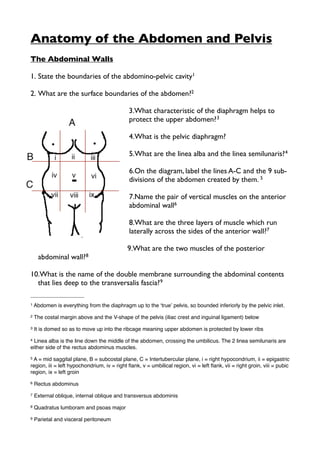
6.On the diagram, label the lines A-C and the 9 sub-
divisions of the abdomen created by them. 5
7.Name the pair of vertical muscles on the anterior
abdominal wall6
8.What are the three layers of muscle which run
laterally across the sides of the anterior wall?7
9.What are the two muscles of the posterior
abdominal wall?8
10.What is the name of the double membrane surrounding the abdominal contents
that lies deep to the transversalis fascia?9
1 Abdomen is everything from the diaphragm up to the ʻtrueʼ pelvis, so bounded inferiorly by the pelvic inlet.
2 The costal margin above and the V-shape of the pelvis (iliac crest and inguinal ligament) below
3 It is domed so as to move up into the ribcage meaning upper abdomen is protected by lower ribs
4 Linea alba is the line down the middle of the abdomen, crossing the umbilicus. The 2 linea semilunaris are
either side of the rectus abdominus muscles.
5 A = mid saggital plane, B = subcostal plane, C = Intertubercular plane, i = right hypocondrium, ii = epigastric
region, iii = left hypochondrium, iv = right flank, v = umbilical region, vi = left flank, vii = right groin, viii = pubic
region, ix = left groin
6 Rectus abdominus
7 External oblique, internal oblique and transversus abdominis
8 Quadratus lumboram and psoas major
9 Parietal and visceral peritoneum
2. 11.Name the three layers of abdominal superficial fascia10
12.Where are the attachments of the Scarpa’s fascia?11
13.What are the two main superficial veins of the abdomen and where are they?12
14.What are the functions and attachments of the psoas major?13
15.Where is the iliacus muscle?14
16.What are the root values of the subcostal, iliohypogastric and ilioinguinal nerves?
15
17.What could cause compression of the nerves in (16)?16
18.Describe the course and distribution of the genitofemoral nerve?17
19.What are the attachments and functions of the external obliques?18
20.What are the attachments and functions of the internal obliques and the
transversus abdominus?19
10 Outside in = Camperʼs fascia, Scarpaʼs fascia. There is a negligible amount of deep fascia below the
11 Bound to the fascia lata of the thigh below the inguinal ligament and to structures in the perineum (region
of the external genetalia). Posteriorly to the throacolumbar fascia and the fascia lata of the buttock.
12 Thoraco-epigastric (run through right and left hypochondrium), superficial epigastric (run up to the
umbilicus from the pubic region
13 Flexes the thigh on the trunk or trunk on thigh. Arises from the lumbar transverse processes and lumbar
vertebral bodies, attaches the illiacus muscle (on the iliac fossa) onto the lesser trochanter of the femur.
14 Inside the iliac fossa on the interior of the hip bone
15 Subcostal nerve = T12, Iliohypogastric = L1, Ilioinguinal = collateral branch of iliohypogastric so also L1
16 Enlargement of the psoas major muscle, which runs alongside these nerves. This can happen following
infection of the psoas fascia, or intra-muscular haematomas.
17 Derived from L1 and 2 and emerges from the anterior surface of psoas major and runs down deep to the
psoas fascia to supply the cremaster muscle in the male, via its genital branch.
18 Pull the trunk downwards and forwards to right, left or if contracted together, forwards. Arises from the
lower ribs 5-12, inserts posteriorly to the iliac crest.
19 Internal oblique begins at ribs 9-12 and inserts into the the iliac crest, thoracolumbar fascia posteriorly and
linea alba anteriorly. It functions to stabilise the lumbar spine. The transversus abdominus originates from the
internal surfaces of the bones and cartilages forming the thoracic outlet and iliac crest, as well as the
thoracolumbar fascia. Its primary role is abdominal compression/breathing, and stabilization.
3. 21.What are the layers of the rectal sheath?20
22.Which dermatomes cover the anterior abdominal wall?21
23.Which nerves (spinal segments) innervate the rectus abdominis muscle?22
Inguinal Region and Hernias
1.Label the locations of hernias A-F23
2.What is the difference between an inguinal
hernia and a femoral hernia?24
3.What stuctures form the inguinal ring?25
4.List the boundaries of the inguinal canal26
5.Most layers of the abdominal wall are brought
down with the testes as they descend to form
the scotum, but which layers must they pass
through?27
6.What is a patent processus vaginalis and how
would you test for it?
20 Linea alba in the centre, either side are each rectus abdominus muscle, the external oblique passes over
the top of this and the transversus behind. Below the arcuate line, the internal oblique passes infront of the
rectus, above it straddles it on both sides.
21 T7-L1
22 T7-T12
23 A = Epigastric, B = Paraumblical, C = Umbilical, D = Spigelian, E = Inguinal, F = Femoral
24 Inguinal is a protrusion through the inguinal canal
25 Medial and lateral margins are formed by the split in the aponeurosis (crura/crus), the lateral crus attached
to the pubic tubercle and the medial to the pubic crest. Intercrucal fibres arising from the inguinal ligament
stop the crura from spreading apart.
26 It is the lower free edge of the external oblique aponeurosis. Openings either end are the deep and
superficial inguinal rings. The deep inguinal ring is the beginning of an invagination in the transversalis fascia
which continues into the canal forming its innermost covering. It passes through all 3 layers of abdominal
muscles, obliquely along the inguinal ligament, the internal oblique gives some slips of muscle covering
known as the cremaster muscle.
27 Under the transversus abdominis tendon and internal oblique. Descended testes leave a trail of
surrounding layers called the vas deferens which forms the spermatic cord, derived from 3 layers of
abdominal wall.
4. 7. Describe the path of the spermatic cord28
8. Describe the coverings of the spermatic cord in relation to the abdominal wall29
9. What is the nerve supply for the cremaster muscle?30
10.What are the boundaries of the inguinal triangle?31
11.Which nerve supplies the muscle fibres of the conjoint tendon?32
12.What is the difference between a direct and indirect inguinal hernia?33
13.What is a hiatus hernia?34
Peritoneum
1. What is a mesentery?35
2. What are the ventral and dorsal mesenteries?36
3. What is mesothelium?37
28 Begins at the deep inguinal ring, lateral to the inferior epigastric artery, ends at the posterior border of the
testis. Passing through the inguinal canal and emerging at the superficial inguinal ring. As the cord leaves
the inguinal canal, it acquires its 3rd covering, the external spermatic fascia.
29 Internal = fascia transversalis, Middle layer = cremaster layer, Lastly = spermatic fascia derived from the
external oblique. (transversalis abdominis does not contribute to the sheath)
30 Genital branch of the genitofemoral nerve, L1-2
31 Medial: The lateral margin of the rectus abdominus muscle (linea semilunaris), Lateral: the inferior
epigastric artery, Inferior: the Inguinal Ligament
32 Supplied by the L1 nerve, loss of this nerve or muscle can lead to direct inguinal hernia
33 A indirect inguinal hernia is where abdominal contents protrude through the deep inguinal ring, direct
inguinal hernias are where the abdominal contents herniate the wall of the inguinal canal without going down
the canal itself.
34 Herniation of stomach up through the hole in the diaphragm through which the oesophagus travels.
35 A double folded membrane which separates the abdominal cavity from the peritoneum. The peritoneal
cavity itself does not contain any organs, rather the gut is trapped within the mesentery in a ʻsandwichʼ.
36 The two sides of the mesentery (either side of the gut organs). The front (ventral) mesentery is shorter
than the dorsal one, so there is continuity beneath it, the mesentery is only a partial septum.
37 The histological term for mesentery, once is has slung around the organs to form the visceral perironeum.
This is a simple squamous columnar epithelium.
5. 4. What is the meaning of the term ligament in context of the peritoneum and what
is the gastro-hepatic ligament?38
5. Explain the difference between an intra-peritoneal and retro-peritoneal organ39
6. Which organs and structures are retro-peritoneal?40
7. What is the caecal bud?41
8. What is the vitelline duct?42
9. What is Meckel’s diverticulum?43
10.What is the origin of the greater omentum?44
11.What is the relation of the greater omentum to the greater and lesser sacs?45
12.What are the functions of the greater omentum?46
13.What is the epiploic foramen?47
14.Name the 4 peritoneal spaces (between the mesenteries)48
38 Ligaments may be formed out of remaining double folds of mesentery, meaning that abdominal organs are
connected to each other in some way. The hepato-gastric ligament is also known as the lesser omentum.
39 As the gut twists and turns in development, some organs lost their mesentery, fusing with the parietal
peritoneum or posterior abdominal wall instead. These are known as retro-peritoneal organs.
40 DUKE CRAPS - Duodenum, Ureters, Kidneys, Espohagus, Colon (ascending and descending), Aorta,
Pancreas, Supraneal Glands
41 Part of the caecum (gut following the stomach) which protrudes into the umbilicus in embreyological
development having been pushed by growth of the liver.During later stages of development, there is anti-
clockwise rotation of the midgut and the caecum retracts back from the umbilicus, so the caecal bud remains
superior to the gut, then as the gut rotates further, it lies inferiorly on the right.
42 Communication between midgut and yolk sac in embreyo
43 The adult remnant of the vitelline duct
44 Expansion of the embryological ʻdorsal mesenteryʼ of the stomach. Greater omentum expands downwards
to cover the small intestine.
45 The lesser sac is the area behind the stomach, the greater sac is everywhere else.
46 The greater omentum is a fat filled apron which folds down over the small intestines. Its function is to
localise infection by sticking to any infected region, trapping the infection and preventing it from spreading.
47 aka Omental foramen, passage of communication between the lesser sac (behind stomach) and greater
sac (everywhere else)
48 Left and right paracolic gutters (between the colon and the abdominal wall) and the left and right
paramesenteric gutters (between the colon and the root of the mesentery)
6. 15.How do the peritoneal folds and spaces differ between male and female?49
Stomach and Spleen
1. What is the approximate position of the stomach in relation to the abdominal
divisions?50
2. What are the main sub-divisions/parts of the stomach, what substances do they
secrete?51
3. What are the main functions of the stomach?52
4. Name the layers of the gut wall53
5. Name the two parts of the enteric nervous system54
6. Name the 3 muscular coats of the stomach in order55
7. Name the sphincters of the stomach and oesophagus56
8. What are the right and left crus?57
9. What are rugae58
49 In males, the peritoneum sweeps forward and around the lateral walls and towards the floor of the pelvis
(levator ani muscle), before ascending up the anterior wall of the abdomen. The fossa between the posterior
and anterior folds is known as the rectovesical pouch. Females have an additional fold of peritoneum
dividing this space into the rectouterine pouch (of Douglas) behind and a vesouterine pouch in front.
50 Occupies parts of the epigastric, umbilical and left hypochondriac regions
51 Cardia (mucus secretion), fundus (storage/gas), body (mucus, HCl, pepsiongen, intrinsic factor), Pyloris
(mixing, gastrin)
52 Storage, secretions, breakdown with enzymes/HCl, absorbtion.
53 From lumen outwards: Epithelium, lamina propria, muscularis mucosae (internal ring of smooth muscle),
submucosa, mucularis externa (1 layer circular, 1 layer longlitudinal), serosa.
54 Submucosal plexus and myenteric plexus
55 Inner oblique layer, middle circular layer, outer longitudinal layer (for peristalsis/churning)
56 Oesophageal sphincter is a layer of muscle but not a true sphincter & pyloric sphincter (exit to duodenum).
Control of gastric reflux is done largely by the muscular fibres of the diaphragm.
57 Tendonous structures which extend from the diaphragm for a short distance down the vertebral column
58 A series of ridges caused by the in-folding of the mucus membrane of the stomach.
7. 10.Which organs and structures are in contact with the stomach?59
11.Which arteries supply the stomach and liver and what are their origins?60
12.Where do all the veins of the stomach ultimately drain into?61
13.Where does the stomach lymph ultimately drain into?62
14.Describe the nerve supply to the stomach63
15.Which dermatomes would be sensitive to foregut pain?64
16.Where is the spleen located in relation to the ribs?65
17.What are the functions of the spleen?66
18.Identify the indentations on the surface of the spleen67
Liver and Hepatobiliary System
1. What is the approximate position of the liver in relation to other abdominal
organs and surface regions?68
2. What are the main functions of the liver?69
59 Superiorly the liver and left diaphragm, laterally the left kidney, supraneal gland and spleen. Splenic artery,
hepatic portal vein and coealiac trunk. The lesser omentum and lesser sac separate the stomach from
adjacent organs inferiorly.
60 The coeliac artery arises from the aorta. It is split into 3 branches, left gastric, splenic and common
hepatic. The common hepatic artery then splits into the proper hepatic and gastroduodenal artery.
61 Hepatic portal vein
62 Coeliac (pre-aortic) nodes and the thoracic duct via the cysterna chyli
63 The right and left vagi (split in the vagus nerve) split anterior and posterior to the stomach.
64 T6-9
65 Immediately beneath ribs 9-10, with ribs 11 and 12 below it. The spleen is highly vascular so a rupture
caused by broken ribs leads to severe haemorrage.
66 Largest lymphoid unit in the body, contains macrophages which destroy old red blood cells, produces
white and red blood cells (in infant), reservoir for 1/3 of platelets, store of blood can be released in response
to adrenaline.
67 The spleen has two surfaces (diaphragmatic and visceral), two borders (one notched and one not). Lower
pole = splenic flexure of the colon, visceral surface = stomach, left kidney and tail of pancreas
68 Lower border corresponds to right costal margin,
69 Produces bile (stored in the gallbladder), glucose into glycogen, production of cholesterol, regulation of
fats and amino acids, stores iron, detoxification, immunity, manufacture of plasma proteins.
8. 3. What are the spaces above and below the liver?70
4. List the lobes of the liver and the ligaments separating them?71
5. Which organs lie on the visceral surface of the liver?72
6. Which structures pass through the porta hepatis/portal triad (on the free edge of
the lesser omentum)?73
7. Which veins form the hepatic portal vein (bearing in mind it drains the whole gut
to the liver)?74
8. Describe the passage of venous blood through porto-systemic anastemosis in the
case of portal hypertension in liver disease?75
9. What clinical symptoms could follow blockage of the portal system?76
10.By contrast, if the IVC becomes blocked, what route will blood be diverted to in
order to reach the heart?77
11.List the functions of the gallbladder78
70 Hepato-renal (inferior/posterior) and sub-phrenic (superior/anterior)
71 Anteriorly: Right lobe and left lobe separated by the falciform ligament. Posteriorly the quadrate lobe and
smaller caudate lobe separated by the coronary ligaments and triangular ligaments
72 Right kidney, hepatic flexure of the colon, oesophagus, stomach and duodenum
73 Portal vein, hepatic artery and the common hepatic duct (into which the bile duct drains from the
gallbladder)
74 The hepatic portal vein is the product of the entire venous drainage of the gut, which includes the superior
and inferior mesenteric veins, the splenic vein, the gastric veins and the cystic vein from the gallbladder.
75 Progressive opening up of pre-existing anastemoses between the systemic and portal venous systems at
one of 4 possible sites: lower oesophagus, anal canal, recanalized umbilical vein to the abdominal wall, or
the posterior abdominal wall.
76 Could result in haemorrhoids, varicose veins on the abdominal wall, diltated superficial veins arising from
the umbilicus (Medusaʼs head).
77 Effectively the reverse of portal hypertension, veins of the anterior abdominal wall enlarge and shunt blood
around the obstruction but in the opposite direction to their normal flow, linking the subclavian/axilliary
system with the iliac/femoral.
78 Reservoir of bile produced by the liver, concentrates the bile and adds mucus, absorbing water out of the
mixture.
9. 12.Name the different bile ducts79
13.What and where is the sphincter of Oddi?80
14.What is the hepatopancreatic ampulla ofVater?81
15.Name the 4 parts of the pancreas82
16.What are the functions of the pancreas83
17.What is the arterial supply of the pancreas84
18.What are the 2 ducts of the pancreas entering the duodenum85
The Intestines
The Intestines
1. Define foregut, midgut and hind gut86
2. What membranous structure is stretched between the liver/gallbladder and
stomach?87
3. What membranous structure is stretched from just below the stomach across the
intestines?88
79 Right and left hepatic ducts drain from right and left lobes of the liver, these merge to form the common
hepatic duct and then combine with the cystic duct from the gallbladder to form the bile duct which enters the
duodenum.
80 Sphincter at the lower end of the common bile duct where it joins the pancreatic duct, controls biliary
secretion
81 The route by which the pancreatic duct enters the duodenum
82 head, body, tail and uncinate process
83 Exocrine gland secreting digestive enzymes, as an endocrine gland producing and secreting insulin and
glucagon
84 Receives blood from the arteria pancreatica magna (from splenic artery), superior pancreatoduodenal
artery (from gastroduodenal), inferior pancreatoduodenal (from superior mesenteric).
85 Main pancreatic duct (joint with the common bile duct), enters the duodenum through the ampulla of Vater.
The accessory pancreatic duct enters the duodenum superior to the main duct.
86 Foregut: ends after entry of common bile duct into duodenum, Midgut: ends 2/3rds of the way along the
transverse colon, Hindgut: Ends halfway down the anal canal.
87 Lesser omentum
88 Greater omentum
10. 4. What are the right and left spaces superficial to the colon?89
5. What are the divisions of the intestine?90
6. Which artery supplies the midgut?91
7. Which artery supplies the hindgut?92
8. What is the suspensory ligament of the duodenum?93
9. What is the most significant difference in the structure of the epithelium between
the small and large intestines?94
10.What are the name of the anatomical folds in the membrane of the duodenum?95
11.Which surface abdominal region does most of the duodenum lie within?96
12.Which organ does the duodenum encircle on three out of four sides?97
13.What is the name of the opening into the duodenum where pancreatic juice and
bile are secreted from the pancreas and gallbladder?98
14.What is the name of the feature between (13) and the hepatopancreatic ampulla?
99
15.What is the main difference between the epithelium of the ileum and
duodenum100
89 Right and left paracolic gutters
90 Small Intestine (6m) (3-6 hours): Duodenum (5%), Jejunum (roughly 40%) and Ileum (roughly 60%). Large
Intestine (20 hours): Caecum, ascending colon, transverse colon, descending colon, sigmoid colon, rectum.
91 Superior mesenteric artery
92 Inferior mesenteric artery
93 Ligament of Treitz
94 Small intestine contains villi whereas large intestine does not
95 Plicae circularis
96 The umbilical (central) region
97 Pancreas
98 Major and minor duodenal papillae
99 Sphincter of Oddi
100 The Ileum lacks the plicae circularis of the duodenum
11. 16.In which surface abdominal regions are the jejunum and ileum located?101
17.Which part of the small intestine contains Peyer’s patches?102
18.What are vasa recta?103
19.What are the three main structural components of the large intestine which are
not found in the small intestine?104
20.What is the difference between appendages and diverticula?105
21.Which organ does the corner of the ascending and transverse colon turn just
below (at the right colic flexure)?106
22.Which part of the large intestine is the appendix attached to?107
23.Which part of the pelvis does the caecum sit within?108
24.What and where (surface location) is McBurney’s point?109
25.Which parts of the the large intestine and small intestine are retroperitoneal and
which are intraperitoneal?110
26.What are the names for the anastemoses of blood vessels that supply the
ascending and descending colon?111
101 Jejunum mostly located in umbilical region, Ileum located in hypogastric/pubic and right inguinal regions
102 Lymph nodules involved in fat absorbtion, found uniquely in ileum
103 Arcades off the mesenteric arteries which run straight to the gut wall
104 Haustra (sac like divisions), Epiploic/Omental appendages (fatty tags on surface), Teniae coli (strips of
longitudinal muscle which contract to produce the haustra)
105 Appendages are normal fatty pouches in the serosa whereas diverticula are pathological pouches of the
whole gut wall and may signify the presence of a blockage or cancer.
106 The right lobe of the liver
107 The caecum
108 The right iliac fossa
109 The tip of the appendix, it lies 2/3rds of the way down a line drawn from the umbilicus to the anterior
superior iliac spine. It is the point of maximum pain in appendicitis.
110 Retroperitoneal: Rectum, ascending and descending portions of the colon, duodenum. Intraperitoneal:
transverse colon, sigmoid colon, caecum, jejunum and ileum.
111 Right colonic or hepatic flexure and left colonic or splenic flexure
12. 27.Which parts of the colon are mobile within
the peritoneum and which are not?112
28.Where does the superior mesenteric
artery arise from?113
29.Where does the inferior mesenteric artery
arise from?114
30.Label arteries A-H on the diagram115
Posterior Abdominal Wall
1.Which structures are retroperitoneal and
therefore lie on the posterior abdominal
wall116
2. Which muscles form the posterior abdominal wall?117
3. Which nerve innervates the psoas major muscle118
4. What are the root values of the iliohypogastric, ilioinguinal and genitofemoral
nerves?119
5. Name the 4 parts of the urinary system120
112 Transverse and sigmoid colon are mobile because they have mesenteries and are within the peritoneum
whereas the descending colon is not because it has no mesentery and is retroperitoneal. The transverse
colon has a mesentery however it is retroperitoneal.
113 The abdominal aorta around L1
114 The abdominal aorta around L3
115 A= Ileocaecal artery, B = right colic artery, C=middle colic artery, D=superior mesenteric artery, E= inferior
mesenteric artery, F=left colic artery, G= sigmoid artery, H=superior rectal artery
116 DUKE CRAPS Duodenum, ureters, kidneys, esophagus, colon (asc. & desc.)
117 Diaphragm, psoas major (longitudinally from lumbar vertebra),
118 Anterior rami of L1-4
119 Iliohypogastric = L1/T12, Ilioingunal = L1, Genitofemoral = L1/2
120 Kidneys (main function to purify blood), ureters, bladder, urethera
13. 6. Describe the position of the kidneys in relation to the anterior abdominal wall
and to the ribs121
7. Name the layers of fat and fascia surrounding the kidney?122
8. Which kidney is higher?123
9. List and describe the locations of the calyxes and pelvis of the kidney124
10.Which vertebral level do the renal arteries arise from?125
11.List the points along the course of the ureter at which it is normally constricted?
126
12.What are the symptoms of renal and ureteric colic and who might be
predisposed to this condition?127
13.Describe the shape and position of each supraneal gland and its main functions128
14.Describe the blood supply and venous drainage of the supraneal gland129
121 Anteriorly, the kidney is related to the spleen, stomach, pancreas, jejunum and descending colon. The
kidneys themselves are in contact with the psoas muscles and fat pads and ribs 11-12 on the posterior
abdominal wall. The centres of each kindey hila lie about 5cm from the medial plane either side of the
transpyloric plane.
122 Surrounded by a thick layer of peri-renal fat and renal fascia, each are contained within a transparent but
tough renal capsule. Outside this is retroperitoneal fat.
123 The left kidney rests on the 11th and 12th ribs, the right just under the 12th
124 The renal pelvis is the flat expansion of the ureter as it passes through the diaphragm. Minor calices are
cup shaped branches of the renal pelvis into individual medullary pyramids. The major calices are formed by
the convergence of several minor calices.
125 Just below the superior mesenteric artery at L1/2
126 1/ The junction with the renal pelvis, 2/ where the ureter crosses the pelvic brim, 3/ where the ureter
passes through the bladder wall
127 Ureteric colic is the precipitation of substances in the urine to form kidney stones. Most common in
chronic dehydration, renal infections and prolonged immobilization. Symptoms normally include severe pain
in the renal angle between the back muscles, erector spinae and 12th rib, or anywhere down the course of
the ureter to the groin.
128 Supraneal (adrenal) glands are positioned on top of each kidney, their functions are control of salt and
water balance, regulation of carbohydrate level and secretion of sex hormones.
129 Receives blood from a number of branches from the renal and inferior phrenic arteries and from the aorta,
the venous drainage is by a large vein into the left renal vein.
14. 15.Name the cortical zones of the supraneal glands130
Abdominal and Pelvic Vasculature
1. At which vertebral levels does the abdominal aorta enter the abdomen and at
which does it end?131
2. Name the branches of the abdominal aorta132
3. Which vertebral level does the inferior vena cava begin?133
4. What are the tributaries of the inferior vena cava?134
5. What is the origin of the superior and inferior epigastric arteries?135
6. Which arteries supply the anterior and lateral abdominal wall?136
7. What are the vertebral levels of the paired visceral and unpaired visceral branches
of the abdominal aorta?137
8. Which arteries supply the foregut, midgut and hindgut?138
9. Which arteries supply the thoracic foregut and proctodaeum?139
130 Zona glomerulosa (salt/water balance), Zona fasciculata (regulates carbohydrates), Zona reticularis (sex
hormones)
131 Enters at T12 and ends at L4
132 1/ Ventral to the gut, 2/ lateral, to the supraneal gland, 3/to the kidneys, 4/ to the gonads (testes or
ovaries), 5/inferior phrenic, 6/inferior phrenic, 7/4th lumbar, 8/median sacral
133 Begins in the body of the 5th lumbar vertebra. It the ascends to the right of the aorta to pierce the central
tendon of the diaphragm
134 Follow the same route as the aortic branches except the anterior ones, whose veins drain into the portal
system
135 The superior epigastric artery a continuation of the internal thoracic artery, it crosses the costal margin.
The inferior epigastric artery is the same artery but further down where the artery anastemoses with an
ascending artery from the internal iliac.
136 Segmental branches from the lower intercostal arteries.
137 Unpaired visceral branches: coeliac (T12), superior mesenteric (L1), inferior mesenteric (L3), Paired
visceral branches: suprarenal (L1), renal (L1/L2) and testicular/overian (L2)
138 Foregut (mouth to 1/3 duodenum) = coeliac trunk, Midgut (duodenum to 2/3 transverse colon) = superior
mesenteric artery, Hindgut (2/3 transverse colon to rectum) = inferior mesenteric artery
139 The early thoracic part of the foregut is supplied by the external carotid and oesophageal arteries, the
proctodaeum (lower anal canal) is supplied by the paired inferior rectal arteries.
15. 10.Why do the gut and the beginning and end of the alimentary tract have different
blood supplies?140
11.Which large vessel does venous blood from the proctodaeum end up in?141
12.Where do the splenic, superior mesenteric and inferior mesenteric veins end up?
142
13.Where does venous drainage from the liver go to?143
14.Where do the ascending lumbar veins and the azygos system of veins drain to?144
15.What are the tributaries of the inferior vena cava?145
16.What is the connection between the SVC and IVC without passing through the
heart?146
17.What is the cisterna chyli?147
18.What are the routes of lymphatic drainage from in the gut, liver and stomach?148
140 Because embreyologically the ends of the alimentary tract are derived from the ectoderm (outside layer)
and the gut from the endoderm (inside layer)
141 The inferior vena cava
142 All the venous drainage of the gut comes together to form the hepatic portal vein.
143 The hepatic vein drains the liver, the hepatic portal vein is a separate vein which brings nutrient rich blood
to the liver from the gut.
144 The azygos veins which are inferior to the ascending lumbar veins drain into them, the ascending lumbar
veins drain into the inferior vena cava. Superiorly, the azygos veins also drain into the superior vena cava
directly.
145 The tributaries of the vena cava correspond to the abdominal aorta i.e. coeliac T12, superior mesenteric
L1 and inferior mesenteric L3 (all paired) and supraneal L1, the difference is that left renal and gonadal
vessels join the IVC via the left renal vein.
146 The azygos vein drains into the SVC and the ascending lumbar veins connect the azygos vein to the
SVC. Should the IVC become blocked then blood can reach the heart via the SVC
147 A sac like expansion at the inferior end of the thoracic duct, not present in all individuals. Located
between the origin of the abdominal aorta and azygos vein. Right of L1 and L2.
148 lacteals drain into larger lymph nodes around the origin of the gut arteries. Gut lymph is divided into pre-
aortic (coeliac, superior and inferior mesenteric nodes (which correspond to arteries of the same name) or
para-aortic (either side of the aorta) and via the neck, which come from the liver and stomach. So enlarged
lymph nodes in the neck can indicade cancer or infection in the stomach, whereas enlarged para-aortic
nodes can indicate cancer or infection in the gut.
16. Pelvis and Perineum
1. What is the pelvic girdle and pelvic inlet?149
2. What are the 3 divisions of the hip bone and the key tuberosities, spines and rami
of each of these bones150
3. What is the function of the sacrotuberous and sacrospinous ligaments?151
4. What are the boundaries of the false and true pelvis152
5. List the muscles of the lateral pelvic wall and pelvic floor/pelvic diaphragm153
6. Name the parts of the levator ani muscle154
7. What is the functional importance of the puborectalis muscle155
8. What nerves innervate the pelvic floor muscle156
9. Define the perineum (not peritoneum) and its boundaries and subdivisions157
149 The pelvic girdle is the left and right hip bones joined together to (pubic symphysis) and the sacrum
(sacroiliac joint). The pelvic inlet is the ring of bone formed in the centre of the pelvic girdle.
150 The hip bones are formed by the fusion of the ilium, ischium and pubis (which fuse in adulthood into the
innominate bone). The ilium has the superior and anterior iliac spine. The pubis has the pubic tubercle and
superior and inferior rami. The ischium has a spine, tuberosity and a ramus which fuses with the pubis called
the ischiopubic ramus.
151 They stabilize the sacrum and prevent tilting
152 False pelvis is the upper part of the pelvis, in the pelvic region but above the pelvic brim. The true pelvis
is inferior to the pelvic brim and bordered inferiorly by the pelvic diaphragm made up of the levator ani
muscles.
153 Iliac fossae are covered by the iliacus muscles. The ʻtrue pelvisʼ contains the piriformis and obturator
internus muscle, the foramen between these is closed by the obturator membrane. The pelvic diaphragm
muscle prevents prolapse of the organs from the cavity.
154 The levator ani is a transeverse sheet of skeletal muscle which forms a support for the pelvic viscera
above. It is divided into 3 parts: puboerectalis (inner ring around anus), pubococcygeus (fans out anteriorly),
iliococcygeus (fans out either side of the pubococcygeus).
155 Main muscle of the levator ani and therefore has control over defecation
156 Mostly innervated by the pudendal, perineal and inferior rectal nerves
157 Diamond shaped region containing the genitals and anus. Divided into perineal pouches (superficial and
deep) and the ischioanal fossa (a fat filled space at the sides of the anal canal.
17. 10.What are the superficial and deep perineal pouches?158
11.How are the fascia of the anterior abdominal wall joined to the fascia of the
peritoneum?159
12.What is the ischio-anal fossae?160
13.Describe the course, origin and main branches of the pudendal nerve161
14.Which artery supplies blood to the perineum?162
15.Which lymph nodes take almost all the lymphatic drainage of the perineum?163
16.Which lymph nodes take the lymphatic drainage from the pelvic viscera?164
Rectum and Anal Canal
1. Where does the rectum begin and end?165
2. Which muscle marks the anorectal junction?166
3. What is the name for the lower expansion of the rectum?167
4. What are the names of the three semicircular folds in the wall of the rectum that
zigzag across the wall acting to slow the passage of faecal material?168
158 Fascia surrounding the inferior boundary of the perineum. In females the superficial layer contains fat, in
males it is called the dartos fascia and contains smooth muscle over the scrotum, giving the scrotum the
ability to contract. The deep perineal pouch is membranous and known as the urogenital diaphragm.
159 The deep, membranous layer of the perineum is continuous with Scarpaʼs fascia of the anterior
abdominal wall.
160 A fat filled space at the sides of the anal canal.
161 Derives from S2,3 and 4, branches into dorsal nerve under the urogenital diaphragm (penis/clitoris), over
the urogenital diaphragm, perineal nerve (scrotum/labia) and inferior rectal nerve (anus).
162 Pudendal artery, a branch of the internal iliac artery.
163 Superficial inguinal nodes
164 Common iliac nodes (apart from ovaries which drain to the para-aortic nodes)
165 Begins at S3 and ends at the pelvic floor
166 Puboerectalis muscle, which is also a pelvic floor muscle and forms part of the anal sphincter
167 Rectal ampulla
168 Valves of Houston/ Rectal Valves/ Plicae Transversalis Recti
18. 5. What is the name for the non-keratinising squamous epithelial lining forming 6-7
ridges along the lower anal canal?169
6. What links the lower ends of these columns?170
7. Describe the structure of the ‘muscularis externa’ of the rectum?171
4. List the parts of the anal sphincters172
5. What are the blood supplies and venous drainage vessels of the upper and lower
anal canal?173
6. What are the 3 main routes of lymphatic spread from the rectum and anal canal?
174
7. Describe the mechanism and control of the defecation reflex175
Bladder, Prostate and Urethra
1. What is the location of the external meatus in males and females?176
2. Name the 4 parts of the male urethera177
169 Anal columns
170 Anal valves, together known as the pectinate line. These are above the Anocutaneous ʻwhite lineʼ.
171 Similar to the structure elsewhere in the GI tract (apart from the colon), there is a complete internal layer
of circular muscle coated in a layer of longitudinal muscle
172 The internal anal sphincter is made up of smooth muscle, ends at the white line. The external sphincter is
made up of striated muscle and consists of 3 parts (subcutanous, superficial, deep), going from the outside
in. These are innervated by S2,3 & 4.
173 Upper = superior and middle rectal vessels, Lower = inferior rectal vessels. All are linked to the inferior
mesenteric.
174 Above the pectinate line, lymph drains into the inferior mesenteric pre-aortic nodes and the internal iliac
nodes. Below the pectinate line it drains into the superficial inguinal nodes.
175 The rectum is a distensible contractile chamber which responds to stretch (S2, 3 & 4) as faeces
accumulates inside it from the sigmoid colon. The defecation reflex is controlled by the internal (smooth
muscle/involuntary) sphincter and external (striated muscle/voluntary). The stretch causes inhibition of the
puboerectalis muscle and anal sphincters. Although the external (voluntary) sphincter remains in a state of
tonic contraction until the point of defecation.
176 The external meatus is the narrowest portion of spongy urethra and therefore the hardest part to
catheterize. It is simply the opening of the urethral orifice.
177 1/ The proximal posterior urethra begins at the interface with the bladder, prostate and urethra, 2/
prostatic urethra is entirely contained within the prostate 3/ membranous urethra is located within the
urogenital diaphragm 4/ the anterior/spongy urethra is anything beyond this including the penis
19. 3. Name the parts of the female urethra178
4. What is the location of the external urethral sphincter in males and females?179
5. How is the female urethra lubricated?180
6. What is the purpose of the internal urethral sphincter in males?181
7. What glands lubricate the male urethra prior to ejaculation?182
8. What is the position and normal size of the prostate gland?183
9. What is the coliculus?184
10.What are the possible routes of spread of infection and cancer from the
prostate?185
11.What does ‘vesical’ refer to?186
12.What are the main differences between the course of the ureter in females as
opposed to males?187
13.Describe the shape of the bladder, its surfaces and borders188
178 The muscular coat is continuous with that of the bladder, it extends the whole length of the tube and
consists of circular fibres. Surrounded by sphincter urethrae between the superior (entrance to the true
pelvis) and inferior urogenital diaphragm.
179 Located on the urogenital diaphragm
180 Mucous coat continuous with that of the vulva. Mucus glands are called glands of Skene.
181 Prevent the reflux of semen into the bladder during ejaculation
182 Bulbouretheral glands & mucous glands
183 Walnut sized and enclosed around the urethra
184 A raised portion of the prostate that contains several openings into the urethra
185 Spreads through lymphatics to the nodes around the internal and common iliac arteries and aorta.
Venous spread through the internal iliac veins and IVC.
186 The bladder, supplied by ʻvesical arteriesʼ, tributaries of the internal iliac
187 In females, the course of the ureter crosses the uterine artery, which is absent in males. In males the
ureter passes under the vas deferens (sperm duct).
188 Pyramid shaped - 2 inferolateral surfaces, superior surface and a base. Ureters enter obliquely either side
of the trigone, other point of the trigone is the urethral opening.
20. 14.What is the median umbilical ligament and where is it derived from?189
15.What are the ‘detrusor muscle’ and trigone?190
16.Which nerve causes detrusor muscle contraction?191
17.Where is the internal urethral sphincter located?192
18.Give a simple account of the neuronal process of micturition involving autonomic
and somatic neurones193
19.How does the position of the bladder change when full?194
20.What are the ligaments which anchor the neck of the bladder in place?195
21.Which parts of the urethra are most susceptible to rupture?196
189 The remnant of the embryonic urachus (which would have drained the bladder to the umbilical cord)
which runs between the bladder and the umbilicus
190 Trigone is the shape formed by the ureters and urethra entering/exiting the bladder. Detrusor muscle
contracts around the bladder when urinating to squeeze the bladder empty.
191 Parasympathetic innervation from S2-4
192 Prominent in males only, on the junction between the bladder and urethra (above the prostate), prevents
reflux of semen and prostate fluid into the bladder.
193 Afferent fibres signal distention through CNS. Parasympathetic efferents from S2-4 inhibit the sphincter
and motor to the detrusor. When holding on, sympathetics from T11-12 and L1-2 cause constriction of
sphincter and inhibit detrusor.
194 When full the bladder becomes an abdominal organ, it can be drained by a suprapubic catheter.
195 In both sexes the pubovesical ligament extends from the pubis to the bladder, in males there is a
puboprostatic (pubis and prostate) ligament, in females there is a pubourethral ligament (pubis and urethra).
196 The urethra above the membranous part can be ruptured by prostatic cancer, in which case the urine will
collect from the membrane upwards. If below it will collect in the superficial pouch. Bulbous (penile) rupture
can be associated with traumatic injury, and in some cases rupture of the membranous urethra can result
from pelvic fracture.