
Recommended
More Related Content
Similar to Intercostal Mz & Nv.pdfKENNEDYLEONARYMUNISI
Similar to Intercostal Mz & Nv.pdfKENNEDYLEONARYMUNISI (20)
More from KennyjrLMunisi
More from KennyjrLMunisi (11)
Recently uploaded
Recently uploaded (20)
Intercostal Mz & Nv.pdfKENNEDYLEONARYMUNISI
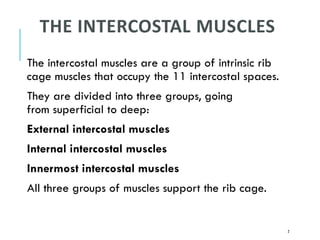
- 1. THE INTERCOSTAL MUSCLES The intercostal muscles are a group of intrinsic rib cage muscles that occupy the 11 intercostal spaces. They are divided into three groups, going from superficial to deep: External intercostal muscles Internal intercostal muscles Innermost intercostal muscles All three groups of muscles support the rib cage. 2
- 2. 3 They are all accessory respiratory muscles that participate in the process of forced breathing. Specifically, the external intercostals facilitate forced inspiration, while the internal and innermost intercostals aid forced expiration There are five muscles that make up the thoracic cage – the intercostals-(external, internal and internal), subcostals and transversus thoracic. The muscles act to change the thoracic volume during respiration. However, there are others attached to thoracic wall but do not constitute the intercostal muscle – pectoralis major, P. minor, serratus anterior and the scalene muscles
- 3. 4 Ext intercostal Mz–fibers run inferoanteriorly Int intercostal Mz –fibers run inferoposteriorly
- 5. TRANSVERSUS THORACIC AND INNERMOST INTERCOSTAL 6
- 6. 7 THE EXTERNAL INTERCOSTAL MUSCLES The intercostal muscles lie in the intercostal spaces between the ribs. They are organized into three layers external intcost. muscle-there are 11 pairs of external Intercostal muscle They run infero-anteriorly from the rib above to the rib below and are continuous with external oblique of the abdomen Attachment Origin: the originated from at the lower border of the rib inserting into the upper of the rib below. Actions- elevate the ribs increasing the thoracic volume Innervation- intercostal nerve T1- T11
- 7. THE INTERNAL INTERCOSTALS These flat muscles lie deep to the external intercostals Run from the rib above to the one below but in an opposite direction, infero-posteriorly, they are continuous with the internal oblique of the abdominal wall Attachment Originate from the lateral edge of the costal groove of the rib and insert into the superior surface of the rib below Actions: interosseous part reduces thoracic volume by depressing the rib cage, the inter-chondral part elevates the rib Innervation- intercostal nerve T1- T11 8
- 8. INNERMOST INTERCOSTALS These muscles are the deepest of the intercostals, have a similar structures to the internal intercostals They are separated from the internal intercostal by the intercostal neurovascular bundle and located in the most lateral portion of the intercostal space Attachment- originate from the medial edge of the costal groove and insert into the superior surface of the rib below Actions: interosseous part reduces thoracic volume by depressing the rib cage, the inter-chondral part elevates the rib Innervation- intercostal nerve T1- T11 9
- 9. TRANSVERSUS THORACIC These muscles are continuous with the transversus thoracic of the abdomen. Attachment- from the posterior surface of the inferior sternum to the internal surfaces of costal cartilages 2-6 Actions: weakly depress the ribs Innervation: Intercostal nerves (T2-T6) 10
- 10. SUBCOSTAL Are found in the various portion of thoracic wall, comprise of thin slips of muscle running from internal surface one rib to that of 2nd-3rd rib below, direction of fibers is parallel to internal intercostal Attachment: this originate from the inf surf of lower rib near the angle of rib and then attach to the sup border of ribs 2-3 below Actions: same as internal intercostals Innervation: intercostal nerves 11
- 11. THE NEUROVASCULAR SPACE The neurovascular space is the plane in which the neurovascular bundle (intercostal vein, artery and nerve) courses. It lies between the internal intercostal and innermost intercostal muscle layers. 12
- 12. 13
- 13. 14
- 14. 15
- 15. ARTERIES OF THORACIC WALL The arterial supply to the thoracic wall. derives from the: Thoracic aorta,- via the posterior intercostal and subcostal arteries. Subclavian artery,- via through the internal thoracic and supreme intercostal arteries. Axillary artery, through the superior and lateral thoracic arteries. 16
- 16. 17
- 17. VEINS OF THORACIC WALL The intercostal veins accompany the intercostal arteries and nerves and lie most superior in the costal grooves . There are 11 posterior intercostal veins and one subcostal vein on each side. The posterior intercostal veins anastomose with the anterior intercostal veins (tributaries of internal thoracic veins). As they approach the vertebral column, the posterior intercostal veins receive a posterior branch, which accompanies the posterior ramus of the spinal nerve of that level, and an intervertebral vein draining the vertebral venous plexuses associated with the vertebral column. 18
- 18. 19 Most posterior intercostal veins (4–11) end in the azygos/hemi-azygos venous system, which conveys venous blood to the superior vena cava (SVC). The posterior intercostal veins of the 1st intercostal space usually enter directly into the right and left brachiocephalic veins. The posterior intercostal veins of the 2nd and 3rd (and occasionally 4th) intercostal spaces unite to form a trunk, the superior intercostal vein
- 19. NERVES OF THORACIC WALL The 12 pairs of thoracic spinal nerves supply the thoracic wall. As soon as they leave the IV foramina in which they are formed, the mixed thoracic spinal nerves divide into anterior and posterior (primary) rami or branches The anterior rami of nerves T1–T11 form the intercostal nerves that run along the extent of the intercostal spaces. The anterior ramus of nerve T12, coursing inferior to the 12th rib, is the subcostal nerve. The posterior rami of thoracic spinal nerves pass posteriorly, immediately lateral to the articular processes of the vertebrae, to supply the joints, deep back muscles, and skin of the back in the thoracic region. 20
- 20. 21 LYMPHATIC DRAINAGE OF THE THORAX A. Sternal or Parasternal (Internal Thoracic) Nodes -Are placed along the internal thoracic artery. -Receive lymph from the medial portion of the breast, intercostal spaces, diaphragm, and supraumbilical region of the abdominal wall. Drain into the junction of the internal jugular and subclavian veins. B. Intercostal Nodes Lie near the heads of the ribs. Receive lymph from the intercostal spaces and the pleura. Drain into the cisterna chyli or the thoracic duct. C. Phrenic Nodes - Lie on the thoracic surface of the diaphragm. -Receive lymph from the pericardium, diaphragm, and liver. -Drain into the sternal and posterior mediastinal nodes-
- 21. APPLIED ANATOMY/CLINCAL CORRELATES 1. Chest Pain -Although chest pain can result from pulmonary disease, it is probably the most important symptom of cardiac disease (Swartz, 2009). However, chest pain may also occur in intestinal, gallbladder, and musculoskeletal disorders. When evaluating a patient with chest pain, the examination is largely concerned with discriminating between serious conditions and the many minor causes of pain. People who have had a heart attack usually describe the associated pain as a “crushing” sub- sternal pain (deep to the sternum) that does not disappear with rest. 22
- 22. 23 Rib Fractures The short, broad 1st rib, posteroinferior to the clavicle, is rarely fractured because of its protected position (it cannot be palpated). When it is broken, however, structures crossing its superior aspect may be injured, including the brachial plexus of nerves and subclavian vessels that serve the upper limb. The middle ribs are most commonly fractured. Rib fractures usually result from blows or crushing injuries. The weakest part of a rib is just anterior to its angle; however, direct violence may fracture a rib anywhere, and its broken end may injure internal organs such as a lung and/or the spleen. Fractures of the lower ribs may tear the diaphragm and result in a diaphragmatic hernia. Rib fractures are painful because the broken parts move during respiration, coughing, laughing, and sneezing.
- 23. 24 Flail Chest Multiple rib fractures may allow a sizable segment of the anterior and/or lateral thoracic wall to move freely. The loose segment of the wall moves paradoxically (inward on inspiration and outward on expiration). Flail chest is an extremely painful injury and impairs ventilation, thereby affecting oxygenation of the blood. During treatment, the loose segment may be fixed by hooks and/or wires so that it cannot move. Rib Excision The surgical creation of an opening through the thoracic wall to enter a pleural cavity is a thoracotomy. An anterior thoracotomy may involve making H-shaped cuts through the perichondrium of one or more costal cartilages and then shelling out segments of costal cartilage to gain entrance to the thoracic cavity.
- 24. 25 Supernumerary Ribs People usually have 12 ribs on each side, but the number is increased by the presence of cervical and/or lumbar ribs, or decreased by failure of the 12th pair to form. Cervical ribs are relatively common (0.5–2%) and may interfere with neurovascular structures exiting the superior thoracic aperture. Lumbar ribs are less common. Supernumerary (extra) ribs also have clinical significance in that they may confuse the identification of vertebral levels in radiographs and other diagnostic images.
- 25. STERNAL ANGLE (ANGLE OF LOUIS) ■ Is the junction between the manubrium and the body of the sternum. ■ Is located at the level where (a) The second ribs articulate with the sternum. (b) The aortic arch begins and ends. (c) The trachea bifurcates into the right and left bronchi at the carina. (d) The inferior border of the superior mediastinum is demarcated. (e) A transverse plane can pass through the intervertebral disk between T4 and T5. 26
- 26. 27 ASSIGNMENT Identify the structures No- 10, 11, 14, 16, 29 and 35 The part of the rib protecting the vessels and nerve is ……………… The part of rib where fractures commonly occur is…………….... Why is the first rib not commonly fractured. How is the rib counted in surgery